A&P 2: Lab Practical 2
1/271
There's no tags or description
Looks like no tags are added yet.
Name | Mastery | Learn | Test | Matching | Spaced | Call with Kai |
|---|
No analytics yet
Send a link to your students to track their progress
272 Terms

1
nasal cavity
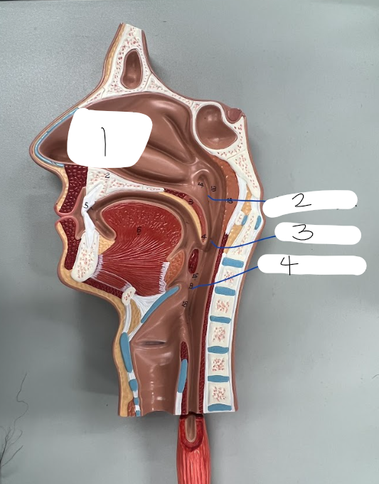
2
nasopharynx

3
oropharynx

4
laryngopharynx

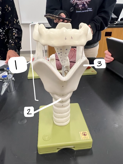
what model is this?
trachea model

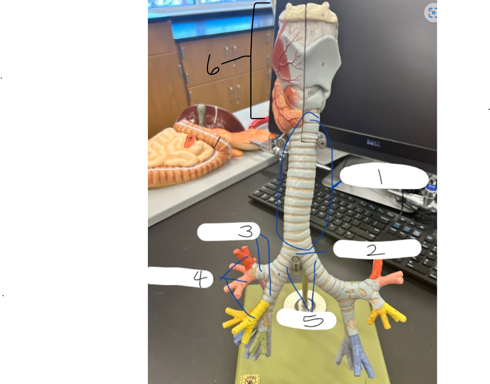
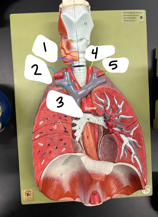
1
trachea

2
carnia

3
secondary/lobar bronchi

4
tertiary/segmental bronchi
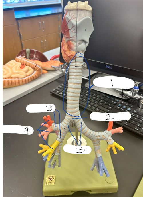
5
primary/main bronchi
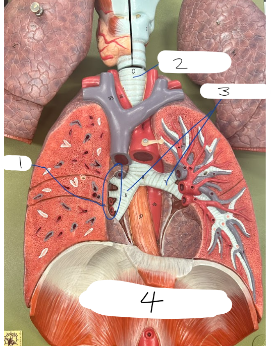
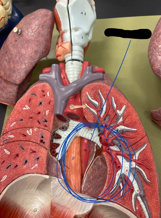
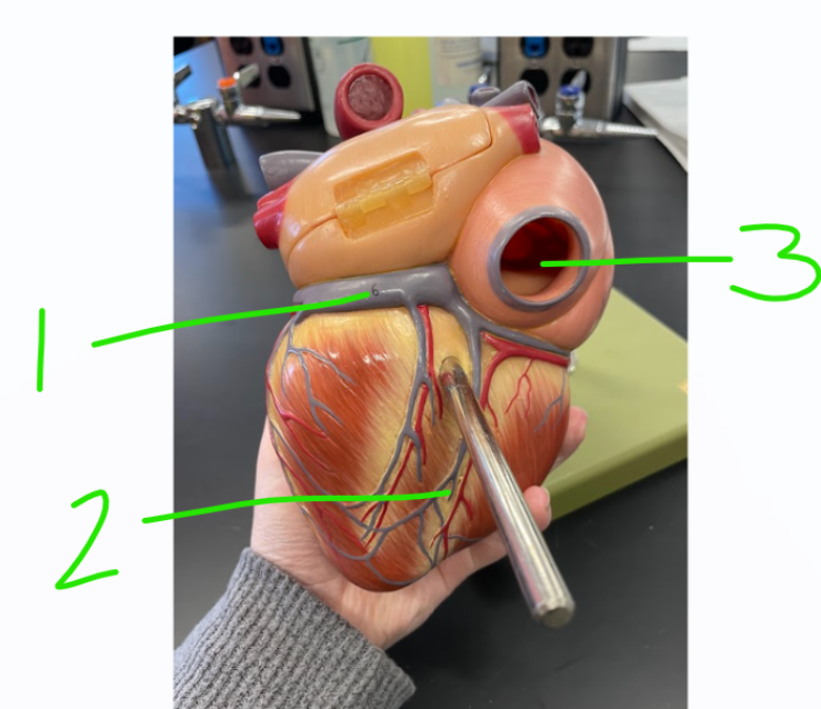
1
lung hilum

2
trachea

3
main/primary bronchi

4
diaphragm
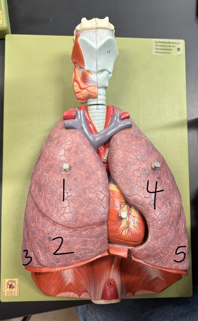
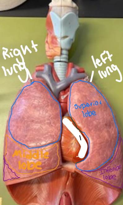
1
superior lobe of right lung

2
middle lobe of right lung

3
inferior lobe of right lung

4
superior lobe of left lung

5
inferior lobe of left lung
cardiac notch/impression
1 (name of the model)
larynx
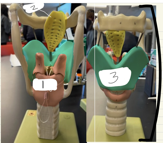
1
cricoid cartilage

3 (green)
thyroid cartilage
1
epiglottis

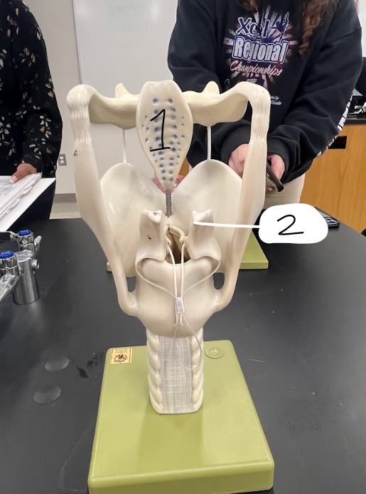
2
corniculate cartilages
6
larynx

3
vestibular fold

4
vocal cord
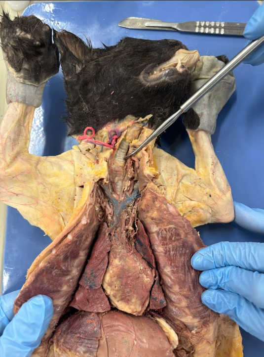
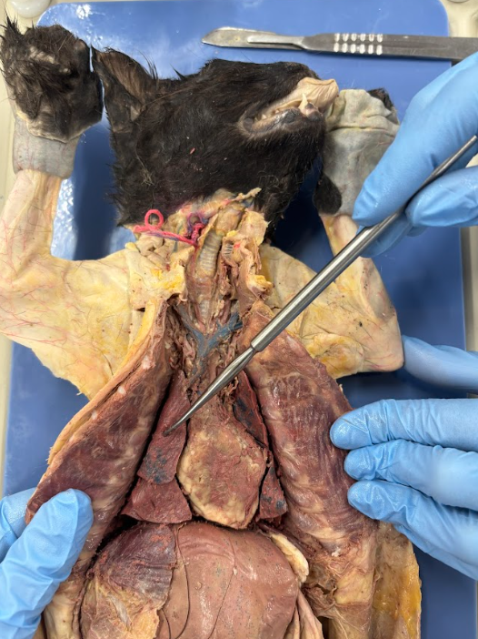
what is the probe pointing to?
trachea

what is the probe pointing to?
larynx
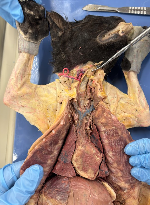
what is the large plate of cartilage you can feel here?
thyroid cartilage
what is the probe pointing to?
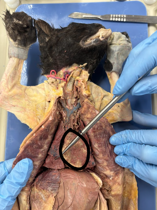
superior lobe of right lung
what is the probe pointing to?
diaphragm
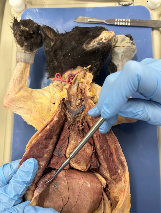
what is circled in the middle?
heart

the lungs are surrounded by a special double layered tissue separating the lungs from other structures in the thoracic cavity called what? (ignore probe)
pleurae

the pleurae is made of 2 layers: which one is very thin layer found on the surface of the lungs? (ignore probe)
visceral pleura

the pleurae is made of 2 layers: which one is a shiny layer found on the inside of the bones and muscles of the thoracic cage? (ignore probe)
parietal pleura

there is a small space between the visceral pleura and the parietal pleura called ___________ and filled with ____________. (ignore probe)
the pleural cavity; serous fluid
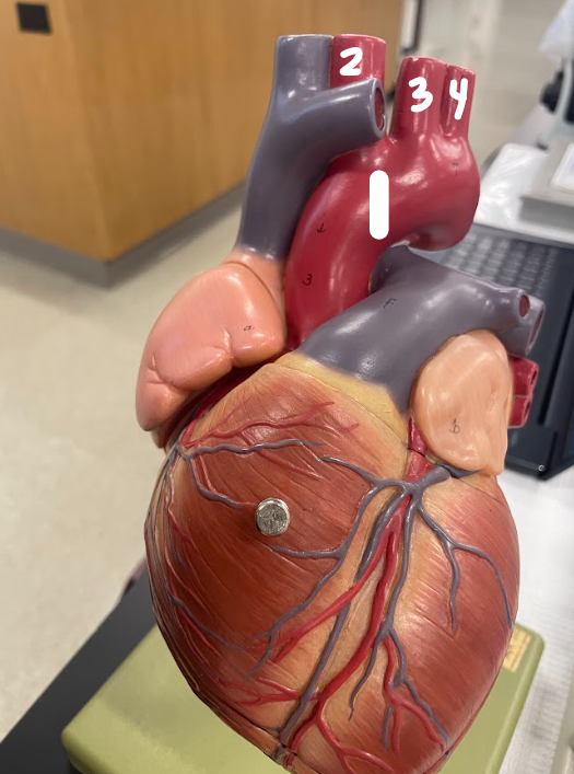
1
right common carotid artery

2
right subclavian artery

3
brachiocephalic trunk

4
left common carotid artery

5
left subclavian artery
2
brachiocephalic trunk

3
left common carotid artery

4
left subclavian artery

5
superior vena cava
3
inferior vena cava
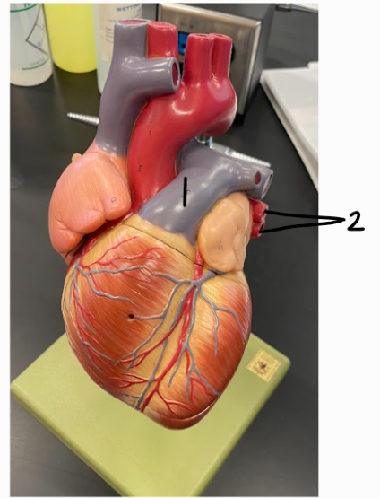
1
pulmonary trunk

2
pulmonary veins
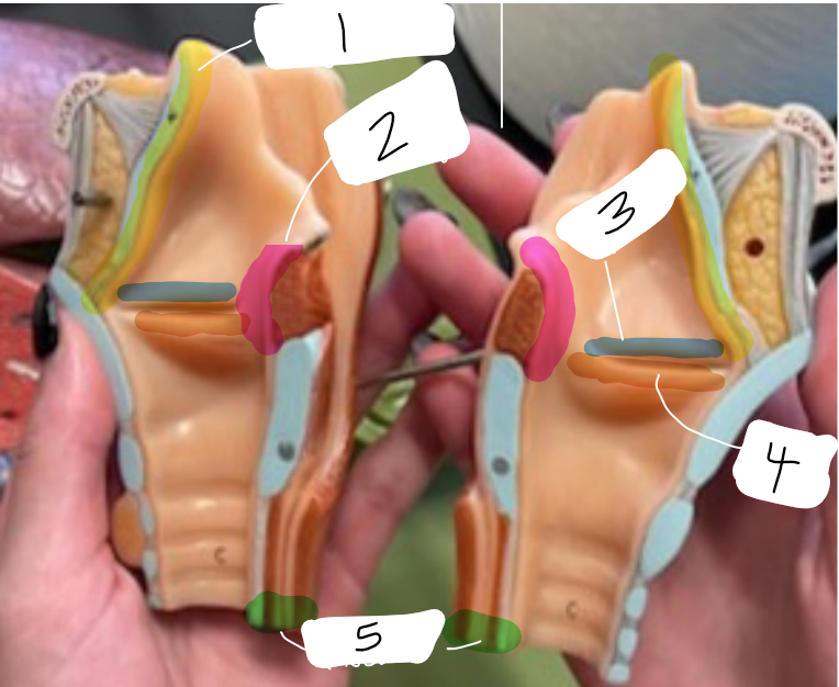
1
epiglottis

2
arytenoid cartilages

5
trachealis muscle
1
cardiac notch

1
internal carotid artery

2
cerebral arterial circle/circle of willis
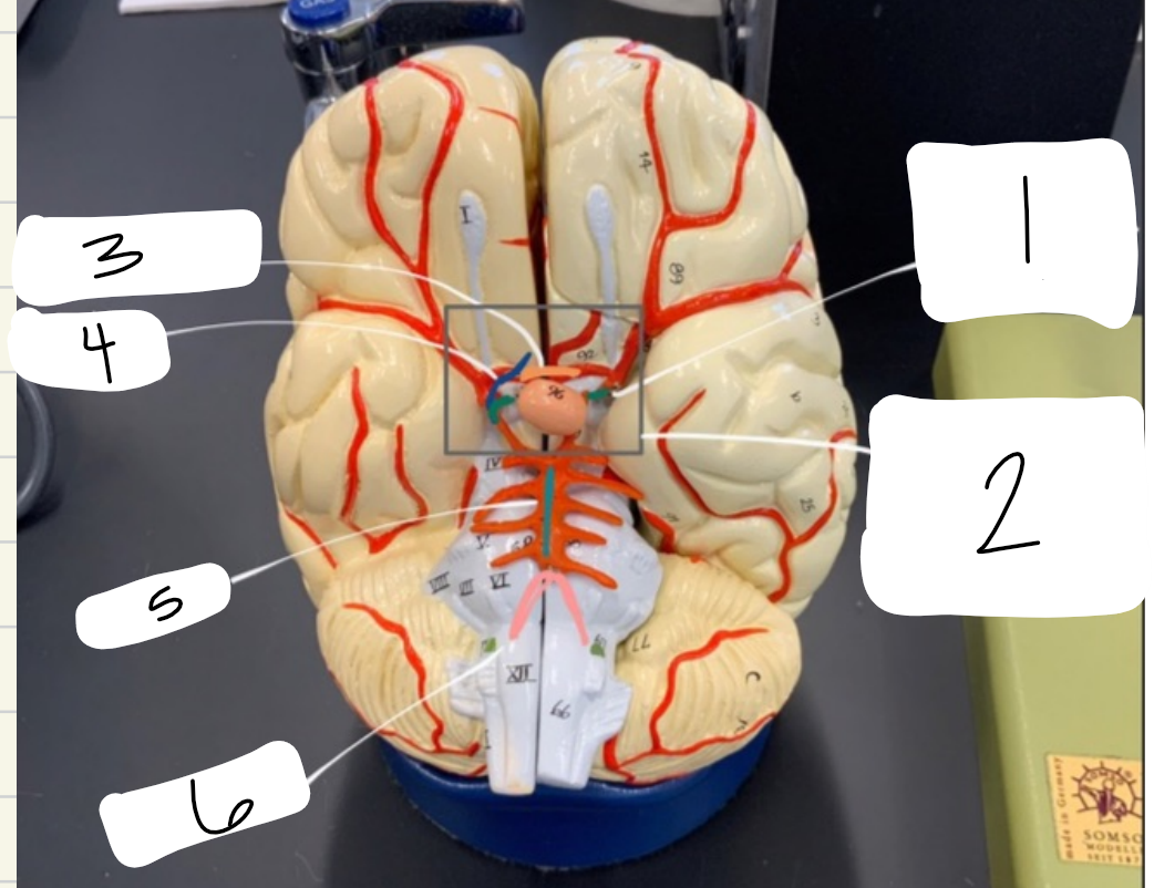
5
basilar artery

6
vertebral arteries

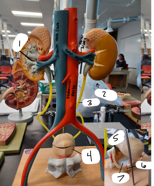
1
inferior vena cava
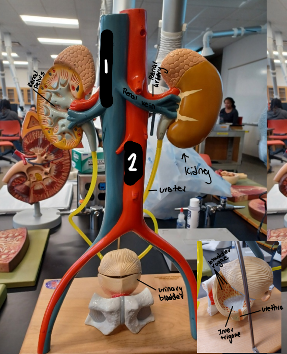
2
abdominal aorta

1
left renal vein

2
right renal vein

3
left renal artery
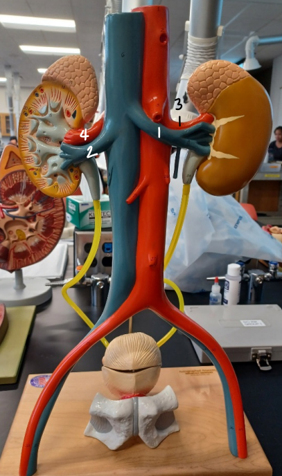
4
right renal artery

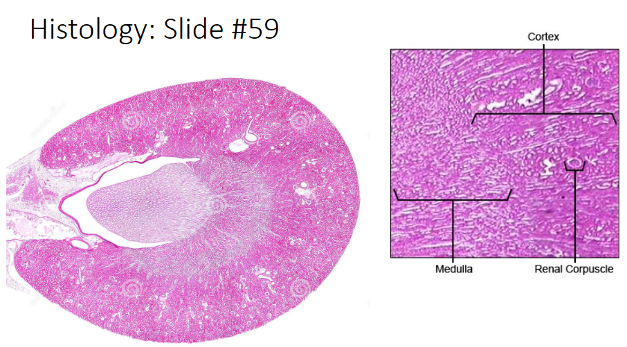
what is this histology slide of?
human kidney

1
renal cortex
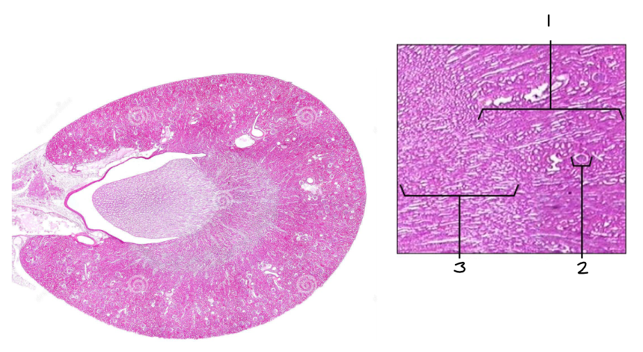
2
renal corpuscle

3
renal medulla

what is this histology slide of?
mammal bladder

1
transitional epithelium
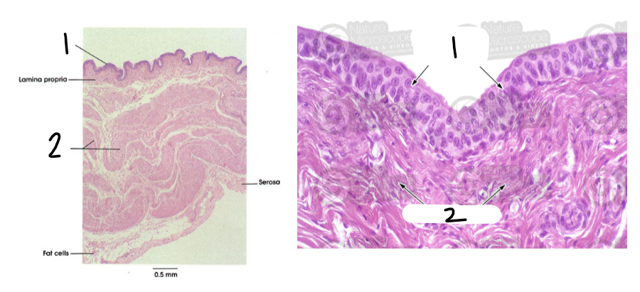
2
detrusor (smooth muscle that contracts the urinary bladder)
longish white lines in the medulla
collecting ducts
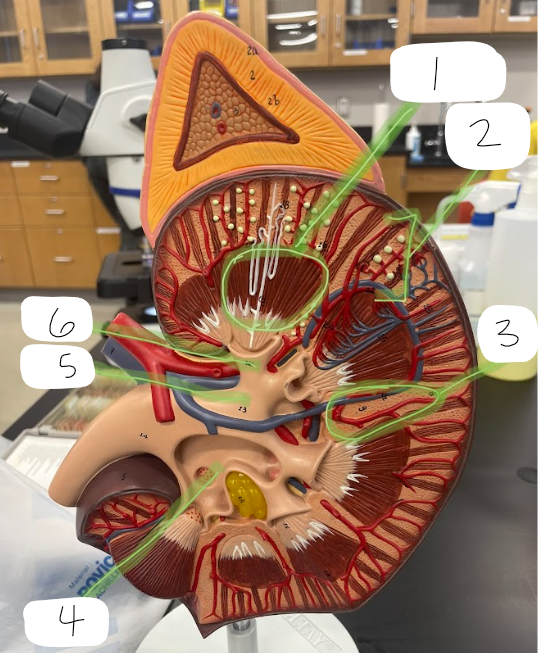
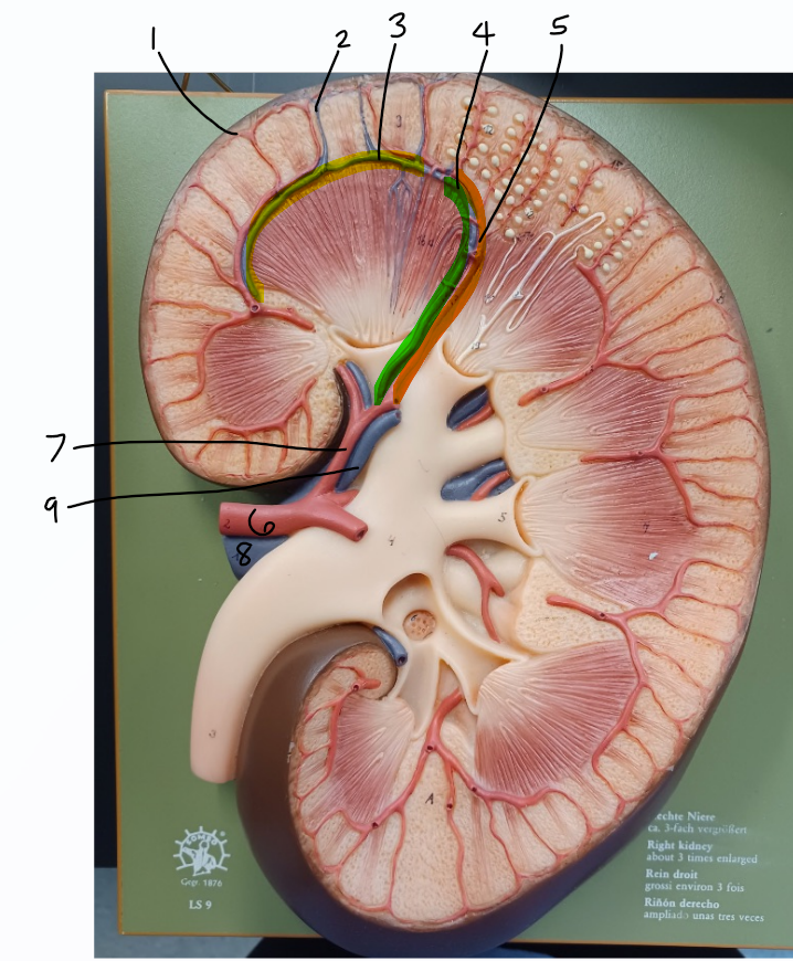
1
renal pyramids

2
renal cortex

3
renal columns

4
renal pelvis

5
major calyx

6
minor calyx
yellow highlight
renal medulla
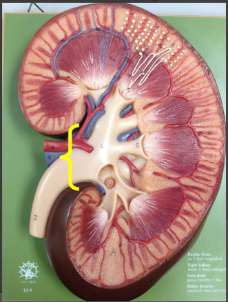
renal hilum

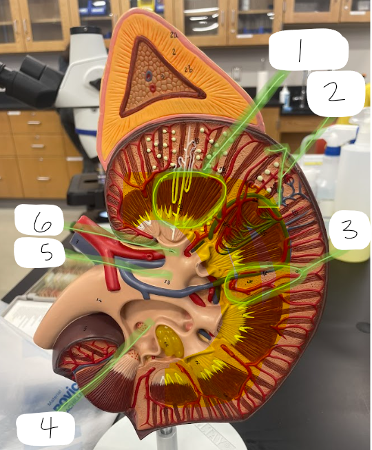
1
cortical radiate artery

2
cortical radiate vein

3
arcuate vein

4
interlobar vein

5
interlobar artery

6
renal artery

7
segmental artery
8
renal vein
9
renal papillae
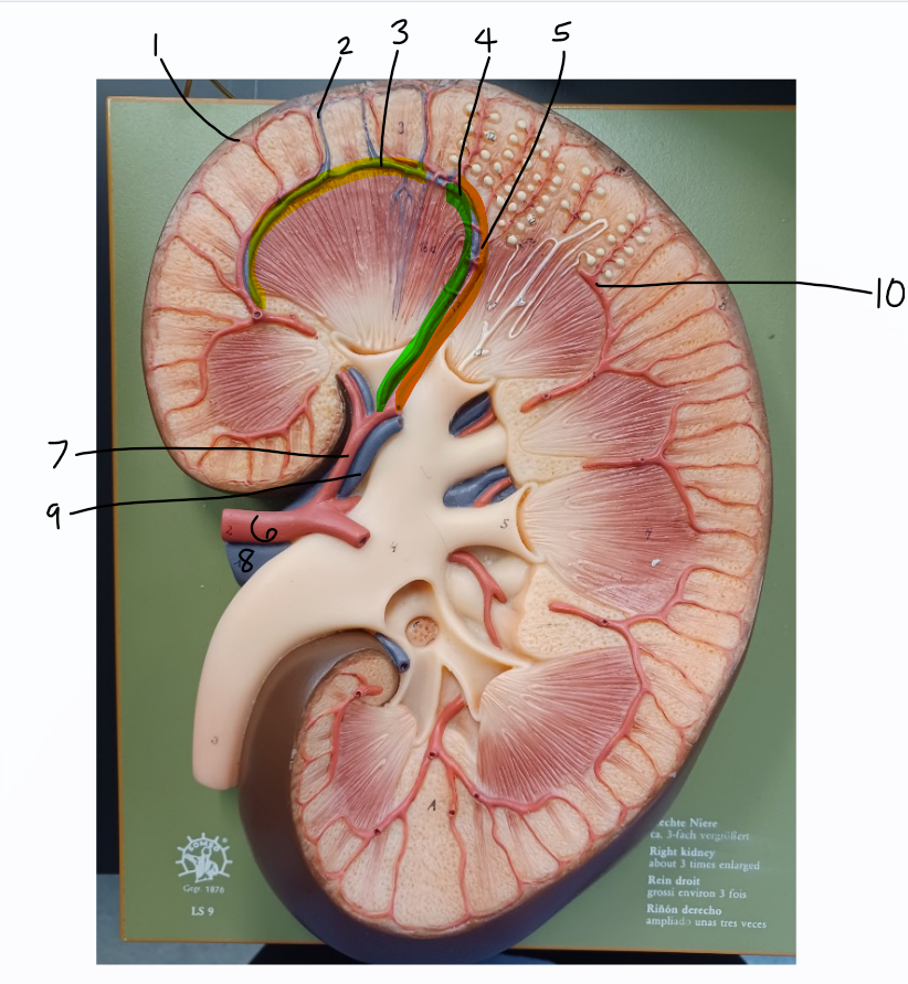
10
arcuate arteries
1
renal pelvis

2
kidney

3
ureter

4
urinary bladder

5
bladder rugae

6
urethra

7
trigone
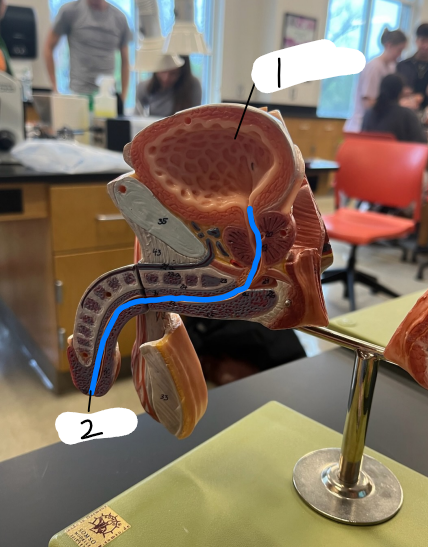
1
male urinary bladder

2
male urethra
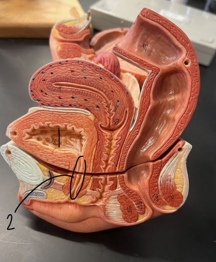
1
female urinary bladder