Bacterial Cell Wall Structure and Composition: Gram-Positive, Gram-Negative, and Acid-Fast Bacteria L2
1/84
There's no tags or description
Looks like no tags are added yet.
Name | Mastery | Learn | Test | Matching | Spaced |
|---|
No study sessions yet.
85 Terms
What is the primary function of the bacterial cell wall?
To provide shape and structure to bacterial cells and protect them from changes in osmotic pressure.
What is the major component of bacterial cell walls?
Peptidoglycan.
What is peptidoglycan also known as?
Murein or mucopeptide.
How does peptidoglycan protect bacteria?
It provides protection from osmotic lysis due to its high tensile strength.
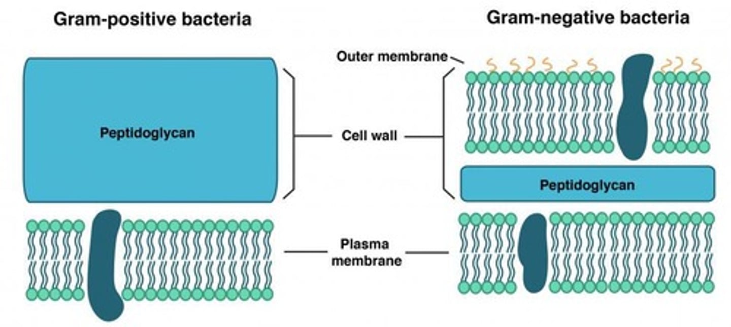
What distinguishes gram-positive bacteria from gram-negative bacteria regarding their cell wall?
Gram-positive bacteria have a thick peptidoglycan layer (approximately 40 layers thick), while gram-negative bacteria have a thin peptidoglycan layer (approximately 1-2 layers thick).
What unique feature do mycoplasmas have regarding their cell wall?
Mycoplasmas are the only bacteria that lack a cell wall and contain sterols in their cytoplasmic membranes.
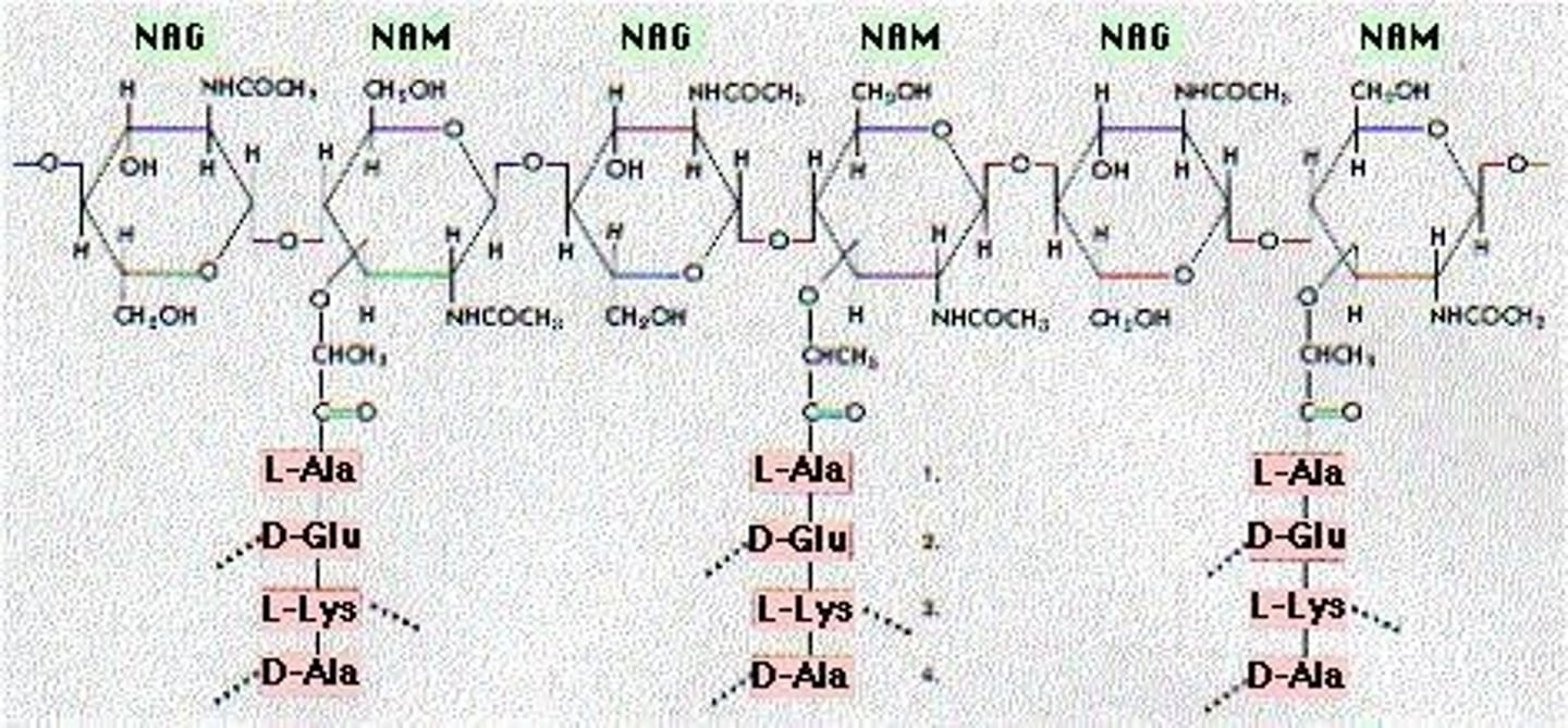
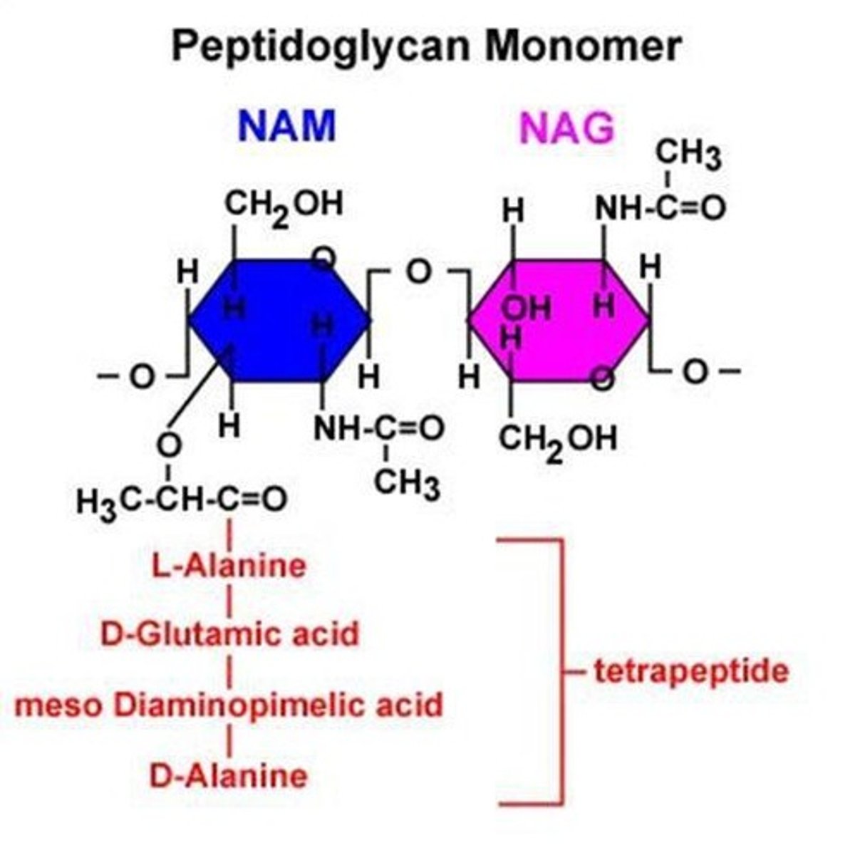
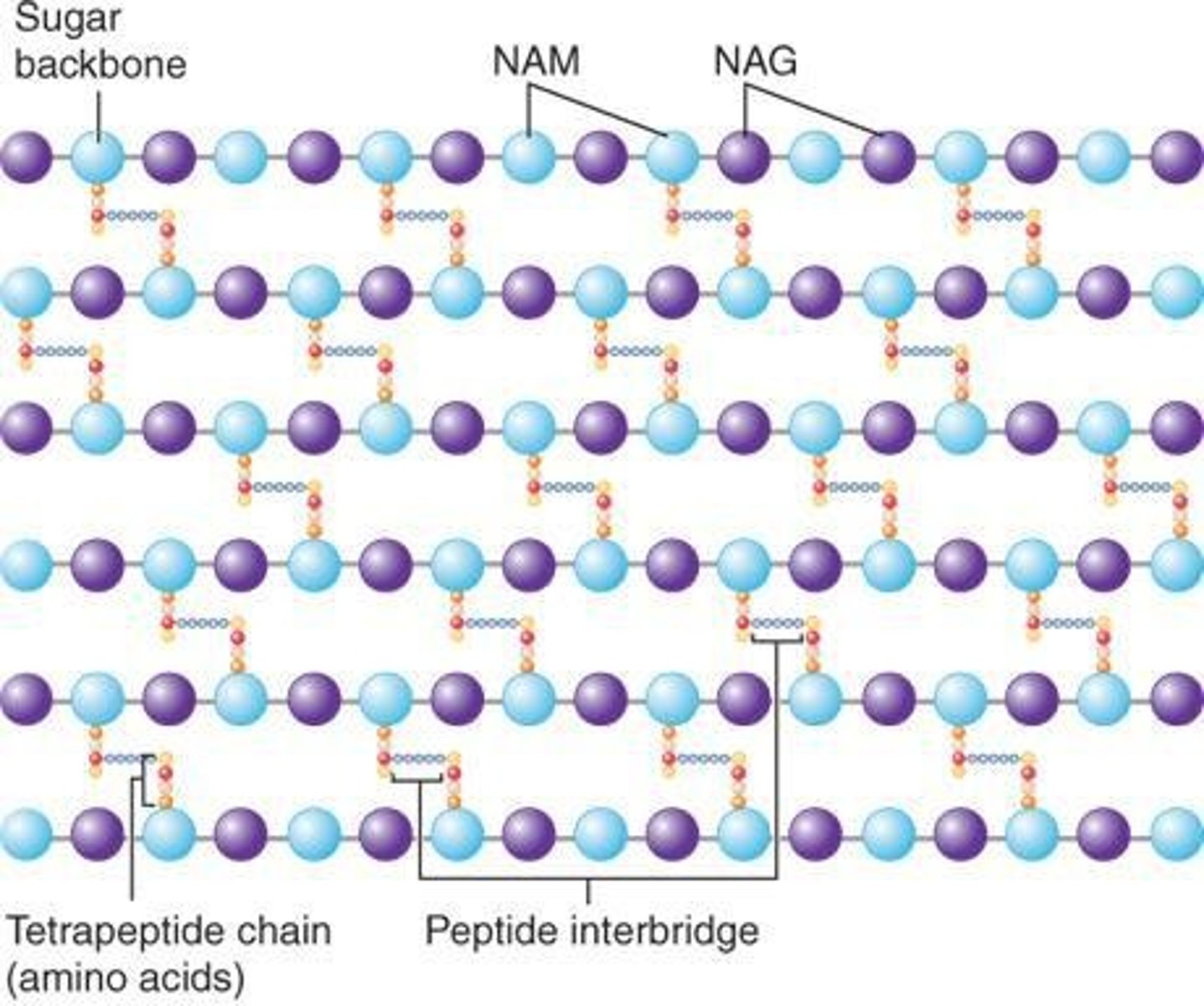
What are the two amino sugars that compose peptidoglycan?
N-acetylglucosamine (NAG) and N-acetylmuramic acid (NAM).
How are NAG and NAM linked in peptidoglycan?
They are linked by β 1-4 linkages forming chains.
What role do diamino amino acids play in peptidoglycan structure?
Diamino amino acids, such as lysine and diaminopimelic acid, are essential for the cross-linking of the peptidoglycan chain.
What is the significance of the mesh-like structure of peptidoglycan?
It gives tremendous strength to the cell wall and is essential for bacterial survival.
What is the clinical relevance of the lipopolysaccharide (LPS) layer in gram-negative bacteria?
The LPS layer is important for eliciting innate immune responses and is unique to gram-negative bacteria.
What is the protective function of the bacterial cell wall against osmotic pressure?
It prevents the cell from bursting or collapsing due to changes in osmotic pressure.
What is the structure of peptidoglycan in terms of its composition?
Peptidoglycan consists of long sugar chains joined by peptide cross-links, forming a mesh-like exoskeleton.
What is the role of peptidoglycan in bacterial cells?
It provides structural integrity and protection, allowing for the diffusion of metabolites to the plasma membrane.
How does the cell wall of bacteria elicit immune responses?
The repetitive structures of the cell wall components bind to pathogen pattern receptors (PRR) on human cells.
What is the significance of the term 'hallmark' in relation to peptidoglycan?
Peptidoglycan is considered a hallmark of true bacteria (eubacteria) because it is found only in bacteria.
What is the relationship between the cell wall and the cytoplasmic membrane in bacteria?
The peptidoglycan forms a mesh-like layer surrounding the cytoplasmic membrane.
What is the importance of the porous nature of peptidoglycan?
It allows for the diffusion of metabolites to the plasma membrane.
What is the overall structure of the bacterial cell wall?
It is a rigid structure that provides shape, protects against osmotic pressure, and is composed of peptidoglycan.
What are the clinical implications of understanding bacterial cell wall structure?
Knowledge of cell wall structure is critical for developing antibiotics and understanding bacterial pathogenesis.
What are the two types of bacteria compared in terms of cell envelope composition?
Gram-positive and gram-negative bacteria.
What are the main components of peptidoglycan?
Peptidoglycan is composed of a glycan chain (N-acetylmuramic acid (NAM) and N-acetylglucosamine (NAG)), a tetrapeptide chain, and a cross-link (peptide interbridge only in gram-positive bacteria).
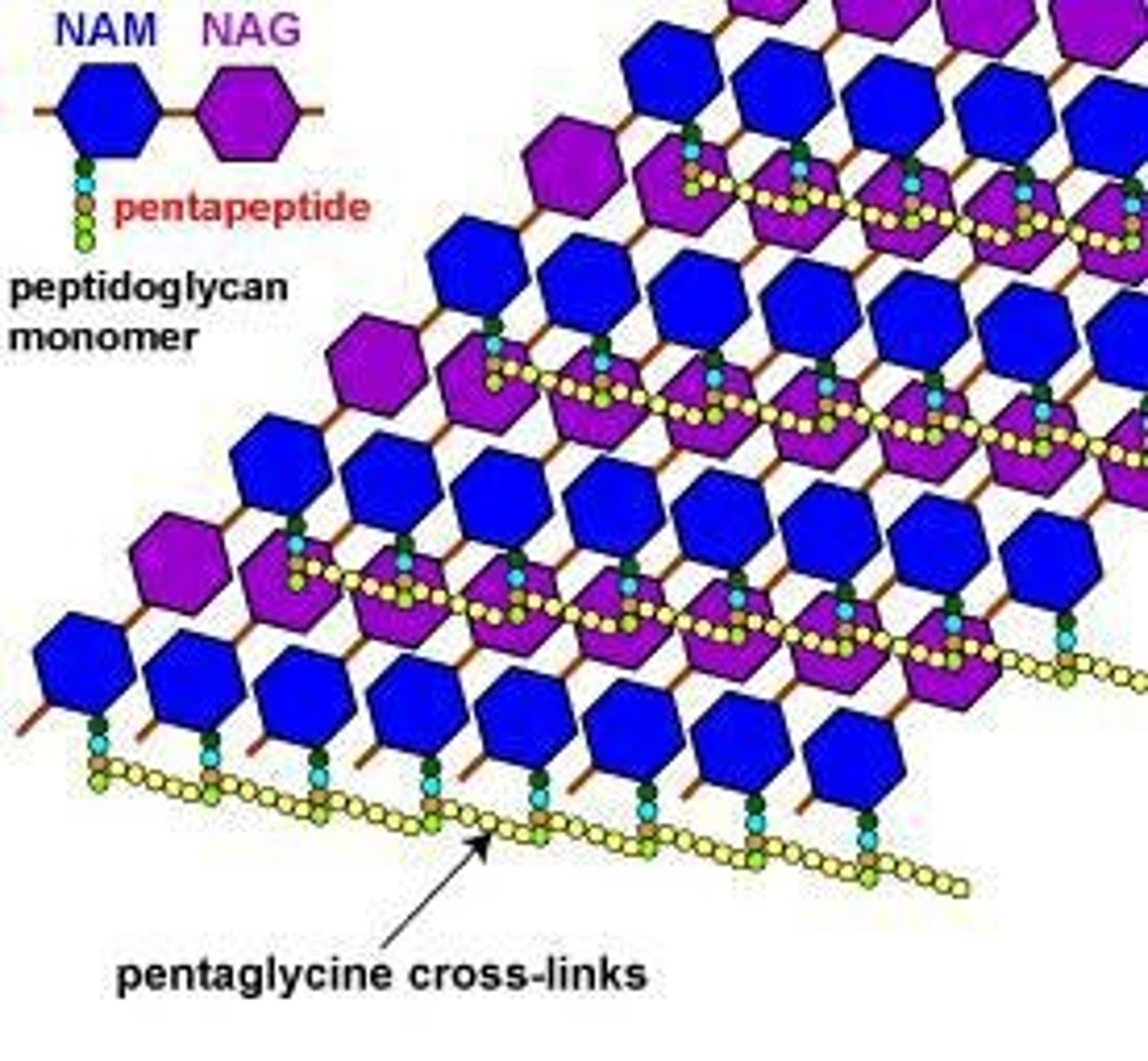
How is the peptidoglycan structure characterized in gram-positive bacteria?
In gram-positive bacteria, the peptidoglycan forms a multilayered, three-dimensional structure.
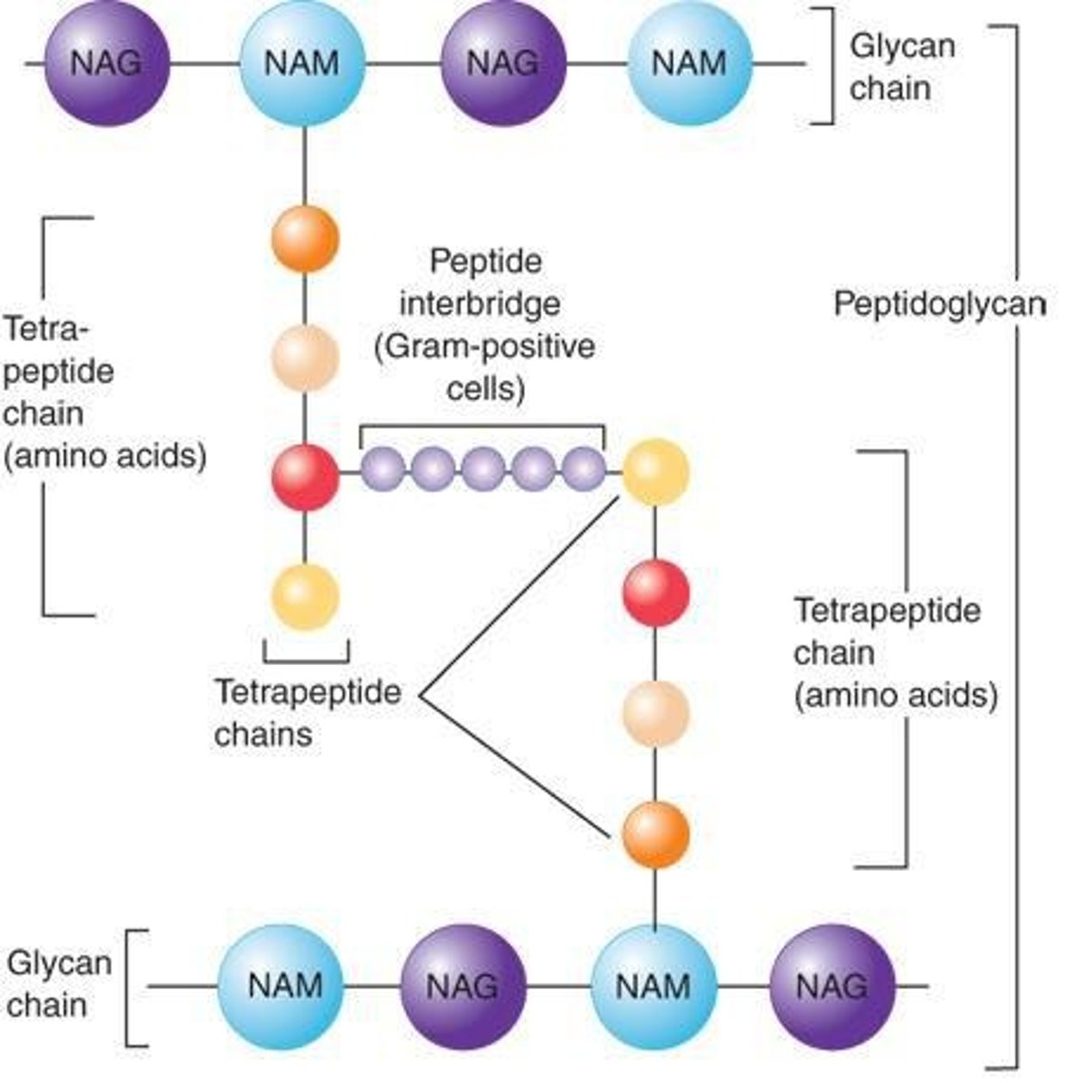
What is the difference in the peptidoglycan structure between gram-positive and gram-negative bacteria?
Gram-positive bacteria have a peptide interbridge in their peptidoglycan, while gram-negative bacteria do not.
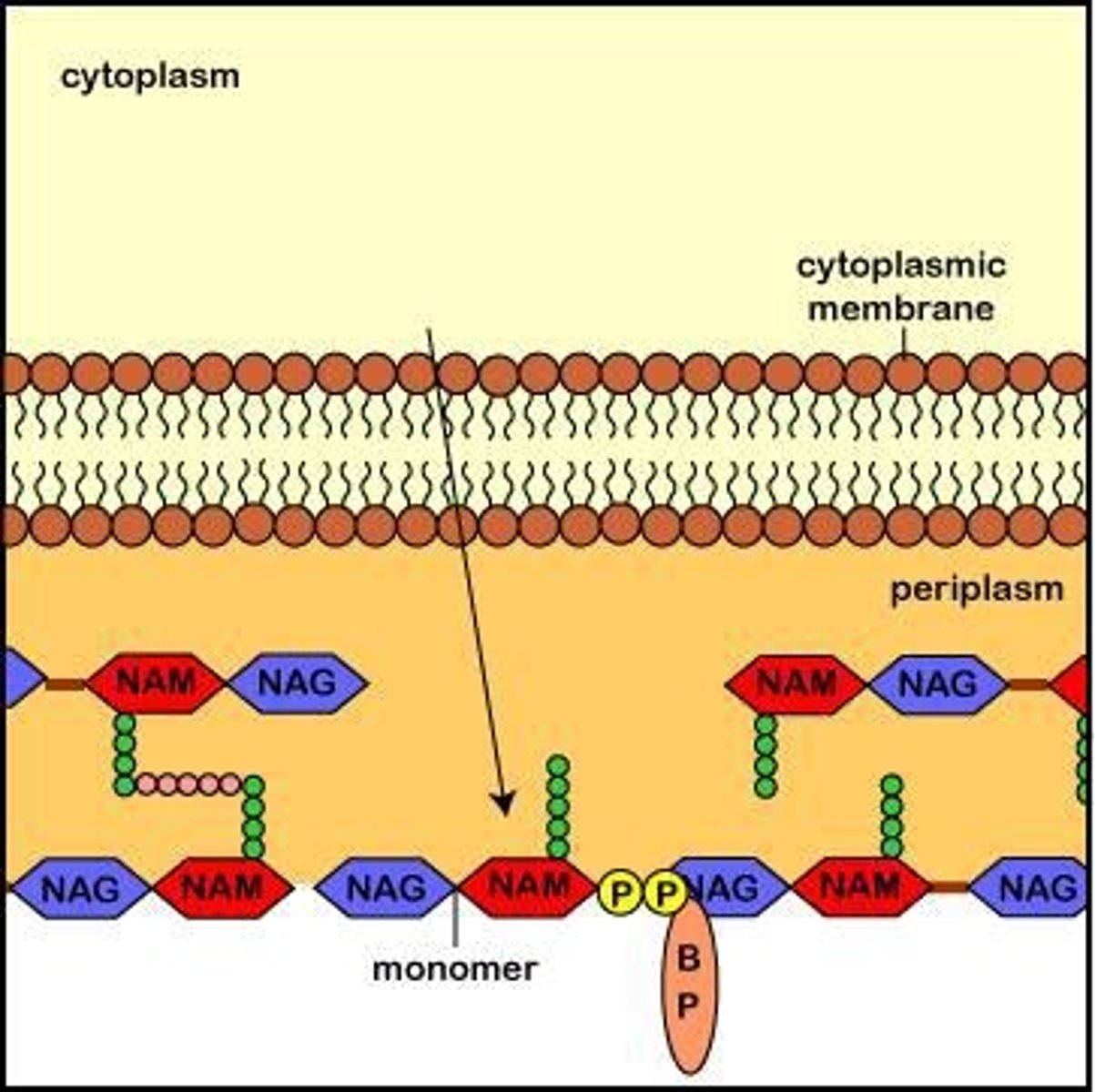
Where does peptidoglycan biosynthesis occur within the bacterial cell?
Peptidoglycan biosynthesis occurs in three compartments: the cytoplasm, cell membrane, and cell wall.
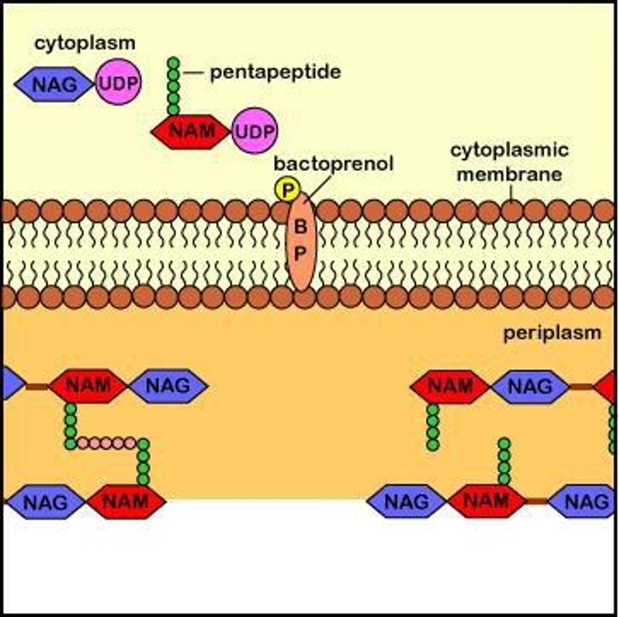
What role does bactoprenol play in peptidoglycan biosynthesis?
Bactoprenol (undecaprenol) transports peptidoglycan monomers across the cytoplasmic membrane.
What happens to peptidoglycan monomers after they are transported by bactoprenol?
The monomers are inserted into the growing peptidoglycan chains in the cell wall and cross-linked to complete synthesis.
What is the function of lysozyme in relation to peptidoglycan?
Lysozyme cleaves the glycan backbone of peptidoglycan by breaking the β-1,4 disaccharide link, creating gaps for new monomers to be added.
How is N-acetylglucosamine (NAG) activated during peptidoglycan biosynthesis?
NAG is activated by attaching uridine diphosphate (UDP) to form UDP-NAG.
What is the process of forming UDP-NAM from UDP-NAG?
Some UDP-NAG is enzymatically converted to N-acetylmuramic acid (NAM), forming UDP-NAM.
What is the structure formed after the addition of five amino acids to UDP-NAM?
A pentapeptide is formed after five amino acids are sequentially added to UDP-NAM.
What is the significance of the pyrophosphate link in peptidoglycan biosynthesis?
The activated NAM-pentapeptide attaches to bactoprenol through a pyrophosphate link, supplying energy for the process.
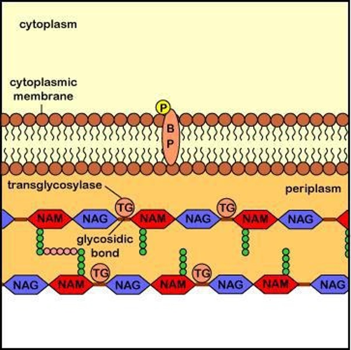
What occurs during transglycosylation in peptidoglycan biosynthesis?
Transglycosylase enzymes catalyze the formation of glycosidic bonds between NAM and NAG of the peptidoglycan monomers and the existing peptidoglycan.
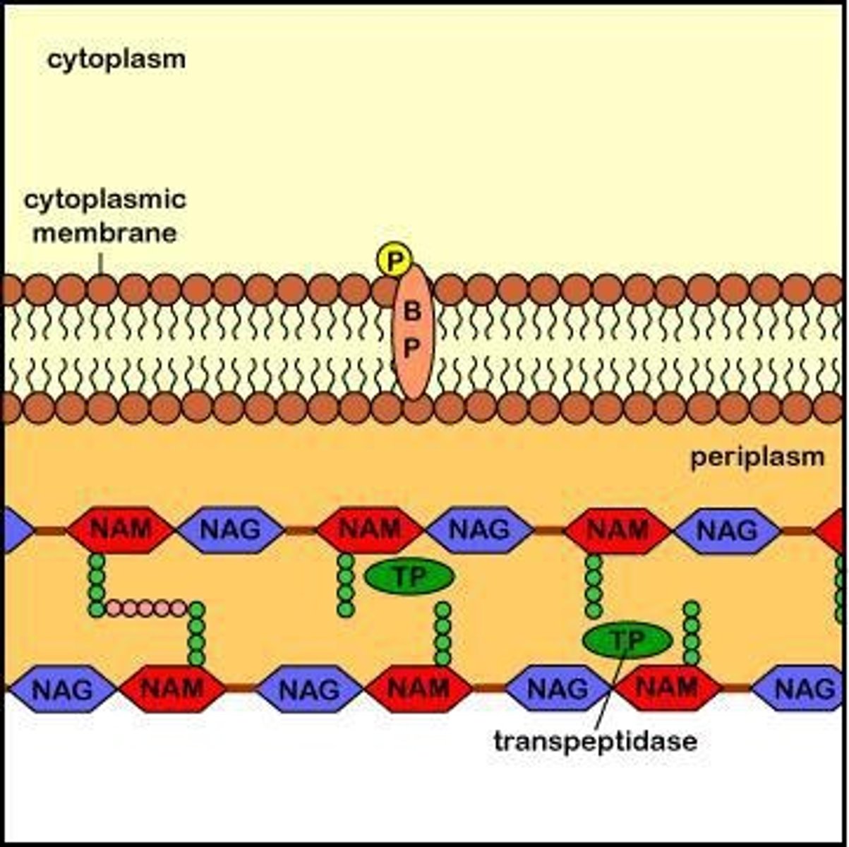
What is the role of transpeptidation in peptidoglycan biosynthesis?
Transpeptidation involves cross-linking carried out by transpeptidases, which are also known as penicillin-binding proteins (PBPs).
How do β-lactam antibiotics affect peptidoglycan biosynthesis?
Penicillins and other β-lactam antibiotics bind to transpeptidase enzymes, blocking the formation of peptide cross-links.
What is the effect of bacitracin on peptidoglycan biosynthesis?
Bacitracin binds to bactoprenol after it inserts the peptidoglycan monomer, preventing its dephosphorylation and subsequent transport across the membrane.
What is the function of autolysins in peptidoglycan biosynthesis?
Autolysins create gaps in the peptidoglycan layer to allow for the insertion of newly formed monomers.
What is the final step after bactoprenol inserts the peptidoglycan monomer?
After insertion, pyrophospho-bactoprenol is converted back to phospho-bactoprenol and recycled.
What is the role of transpeptidases in the cell wall?
Transpeptidases facilitate the cross-linking of peptidoglycan layers, which is essential for cell wall integrity.
Where is lysozyme found in the human body?
Lysozyme is found in human tears and mucus.
What is the significance of the energy supplied by UDP in peptidoglycan biosynthesis?
The energy from one of the high-energy phosphate groups of UDP is used to attach the NAM-pentapeptide to bactoprenol.
What are the two main enzymatic processes involved in peptidoglycan biosynthesis?
Transglycosylation and transpeptidation are the two main processes involved.
What results from a weak cell wall in bacteria?
Osmotic lysis of the bacterium.
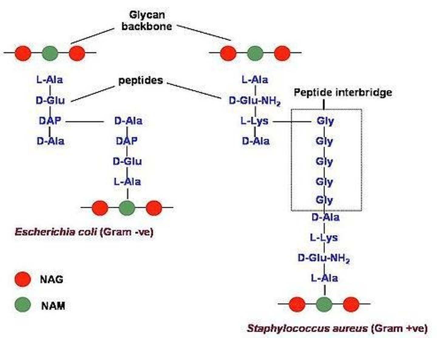
What amino acids compose the pentapeptide in the peptidoglycan monomer of gram-positive bacteria?
L-alanine, D-glutamine, L-lysine, and two D-alanines.
How is the peptide cross-link formed in gram-positive bacteria?
By formation of a short peptide interbridge consisting of 5 glycines.
What happens to the terminal D-alanine during the formation of the tetrapeptide in gram-positive bacteria?
It is cleaved from the pentapeptide.
Give an example of a gram-positive bacterium.
Staphylococcus aureus (S. aureus).
What amino acids compose the pentapeptide in the peptidoglycan monomer of gram-negative bacteria?
L-alanine, D-glutamic acid, meso-diaminopimelic acid, and two D-alanines.
How is the peptide cross-link formed in gram-negative bacteria?
Between the diaminopimelic acid of one peptide chain and the D-alanine of another.
What happens to the terminal D-alanine during the formation of the tetrapeptide in gram-negative bacteria?
It is cleaved from the pentapeptide.
Give an example of a gram-negative bacterium.
Escherichia coli (E. coli).
What characterizes the gram-positive cell wall structure?
A dense layer of numerous rows of peptidoglycan, teichoic acids, lipoteichoic acids, and surface proteins.
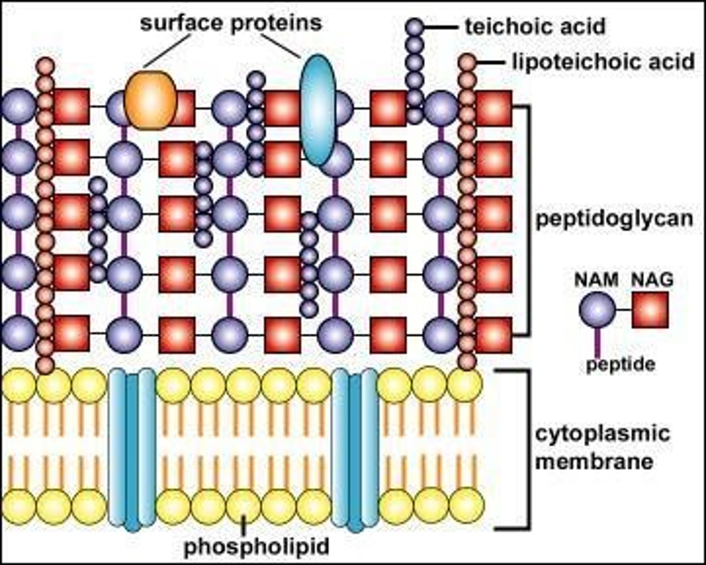
What are teichoic acids and their function in gram-positive bacteria?
Polymers of glycerol phosphate and ribitol phosphate that extend through the cell wall, covalently bound to peptidoglycan.
What are lipoteichoic acids (LTA) and their role?
Teichoic acids covalently bound to cytoplasmic membrane lipids that can bind to pathogen pattern receptors and initiate innate immune responses.
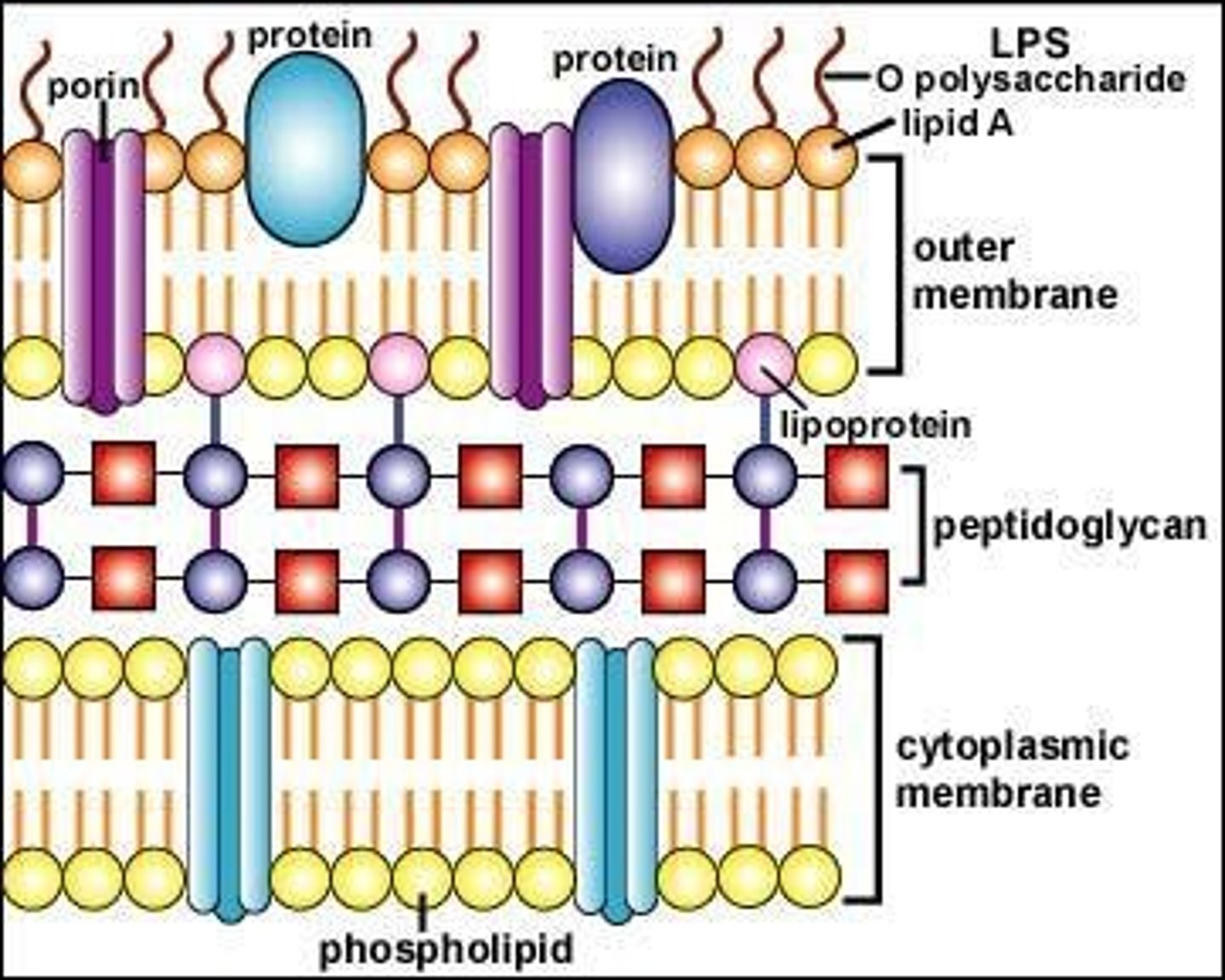
What is the composition of the gram-negative cell wall?
A thin inner layer of peptidoglycan and an outer membrane made of phospholipids, lipopolysaccharide (LPS), lipoproteins, and proteins.
What is the periplasmic space in gram-negative bacteria?
The space between the cytoplasmic membrane and the outer membrane containing hydrolytic enzymes and transport proteins.
What is unique about the outer membrane of gram-negative bacteria?
It is an asymmetric bilayer composed of phospholipids, lipoproteins, LPS, and proteins.
What is the function of lipoproteins in the outer membrane of gram-negative bacteria?
They covalently connect the outer membrane to the peptidoglycan.
What is the role of porins in the outer membrane of gram-negative bacteria?
Porins are pore-forming proteins that function as channels for the entry and exit of chemicals and nutrients.
How do porins restrict the entry of substances in gram-negative bacteria?
They restrict entry of large hydrophilic and hydrophobic molecules, including many antimicrobials.
What is the diameter of the pores formed by trimeric porins?
1 nm.
What is the significance of the outer membrane in maintaining bacterial structure?
It acts as a permeability barrier to large molecules.
What are the two major porins found in Gram-negative bacteria?
OmpC and OmpF.
What is the outer membrane of Gram-negative bacteria primarily composed of?
Lipopolysaccharide (LPS).
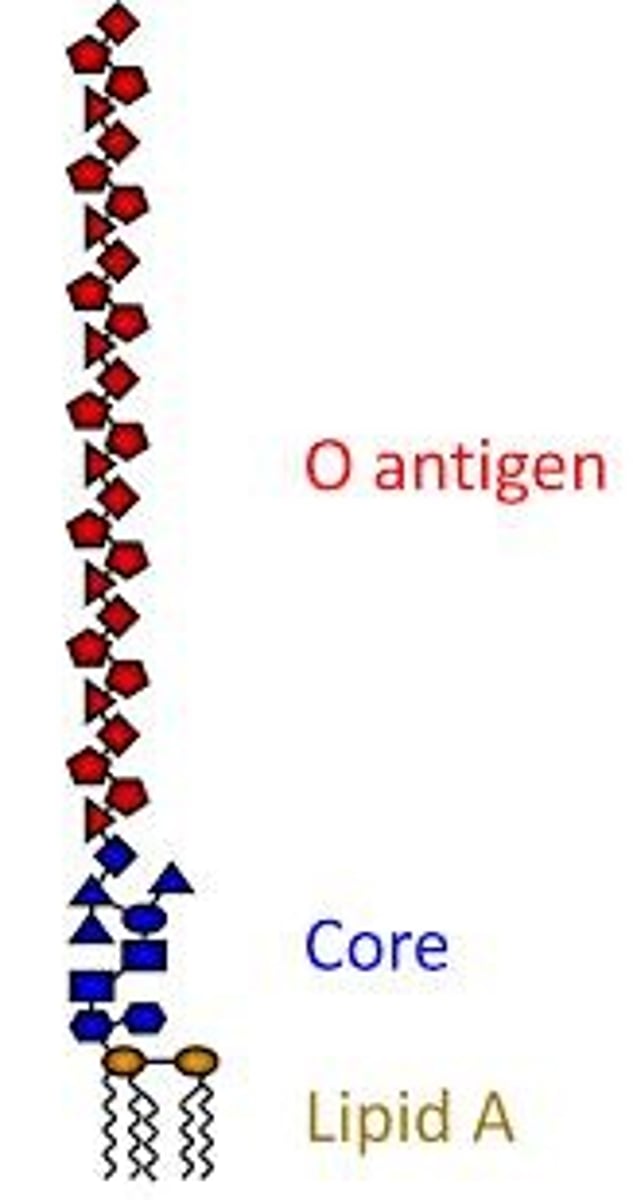
What are the three basic components of Lipopolysaccharide (LPS)?
1) Lipid A, 2) Core polysaccharide, 3) O antigen.
What is the function of Lipid A in LPS?
Responsible for the endotoxin activity of LPS and essential for bacterial viability.
What unique structural feature does the core polysaccharide of LPS contain?
It contains a unique octose known as 2-keto-3-deoxyoctanoate (KDO).
What is the role of the O antigen in LPS?
It is a long, linear polysaccharide consisting of repeating saccharide units, contributing to antigenic diversity.
How does LPS affect the immune system at low concentrations?
It stimulates protective responses such as fever, vasodilation, and activation of immune and inflammatory responses.
What can happen at high concentrations of LPS in the blood?
It can cause a systemic response resulting in sepsis, shock, and possibly death.
What is disseminated intravascular coagulation (DIC)?
A condition where blood coagulates throughout the entire body, potentially leading to organ dysfunction and multi-organ failure.
What is the difference between Lipopolysaccharide (LPS) and Lipooligosaccharide (LOS)?
LOS lacks the O-antigen polysaccharide found in LPS and is a low molecular weight form of bacterial LPS.
What is the effect of lysozyme on bacterial cell walls?
It hydrolyzes the bond between N-acetyl glucosamine and N-acetylmuramic acid, leading to wall-less bacteria.
What are protoplasts and spheroplasts?
Protoplasts are wall-less forms of Gram-positive bacteria, while spheroplasts are wall-less forms of Gram-negative bacteria.
What distinguishes acid-fast bacteria from other bacteria?
They have a waxy outer layer containing mycolic acids, making them resistant to Gram staining.
What is the significance of the Ziehl-Neelsen stain?
It is used to stain acid-fast bacteria, allowing them to be visualized against a light blue background.
What are the components of the complex cell wall of Mycobacterium?
It includes a peptidoglycan layer linked to arabinogalactan, surrounded by a waxy lipid coat of mycolic acids.
What is the morphology of Mycoplasma?
Mycoplasma have no cell wall and exhibit variable morphology.
What is the role of sterols in Mycoplasma?
Sterols are present in the cytoplasmic membrane, providing stability.
What is the function of porins in Gram-negative bacteria?
Porins allow the passage of small molecules through the outer membrane.
What are acute-phase cytokines stimulated by LPS?
IL-1, TNF-α, IL-6, and prostaglandins.
What is the relationship between LPS and Gram-negative bacteremia?
LPS can cause severe systemic responses in patients with Gram-negative bacteremia.
What are the consequences of high levels of LPS in the bloodstream?
They can lead to sepsis, shock, and multi-organ failure.
What is the role of pathogen pattern receptors (PRR) in the immune response to LPS?
PRRs like CD14 and TLR4 bind LPS, triggering immune responses.
What are the implications of structural variability in the outer core of the core polysaccharide?
It contributes to the diversity of strains among bacteria.