13 Topography of the ruminant abdomen
 Call Kai
 Learn
Learn Practice Test
 Spaced Repetition
 Match
Match Flashcards
 Knowt Play
1/69

Earn XP
Description and Tags
W4 Elnady
Name | Mastery | Learn | Test | Matching | Spaced | Call with Kai | Chat |
|---|
No analytics yet
Send a link to your students to track their progress
70 Terms
What is present in the abdominal wall?
1. superficial fascia (contains cutaneous trunci muscle and subiliac lymph node)
2. tunica flava abdominis
3. wide linea alba
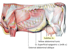
Where is the subiliac lymph node?
below the iliac or in front of the femur (which is why it is also called prefemoral lymph node)
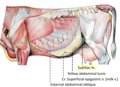
What are the boundaries of the paralumbar fossa?
-cranial: last rib
-caudal: origin of the internal abdominal oblique on the tubar coxae
-dorsal: lumbar vertebrae

why is the paralumbar fossa smaller in the horse than the ox?
because they have more ribs (18 pairs in horse and 13 pairs in ox)
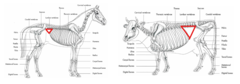
What is the clinical significance of the paralumbar fossa?
you can access the rumen of bovine in the left paralumbar fossa
we can do surgery in paralumbar fossa in ox more than in horse

true/false- ruminants have 4 stomachs
FALSE- they have a four CHAMBERED stomach = Multilocular stomach

What are the components of the ruminant stomach?
-3 non-glandular forestomachs (proventriculus) = rumen, reticulum, omasum
-1 glandular = abomasum (true stomach)
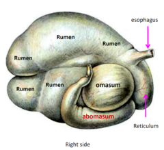
what type of stomach do ruminants have?
complex composite
(compared to simple in equine and K9)

The rumen fills all of the ____________ side
LEFT (from top to bottom)
What is the first chamber of the ruminant stomach?
RUMEN
If the esophagus opens into the reticulum, how come the rumen is considered the first chamber?
the esophagus opens at the cardia in the reticulum, but its mucosa protrudes forcing the food into the cranial ruminal sac.
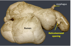
Where is the rumen located?
from the 8th ICS to the pelvic inlet

what is a normal paralumbar fossa supposed to look like?
It is supposed to look very sunken, however, if filled with gas it can look shallow (which is abnormal)

What procedures can be performed in the left paralumbar fossa?
1. Rumen trocarization: a procedure that involves inserting a trocar and cannula into the rumen of a cow to treat bloat or other conditions
2. rumenotomy
3. intraperitoneal injections
4. cesarean section

true/false- the rumen extends slightly to the right of the midline
TRUE
it disturbs the regular arrangement of the viscera

What are ruminal grooves?
a series of grooves that divide the rumen of a ruminant animal into different parts
they are external, filled with fat, and covered with peritoneum

Why should you avoid cutting ruminal grooves?
they lodge the neurovascular supply so if you cut into them you will damage blood vessels and nerves

What are the names of the ruminal grooves?
(left and right) longitudinal groove,
(cranial and caudal) transverse groove,
(dorsal and ventral) coronary groove

What are ruminal pillars?
projections of the rumen wall
contract to aid in mixing and digestion
increase surface area for the absorption of VFAs into the blood to use for energy
They correspond to the ruminal grooves
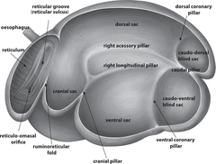
what is inside the rumen interior?
ruminal papillae

what is the difference between the rumen of the small and large ruminant?
the small ruminant has a smaller caudo-dorsal sac and they have rumen papillae on the dorsal sac with indistinct ruminal pillars
the large ruminant has a larger caudal sac and sparse rumen papillae on their dorsal sac. They have distinct ruminal pillars

the reticulum is the most _______________ compartment
Cranial
BUT, it is the second compartment (the rumen is the first)
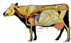
Where is the reticulum located?
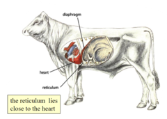
From the 6th to 8th ICS. It contacts the diaphragm and is close to the heart. it lies over the xiphoid cartilage
it is in the midline (slightly to the LEFT)
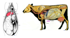
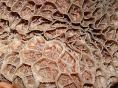
what does the interior of the reticulum look like?
honeycomb appearance
intersecting mucosal crests
What are the parts of the ruminoreticular compartment?
(1) ruminoreticular opening
(2) ruminoreticular groove
(3) ruminoreticular fold (U-shaped)

Where does milk enter the digestive tract?
there is a reticular groove/gastric groove that prevents milk from going into the rumen

What is traumatic reticulopericarditis?
perforation of the heart. usually occurs in ruminants after eating metal objects
sharp metallic object --> reticulum (cranial wall) --> diaphragm --> pericardium --> pericarditis

What is traumatic reticuloperitonitis?
sharp objects --> reticulum (right wall) --> liver --> hepatitis and live abscess
what can be used as a preventative method for traumatic reticulopericarditis and reticuloperitonitis?
introduce magnets into the reticulum

During a rumenotomy (paralumbar fossa --> reticulum), how can the cranial sac of the rumen be differentiated from the reticulum?
the rumen can be differentiated from the reticulum by the absence of the honeycomb mucosa

why do small ruminants have a lower incidence of traumatic pericarditis?
because they are selective grazers, they do not vacuum the ground as cattle do

Where is the omasum located?
on the RIGHT side entirely in the intrathoracic part of the abdominal cavity (8-11 ribs and ~10 cm protrudes below the costal arch)

what is the clinical significance of the omasum?
The omasum grinds food and sometimes gets impacted.
In the case of impaction, pushing on the omasum through the body wall below the costal arch will elicit pain.
HOWEVER, in small ruminants, the omasum does not protrude under the costal arch and is covered by the liver so it has no contact with the body wall in the small ruminant.

where is the abomasum located?
on the RIGHT side (7-11 rib)
it lies just caudal to the reticulum and under the omasum

what is the abomasum?
the 4th (and last chamber). it is the glandular/true stomach

true/false- the abomasum directly contacts the abdominal floor
TRUE

where is the abomasum when there is a left displaced abomasum?
it is between the rumen and the left abdominal wall
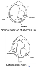
What is a right displaced abomasum?
Associated with torsion of the pylorus --> twist and damage of vagal nn. (which are located along the lesser curvature)
Abomasum fills with gas, ascends cranial to omasum. More dangerous as can lead to torsion. Cuts off blood supply.
true/false- the dorsal and ventral vagal trunks run along the lesser curvature of the abomasum
TRUE

Where is the greater omentum attached in the ruminant?
to the left longitudinal groove
What are the parts of the greater omentum in the ruminant?
(1) superficial layer
(2) deep layer
(3) omental bursa
(4) supraomental recess = superficial and deep layers fold to form supraomental bursa
epiploic foramen = connects omental bursa with the peritoneal cavity

what is the supraomental recess?
the intestinal pouch (V-shaped hammock) formed by the deep wall of the greater omentum
it is closed cranially and open caudally

What is the largest chamber in neonates?
the abomasum because milk needs no fermentation

where is the spleen located?
entirely on the left side. it is firmly attached to the craniodorsal aspect of the rumen and is compressed between the rumen wall and left body wall
it extends from the 7th or 8th CCJ to the top of the last ICS
True/false- ruminants have no gastrosplenic ligament
TRUE

what is the shape of the spleen in large versus small ruminants?
in cattle it is elliptical-shaped (rectangular) in sheep and goats it is somewhat triangular

what are Hemal nodes?
they are lymphatic structures with blood circulation only (they receive and filter blood instead of lymph)
Where are hemal nodes found in ruminants?
along the great vessels (aorta), mesenteric regions, perineal fat, subcutaneous tissue
they are distinguished from lymph nodes by their dark brown color
Where is the liver in the ruminant?
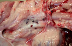
the rumen pushes it to the right so the right lobe is dorsal and the left lobe is ventral
it extends from the 6th ICS to the last rib

what are the parts of the pancreas?
1. left lobe
2. right lobe (in mesoduodenum)
3. body (joins the 2 lobes)
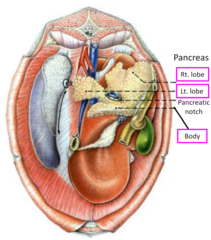
What are the parts of the small intestine?
1. duodenum
2. jejunum
3. ileum

What are the parts of the duodenum?
cranial part --> sigmoid loop --> cranial flexure --> descending duodenum --> caudal flexure --> ascending duodenum --> duodenojejunal flexure

Where is the jejunum located?
in the supraomental recess
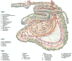
what are the parts of the ileum?
ileocecal fold and opening

where are the mesenteric lymph nodes located?
along the attached border of the mesojejunum and not at the root as in dogs and horses. the nodes are located in the mesojejunum, between the jejunum and the outermost coil of the spiral colon.

where is the small intestine located?
on the right side in the supraomental recess

what are the parts of the cecum?
base, body, apex
the apex is directed caudally towards the pelvic inlet

what are the characteristics of the cecum?
straight, cylindrical, no tenia, no haustra
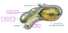
Where is the cecum located?
dorsally in the supraomental recess in the right paralumbar fossa

what are the parts of the colon?
1. ascending
2. transverse (1)
3. descending (2)

What are the parts of the ascending colon?
1. proximal loop (pink)
2. spiral loop (blue)
3. distal loop (green)
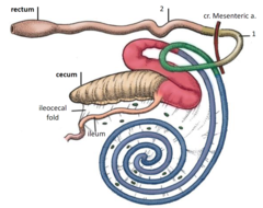
the spiral colon is on the _____________ of the mesoejunum
it is on the left of the mesojejunum therefore it is not visible

where is the distal loop of the ascending colon located?
within the duodenal loop. It is attached to the duodenal loop by the duodenocolic fold

what structure should you listen for in the right paralumbar fossa?
the cecum and spiral colon

distension of the abdominal wall is best viewed from the __________
REAR

What does the bovine kidney look like?
it is lobated (NOT LOBULATED)
the divisions we see on the surface are lobes, no lubules

true/false- the bovine kidney has no renal pelvis
TRUE
what is going on with the left kidney?
it is pendulous/floating/physiologically wandering kidney.
it is pushed by the rumen to the midline (caudal to the right kidney).
the hilus of the left kidney lies dorsally and the left ureter begins in the midline and inclines back to the left side of the body

small ruminant kidneys resembles _____________ kidneys
Canine (smooth unipapillary/unipyramidal)
they have a renal pelvis

what accessory glands are present in ruminants?
prostate, ampulla, vesicular gland, bulbourethral gland