Apical Study Guide
 Call Kai
 Learn
Learn Practice Test
 Spaced Repetition
 Match
Match Flashcards
 Knowt Play
1/20

Earn XP
Description and Tags
Cardiac Sonography
Name | Mastery | Learn | Test | Matching | Spaced | Call with Kai | Chat |
|---|
No analytics yet
Send a link to your students to track their progress
21 Terms
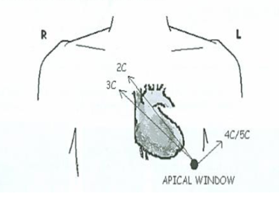
The Apical Window
is identified initially by palpating of the LV apex with the patient in steep LLD position. Placement of the probe on the Apical Impulse (PMI)- The most Lateral and Inferior point over the Apex where the Cardiac Impulse can be felt.
(Then angle Superior and Medial toward the Heart).
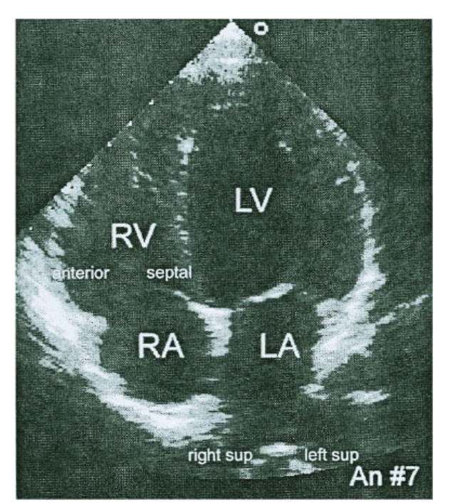
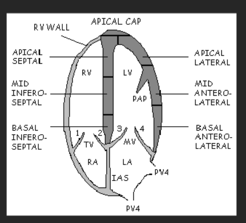
Apical 4chamber (AP4ch)
Chambers visualized: RV, RA, LV, LA
Valves visualized: TV, MV
Walls visualized: RVFW, Inferoseptal, and Anterolateral walls.
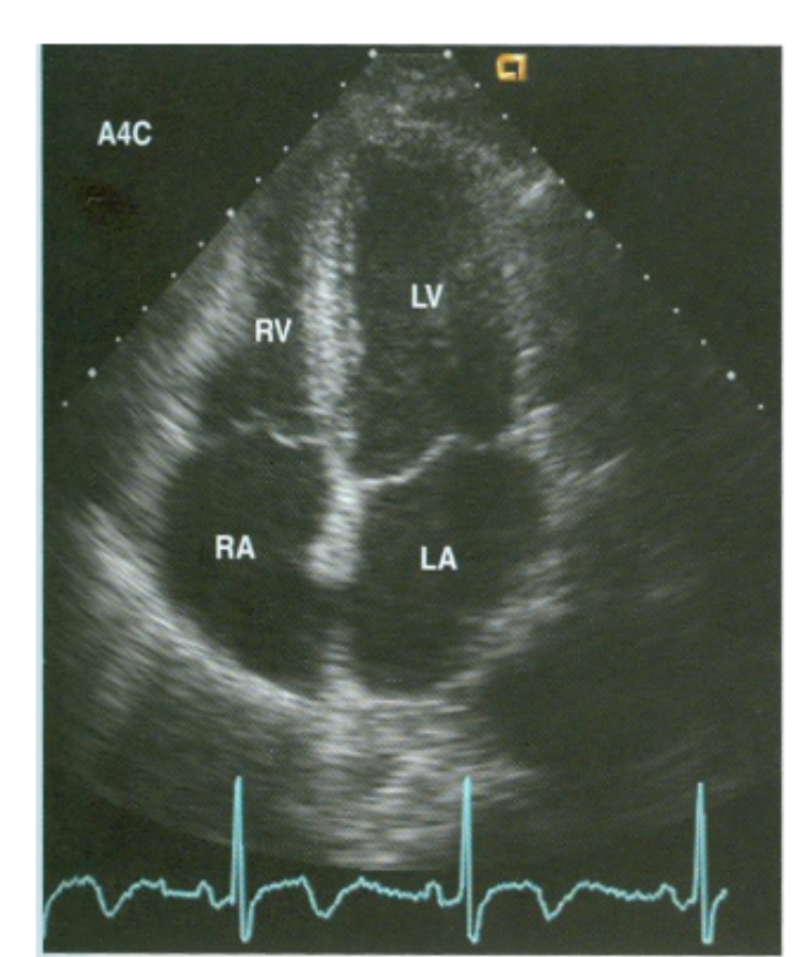
Probe Positioning: AP4ch
Angle Anterior and Posterior to scan through the LV. The LV appears as a truncated ellipse with a longer length than width and a tapered but rounded apex.
MV in Ap4ch
In Diastole the MV leaflets open.
The AML is adjacent to the Septum
The PML is adjacent to the Lateral wall.
TV in Ap4ch
In Diastole the TV Leaflets open.
The TV annulus is closer to the apex (up to 1 cm).
Septal leaflet – is located adjacent to the septum.
The Anterior Leaflet is the other leaflet seen adjacent to the free wall.
Other anatomy seen in Ap4ch
The DAO may be seen Lateral to the LA.
The PV4 enter the LA Posteriorly but may be difficult to see at this depth in adults.
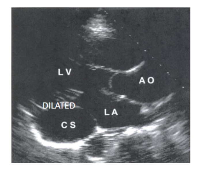
If probe is angled posteriorly from the 4CH view, the Coronary Sinus is seen in the Atrioventricular groove.
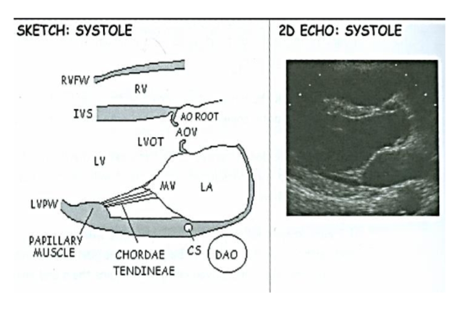
Coronary Sinus Diagram/ Dilated CS
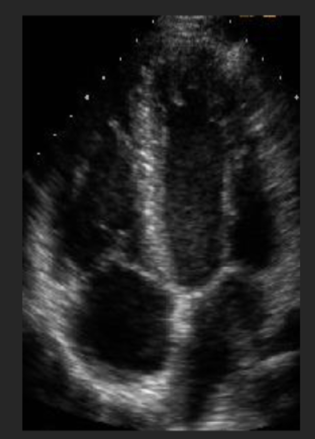
Ap4ch Diagram
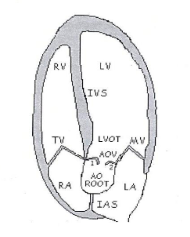
Apical 5 Chamber (Ap5ch) Diagram
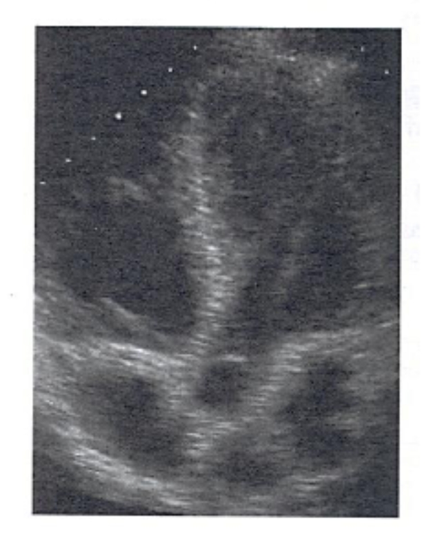
Probe Positioning: Ap5ch
Angle the transducer more Anterior from the Ap4ch to acquire the apical 5 chamber (Ap5ch) view.
This opens up the LVOT and Aortic Valve. (hence the 5th chamber)
Ap5ch
Chambers visualized: RV, RA, LV, LA (including LVOT)
Valves visualized: AoV, TV (MV should be closed).
Probe Positioning: Ap2ch
Rotate the transducer approximately 60 - 90˚ counterclockwise from the Ap4ch and 5ch views to acquire the apical 2 chamber (Ap2ch) view.
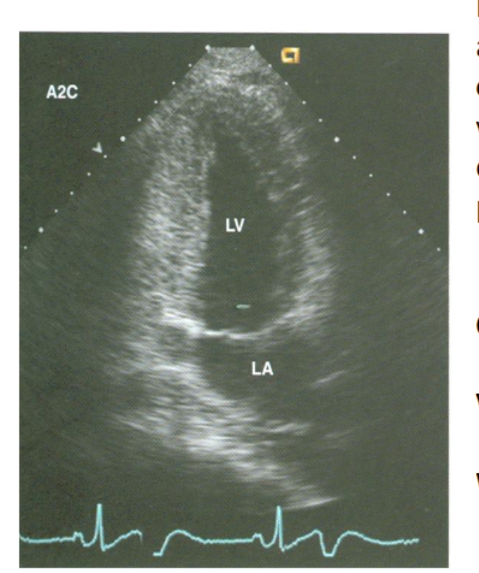
Apical 2 Chamber (Ap2ch)
Chambers visualized: LV, LA
Valve visualized: MV
Walls visualized: Anterior and inferior walls.
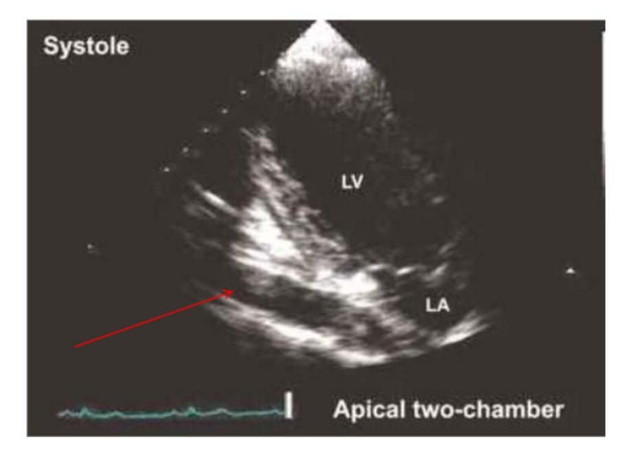
DAO shown in Ap2ch
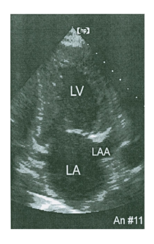
The LAA in Ap2ch
The left atrial appendage may be visualized adjacent to the anterior wall – by angling the transducer posteriorly and rotating counterclockwise from
the Ap2Ch view – standard view of LAA.
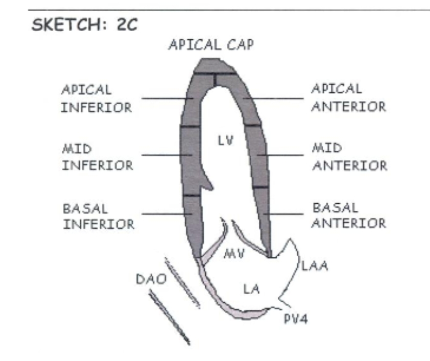
Ap2ch Diagram
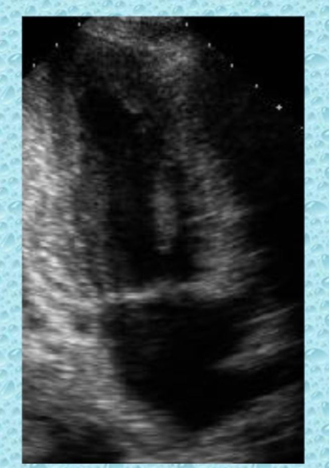
Probe Positioning for Ap3ch *The 3rd Chamber is the LVOT*
Rotating the transducer another 30° - 60˚ from the apical 2 chamber view (120° from the Ap4Ch).
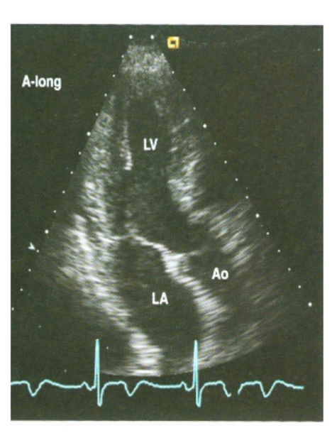
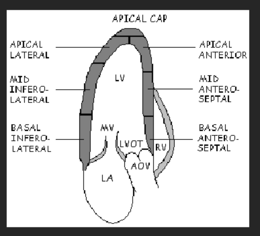
Ap3Ch or Apical Long Axis View
Chambers visualized: LV, LA, a portion of the RV
Valves visualized: MV, AoV
Walls visualized: Inferolateral wall (LVPW), Anteroseptal wall.
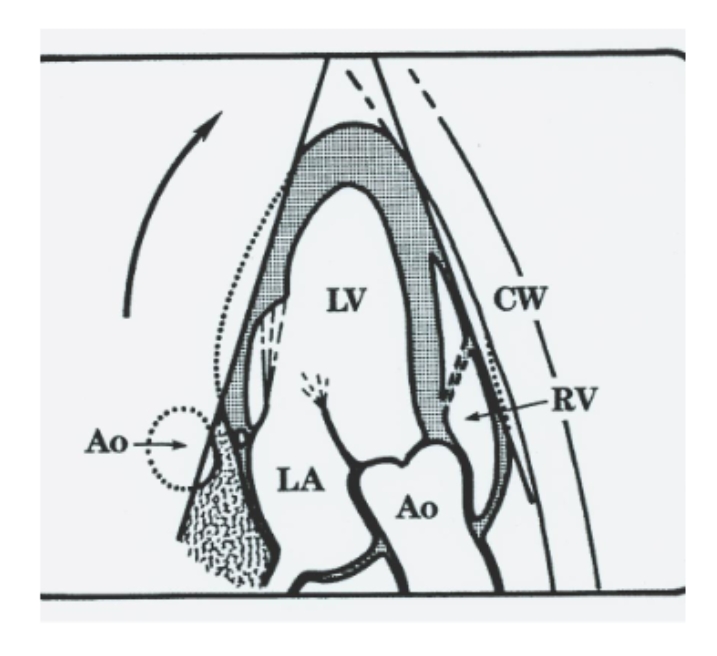
Ap3ch Diagrams
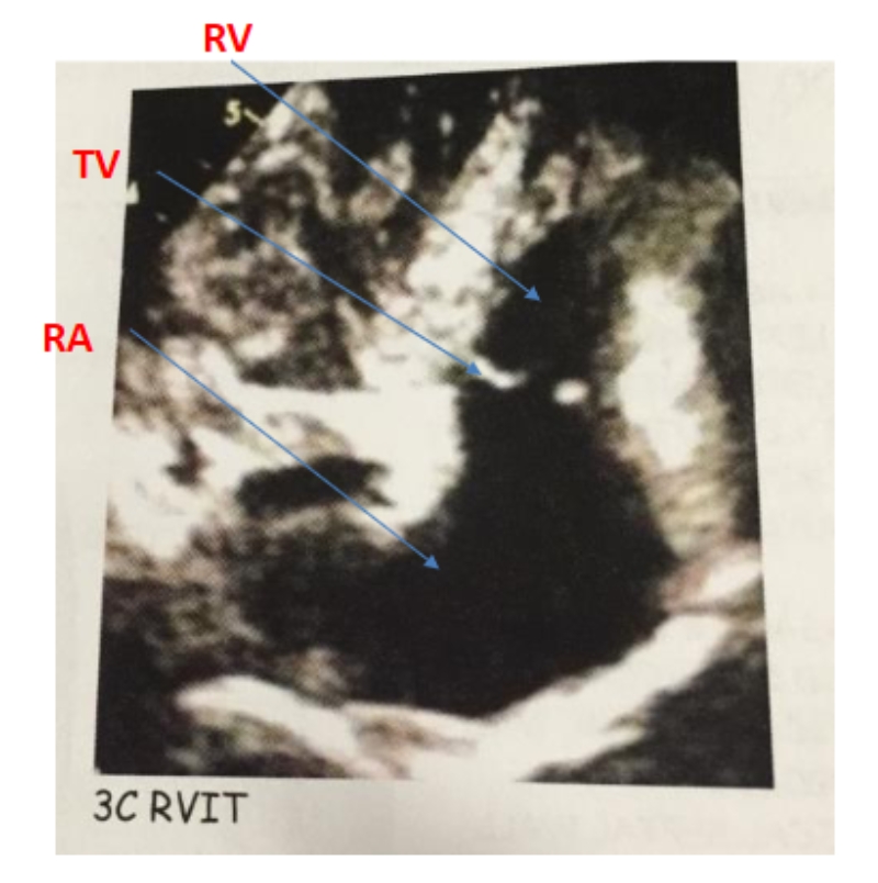
Probe Positioning: Ap3ch RVIT
Angle the transducer more Medially and Inferior from the 3C.
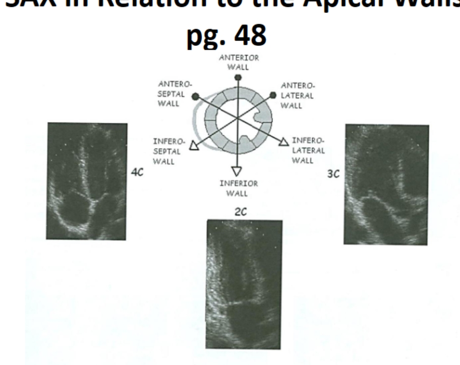
The order to Visualize all walls in the Apical Window
4, 2 and 3.