Chapter 7: Bone Tissue
 Call Kai
 Learn
Learn Practice Test
 Spaced Repetition
 Match
Match Flashcards
 Knowt Play
1/65

There's no tags or description
Looks like no tags are added yet.
Name | Mastery | Learn | Test | Matching | Spaced | Call with Kai | Chat |
|---|
No analytics yet
Send a link to your students to track their progress
66 Terms
Skeletal system?
Composed of bones, cartilages, and ligaments
Cartilage?
Covers many joint surfaces of mature bone
Ligaments?
Hold bones together at joints
Tendons?
Attach muscle to bone
Functions of the Skeletal System?
Support
Protection
Movement
Electrolyte balance
Acid-Base balance
Blood formation
Support?
Our bones act as the body’s structural framework and as a point of attachment
Protection?
Internal organs are protected from injury
Ex. Skull and ribs
Movement?
Most muscles of the skeletal system are attached to bone
When muscles contract, bones are pulled to produce → movement
Electrolyte balance?
Bones store calcium (99%) and phosphorous
Give bones → strength
Bones release minerals to be distributed throughout the body and maintain → homeostasis
Acid-Base balance?
Bone tissue can buffer the blood and prevent major pH changes
Absorbs or releases phosphate and carbonate salts
Blood formation?
Red bone marrow – produces all major blood cells and the cells of the immune system
Bone (osseous tissue)?
A connective tissue with a hardened matrix of calcium phosphate and other ions
Hardened during mineralization or calcification
Most of the long bone outer shell structure is composed of?
Compact (dense) bone
Shell encloses the?
Medullary cavity (marrow cavity), which contains → bone marrow
At the ends of the bones the central space contains?
Spongy (cancellous) bone tissue that is loosely organized
Diaphysis?
Shaft or body of the long bone
Epiphyses?
The proximal and distal ends of the long bone
Strengthen joints and anchor ligaments and tendons
Mature bones have an?
Epiphyseal line, which contains denser spongy bone between epiphysis and diaphysis
Contains the epiphyseal → plate (growth plate)
Present until bones → stop growing (ages 14-24)

Articular cartilage?
Thin layer of hyaline cartilage that covers the → epiphyses
Absorbs shock and reduces friction for free moving joints
No blood vessels (avascular), slow repair time
Periosteum?
Tough cover of connective tissue covering the → diaphysis
Associated with the blood supply surrounding the bone surface
Offers protection, assists in fracture repair, bone tissue nourishment, and as attachment for tendons and ligaments
Medullary cavity (marrow cavity)?
Hallow cylindrical space found in the → diaphysis
Contains yellow bone marrow and → blood vessels

Endosteum?
Thin membrane that lines the → marrow cavity
Osteogenic cells?
Are stem cells that can become the other bone cell types
Found in the endosteum and the inner periosteum
Multiple continually into more stem cells or become (differentiate into) → osteoblasts

Osteoblast?
Cells that → build bone
Line up under the endosteum and periosteum
They do not undergo mitosis, only made from osteogenic cells
They secrete bone extracellular matrix, which is hardened by mineral deposits

Osteocytes?
Are former osteoblasts that had gotten stuck in the bone extracellular matrix (ECM)
They are found in cavities called lacunae, which connect through channels called canaliculi
Contribute to bone maintenance and sense bone strain

Osteoclasts?
Bone-carver, dissolve bone on the surface
Develop from bone marrow → stem cells
Perform bone resorption, which is key in bone remodeling
This is normal and part of bone development and growth (osteolysis)

Osteoblasts?
Build bone
Osteoclasts?
Carve out bone
Compact bone tissue?
Is the strongest form of bone with few spaces
Found beneath the periosteum in all bones and makes up most of the diaphysis of long bones
Provides protection, support, and stress → resistance
Osteon?
Is a structural unit of compact bone and consists of:
Repeating circular units of concentric lamellae, which is layers of the bone hardened bone matrix
The layers of lamellae surround a?
Central canal - which contains blood vessels and nerves
The vessels in the central canal are fed by → perforating canals
Between layers of lamellae are?
Tiny cavities called lacunae (contain osteocytes), which are all interconnected by canaliculi
Circumferential lamellae?
Are found in the inner and outer boundaries and run parallel to osteons and the bone surface for → support
Old osteons that have been broken down for → regrowth
Osteons are aligned and parallel with?
The diaphysis → length
Allow for resistance to bending or fracturing
Spongy bone tissue?
Does not have osteons
Consist of trabeculae – lamellae arranged in an irregular pattern
Located in the interior of the bone and is protected by compact bone → covering
What is Bone Marrow?
A soft tissue that is found in the marrow cavity and between the trabeculae of → spongy bone
What are the two types of bone marrow?
Red bone marrow(myeloid tissue)
Yellow bone marrow
Red bone marrow(myeloid tissue)?
Produces blood cells (hemopoiesis)
Found in almost every bone of a child
In adults, the skull, vertebrae, ribs, sternum, pelvic girdle, and the proximal epiphyses of the humerus and femur
Yellow bone marrow?
Fatty marrow that was red bone marrow but no longer produces blood cells
Bone is formed through?
Ossification (osteogenesis)
The embryo skeleton is initially in the what?
General shape of bones and is made of → mesenchyme (embryonic tissue)
Cartilage formation and ossification start in the 6th week of development
Intramembranous ossification?
Bone forms within mesenchyme in sheet-like layers
Forms flat bones in the skull, facial bones, mandible, and part of the clavicle
Also forms the “soft spots” (fontanel) that help the
fetus pass through the birth canal
Mesenchyme is a what?
An embryonic tissue which develops into connective and skeletal tissues, including blood and lymph
Endochondral ossification?
Bone forms within → hyaline cartilage
This also starts at 6 weeks of development and continues into your 20s (stops sometime between ages 14-24...)
Cartilage forms what?
A bone-like structure, called a → cartilage model
Bone will initially form in the what?
In the diaphysis then → hallow out
Forms the primary ossification center and primary marrow cavity
Bone will then develop in what?
In proximal epiphysis then → hallow out
Forms the secondary ossification center and secondary marrow cavity
Epiphyseal plate?
Cartilage left over from endochondral ossification is → turned to bone
Functions as growth zone where bone → elongates
Has hyaline cartilage in the middle with transition zones on each side where cartilage is replaced by bone
Metaphysis is zone of transition facing the marrow cavity
Interstitial growth?
Growth from within
Bone elongation is a result of cartilage growth within the epiphyseal plate
Epiphyses close when cartilage is gone, epiphyseal line of spongy bone marks site of former epiphyseal plate
Lengthwise growth is → finished
Occurs at different ages in different bones (14-24)
Appositional growth?
Occurs at bone → surface
Continual growth in diameter and thickness
Similar to intramembranous ossification
Osteoblasts (bone builders) of inner periosteum deposit?
Osteoid tissue (prebone)
Become trapped as tissue calcifies and become osteocytes
Osteocytes lay down what?
Matrix in layers parallel to surface
Forms circumferential lamellae
Osteoclasts (bone carvers) of endosteum enlarge marrow cavity
Ossification continues throughout life with what?
The growth and remodeling of bones
Bone remodeling?
Replacement of old bone with new bone tissue
Includes bone reabsorption – minerals and collagen fibers are removed by → osteoclasts
Followed by bone deposition – the addition of minerals and collagen fibers by → osteoblasts
Mineral deposition (mineralization)?
Occurs when calcium, phosphate, and other ions are taken from blood and deposited in → bone
Osteoblasts produce collagen fibers that spiral the length of the osteon
Fibers become encrusted with minerals
Mineral resorption?
Process of dissolving bone and releasing minerals into blood
Performed by → osteoclasts
Dissolve bone and digest collagen
Calcium homeostasis depends on what?
A balance between dietary intake, urinary and fecal losses, and exchanges between osseous tissue
Calcium homeostasis is regulated by three hormones:
Calcitriol, calcitonin, and parathyroid hormone
What is Calcitriol?
A hormone that raises blood calcium level
Is also necessary for bone resorption, helping provide calcium and phosphate
Calcitonin?
Secreted by the thyroid gland when blood calcium levels rise too → high
Lowers blood calcium concentration in two ways:
Inhibits osteoclasts thereby reducing bone resorption
Stimulates osteoblasts to deposit calcium into bone
Parathyroid hormone (PTH)?
Secreted by parathyroid glands
PTH released when calcium levels are low in blood
Acts to raise blood calcium levels
Causes bone → resorption
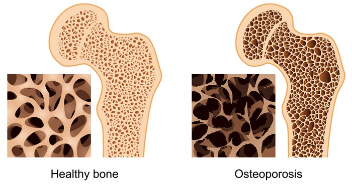
Osteoporosis?
Porous bone condition
Bone resorption (breakdown) out does bone deposition (formation)
Mostly caused by calcium depletion in the body
Fracture?
Is a break in the bone
Formation of hematoma and granulation tissue?
Blood vessels rupture across the fracture line and stimulate → inflammation
A fracture hematoma – blood clots in a mass (6-8 hours)
Clot is converted to granulation tissue, a soft fibrous mass
Bone cells die around the fracture due to lack of circulation
Causes more inflammation and → swelling
Phagocytes (part of our immune cells) and osteoclasts remove dead or damaged tissue around the hematoma (several weeks)
Formation of a soft callus?
Soft callus - Osteogenic cells become chondroblasts (cartilage builder) that produce patch of fibrocartilage and fibroblasts depositing collagen in granulation tissue
Conversion to hard callus?
Osteogenic cells differentiate into osteoblasts, which produces a bony collar called the hard callus around the fracture
Acts as a temporary splint around the fracture to join the two broken ends together
Takes 4-6 weeks to form
Remodeling?
Hard callus lasts for 3-4 → months
Osteoclasts dissolve small fragments of bone
Osteoblasts deposit spongy bone between the broken ends
Spongy bone gradually fills to become compact bone (like intermembranous ossification)