LAB MIDTERM MASTER LIST
1/254
There's no tags or description
Looks like no tags are added yet.
Name | Mastery | Learn | Test | Matching | Spaced |
|---|
No study sessions yet.
255 Terms
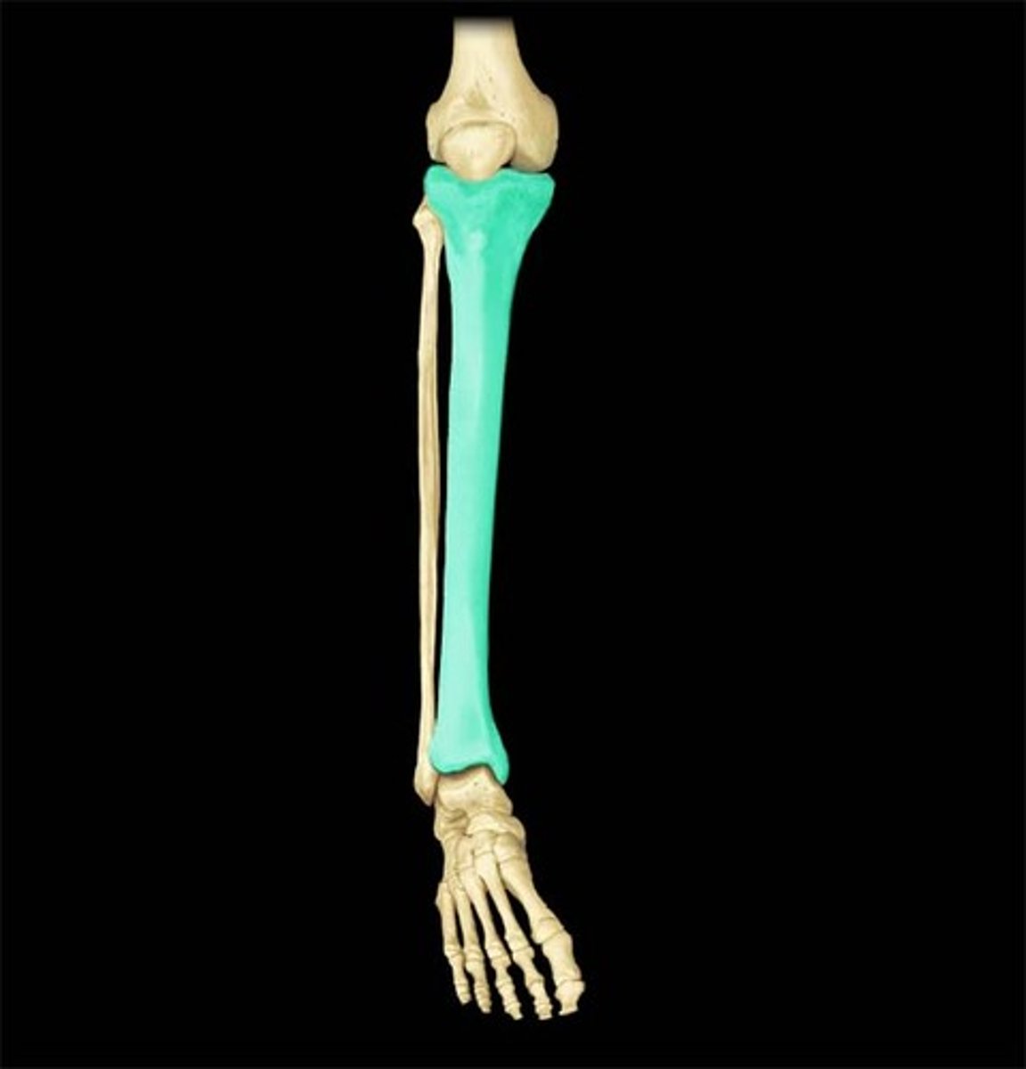
Tibia
Large bone in the lower leg.
Anterior border of the tibia
Ridge that runs down the anterior of the bone (the shin)
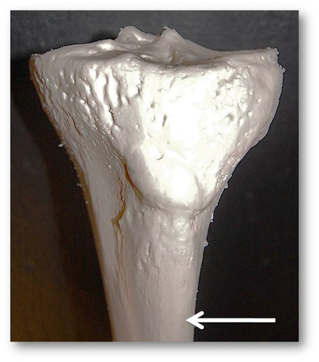
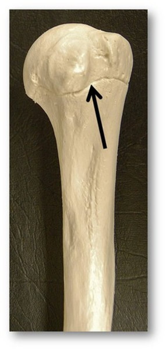
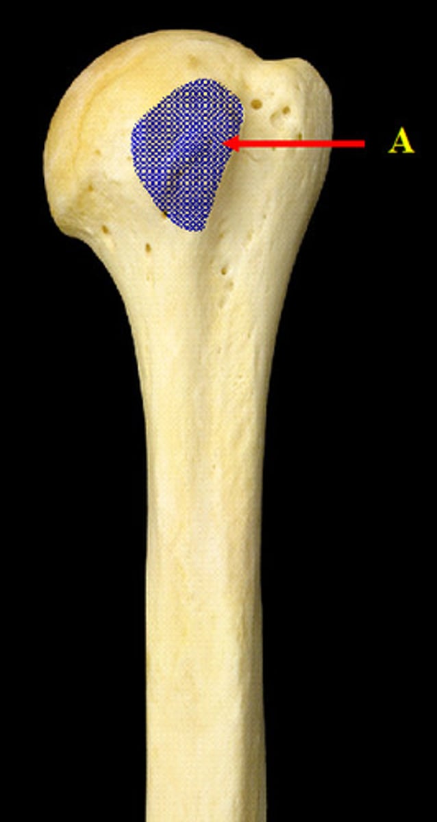
Tibial plateau
The flat portion of the tibia that articulates with the femur
Medial condyle of the tibia
The medial side of the tibial plateau
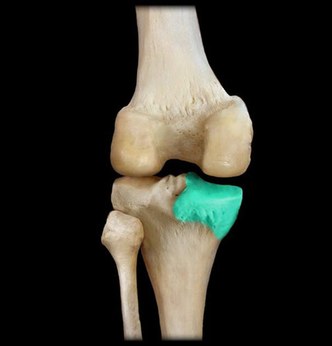
Lateral condyle of the tibia
The lateral side of the tibial plateau ( the side were the fibula is located.
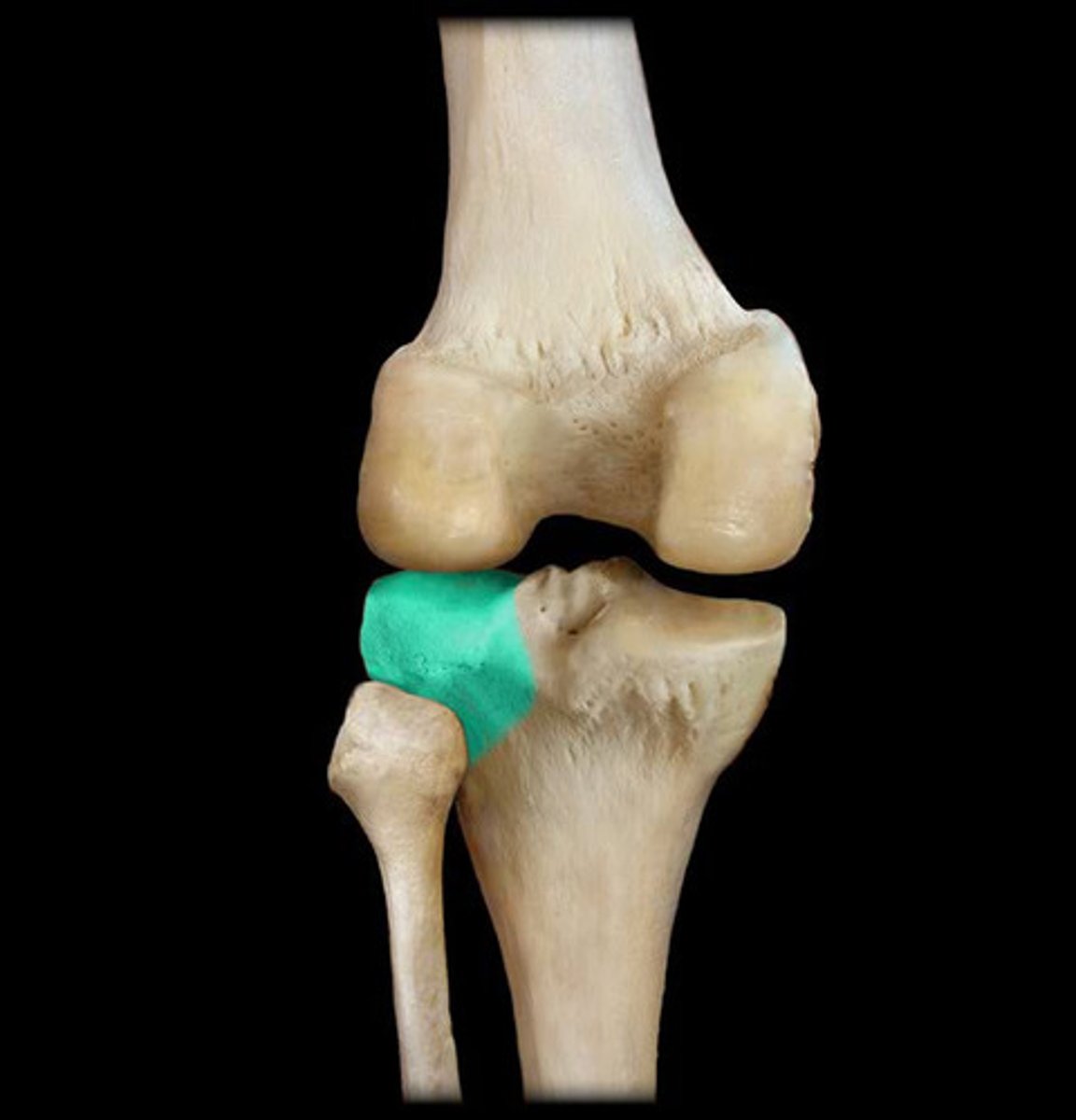
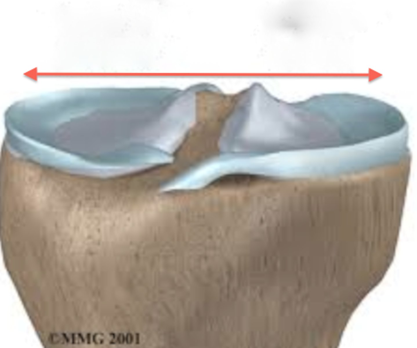
Intercondylar eminence
The protrusions on the top of the tibial plateau.
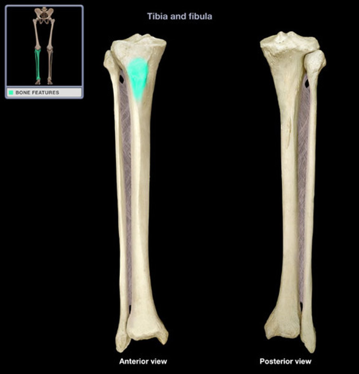
Tibial tuberosity
The bump at the top of the anterior border
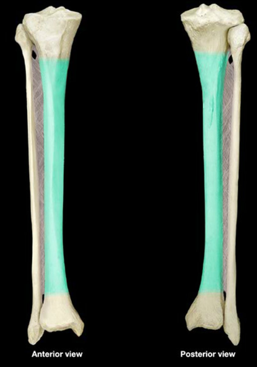
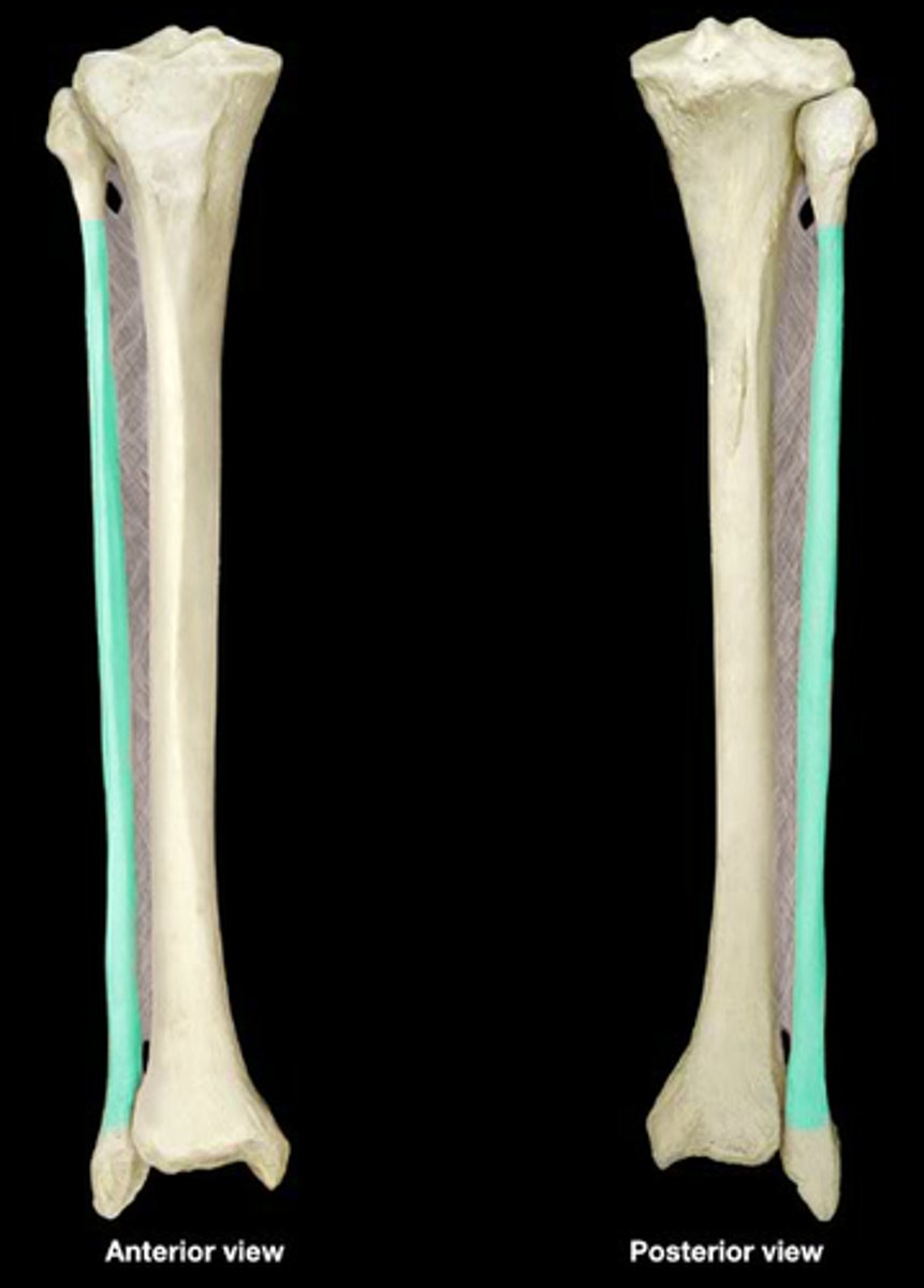
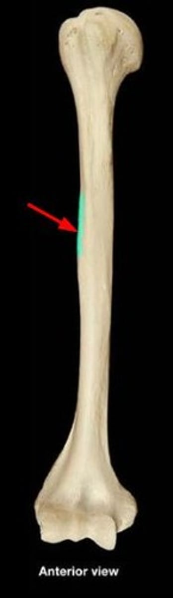
Shaft (body) of the tibia
The length of the bone excluding the two ends
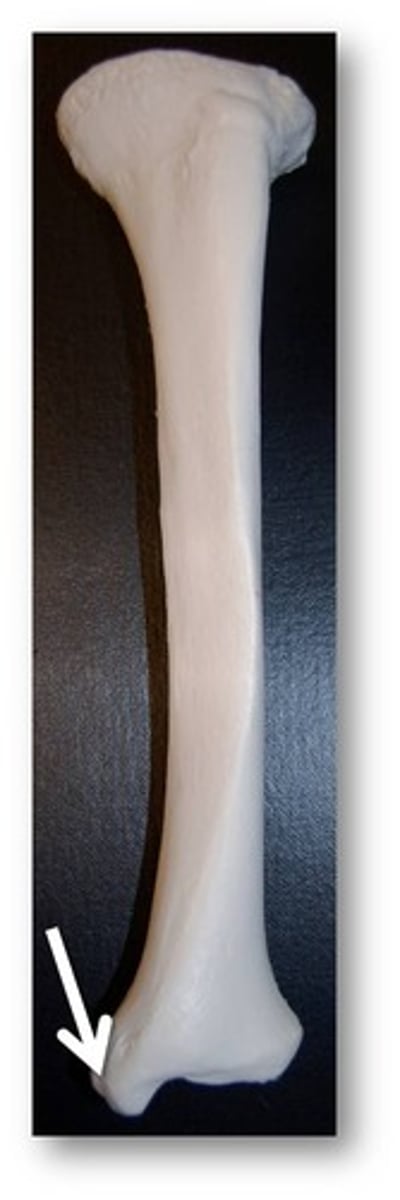
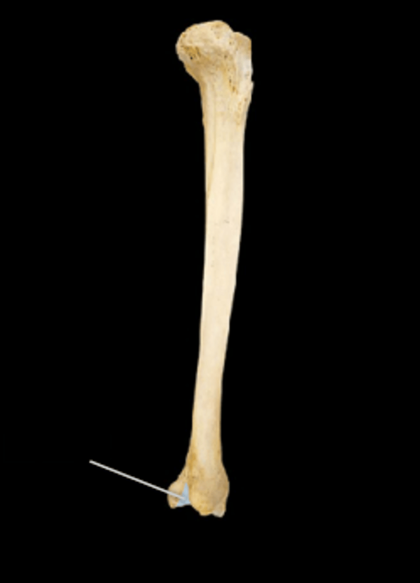
Medial malleolus
The protrusion on the medial side of the distal end of the Tibia ( bump on the inside of the ankle)
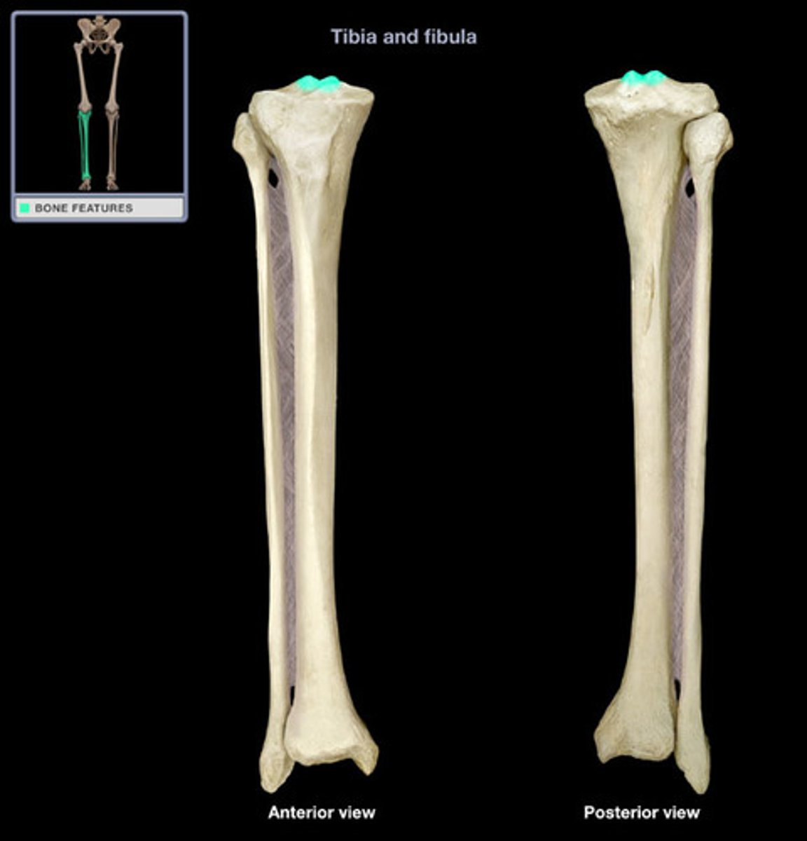
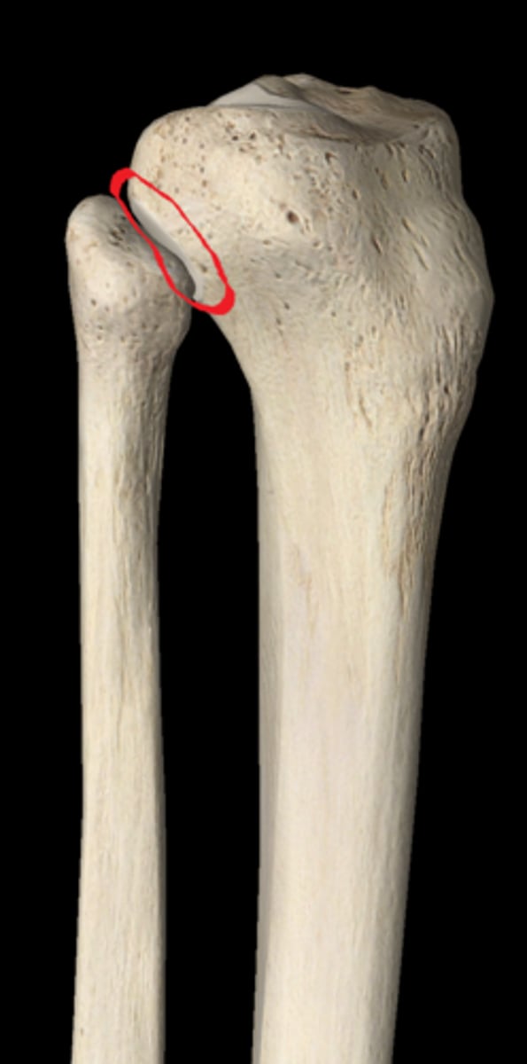
Proximal articular facet
Small indent on the proximal end by the lateral condyle that articulates with the fibula.
Distal articular facet (fibular notch)
Small notch at the distal end on the lateral side of the tibia that articulates with the fibula
Fibula
Small thinner bone on the lateral side of the lower leg.
Head of fibula
Medial end of the fibula
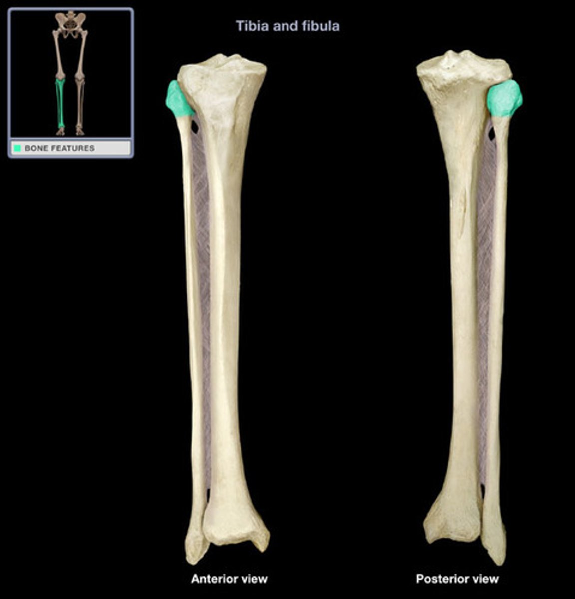
Shaft (body) of fibula
Length of the fibula excluding the ends
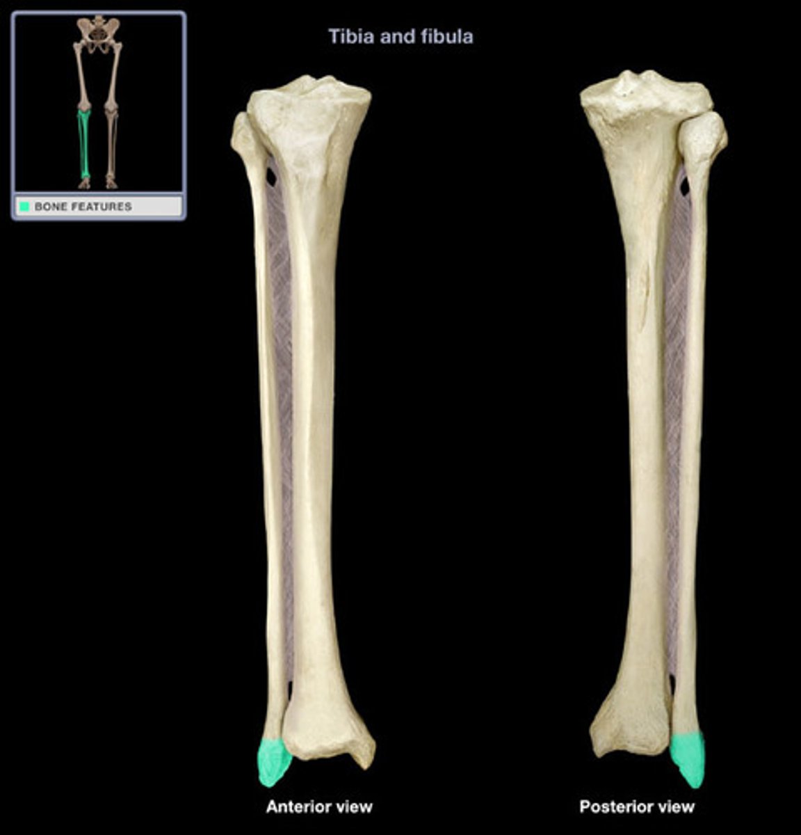
Lateral malleolus
Protrusion on the distal end of the fibula (bump on the outside of the ankle)
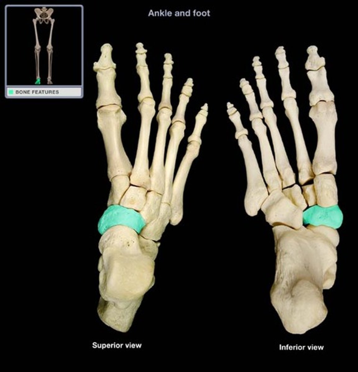
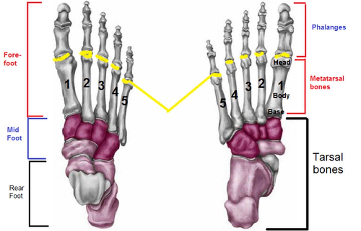
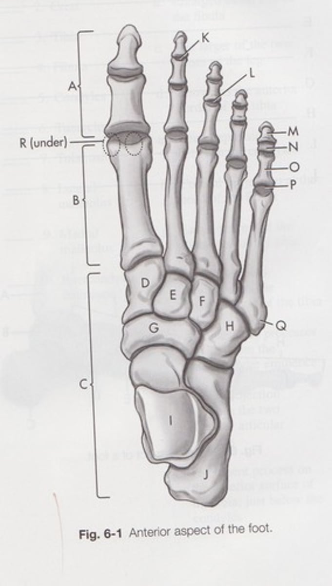
Tarsals
Ankle bones
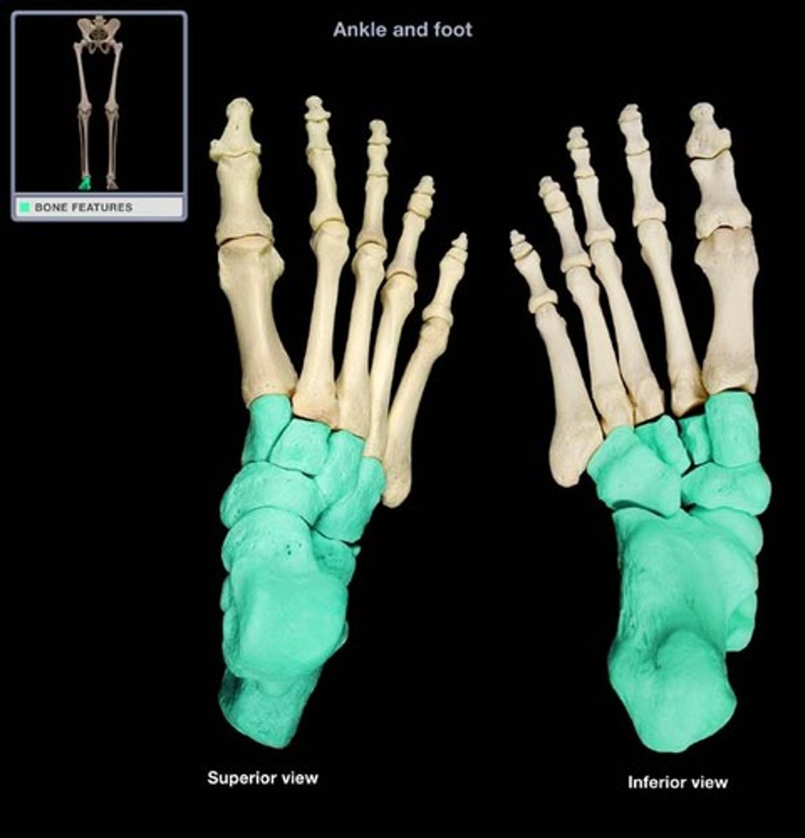
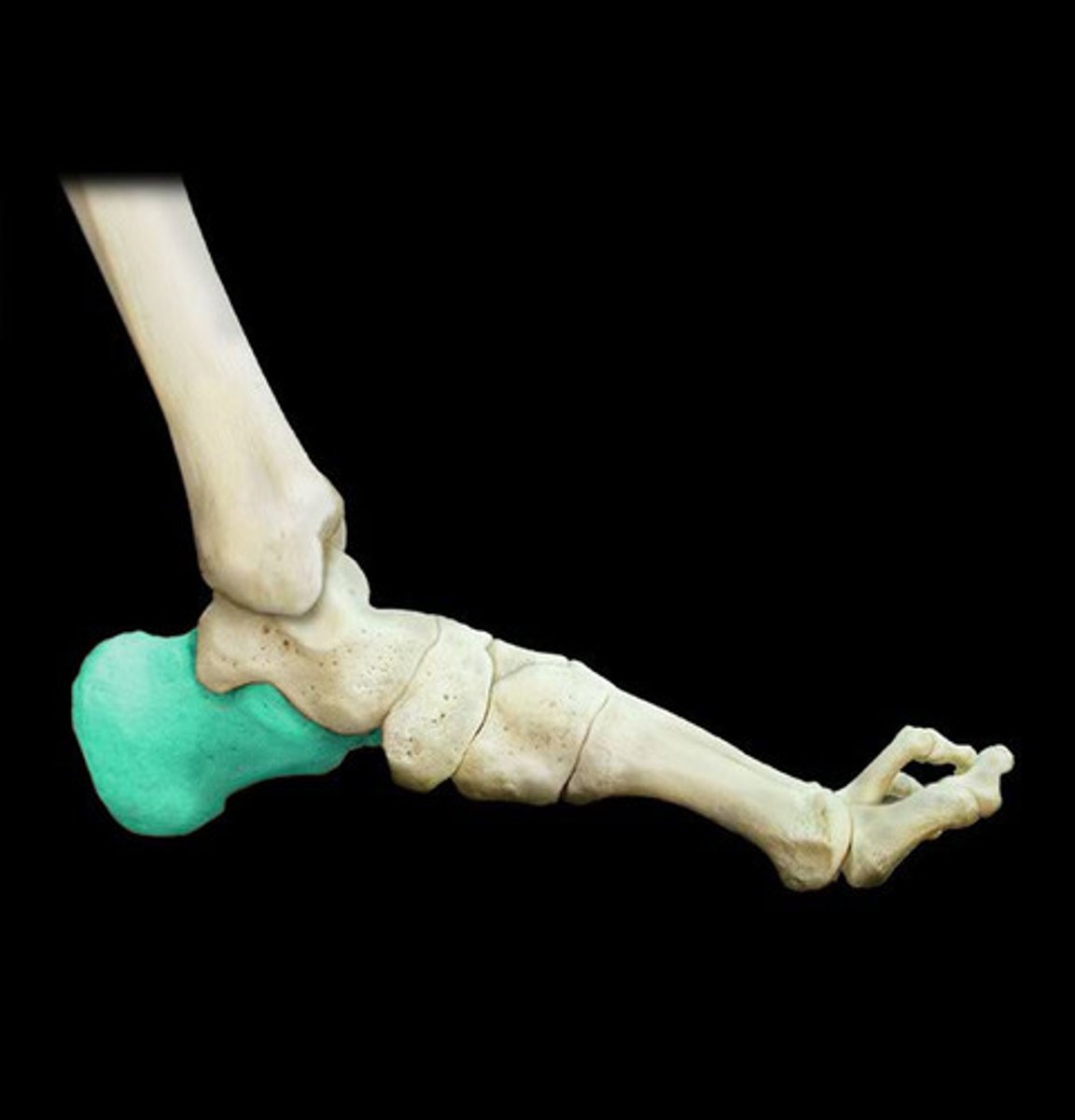
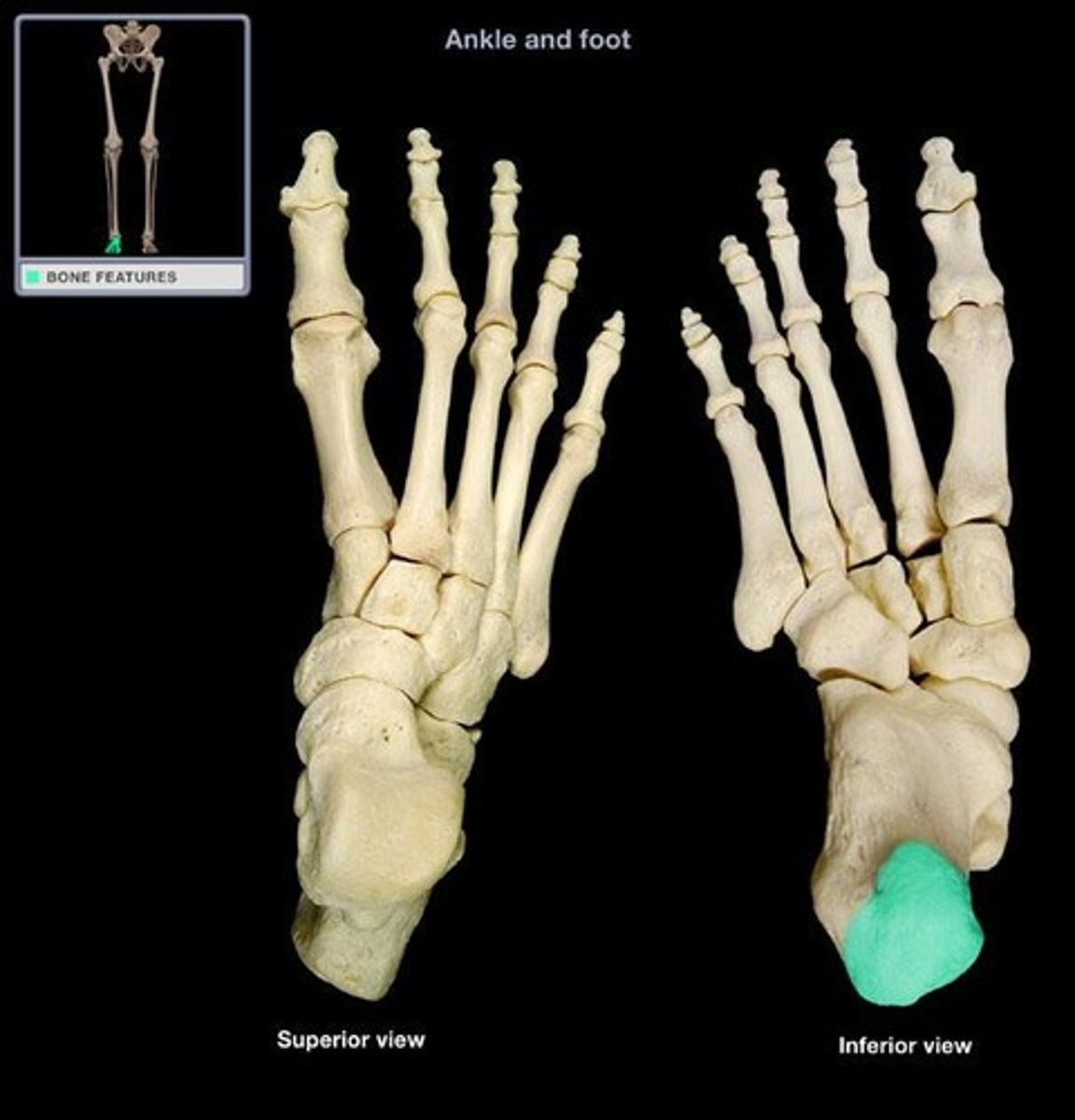
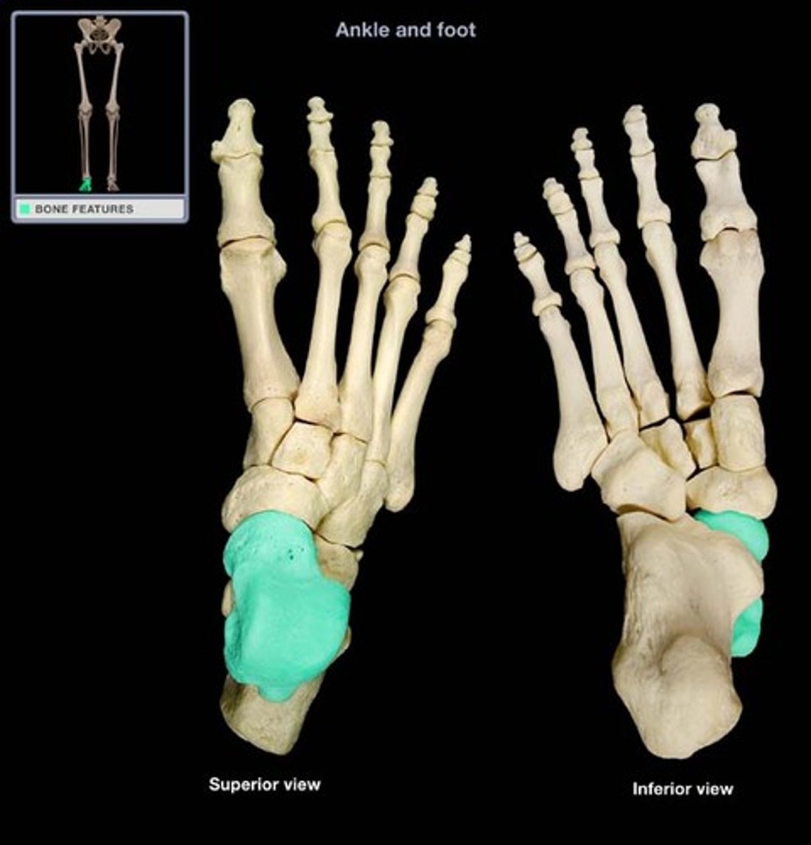
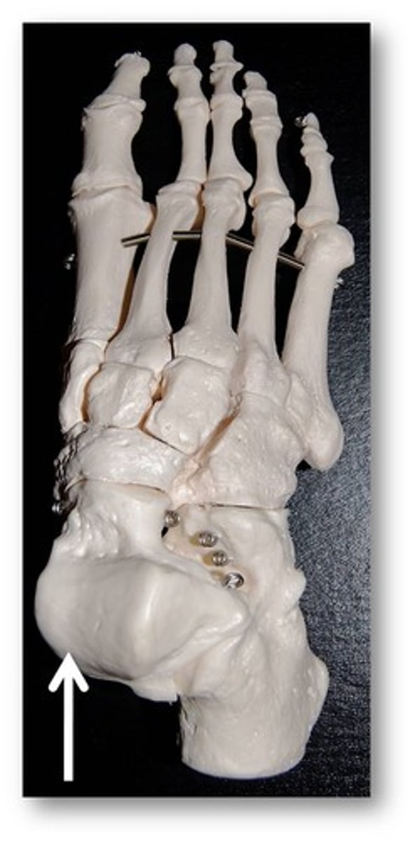
Calcaneus
Largest bone in the foot
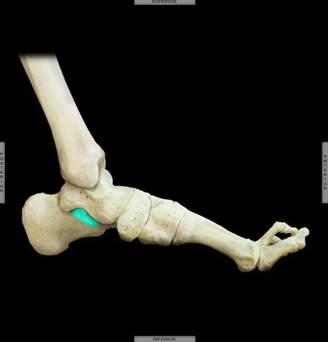
Sustentaculum tali
small protrusion on the medial side of the calcaneus.
Calcaneal tuberosity
The heel of the foot
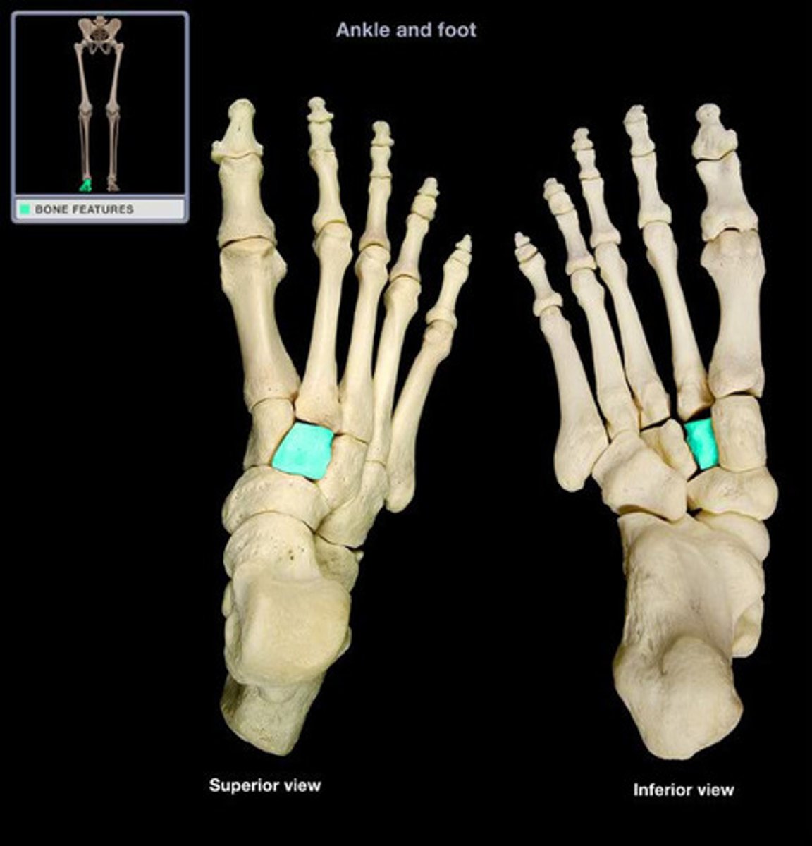
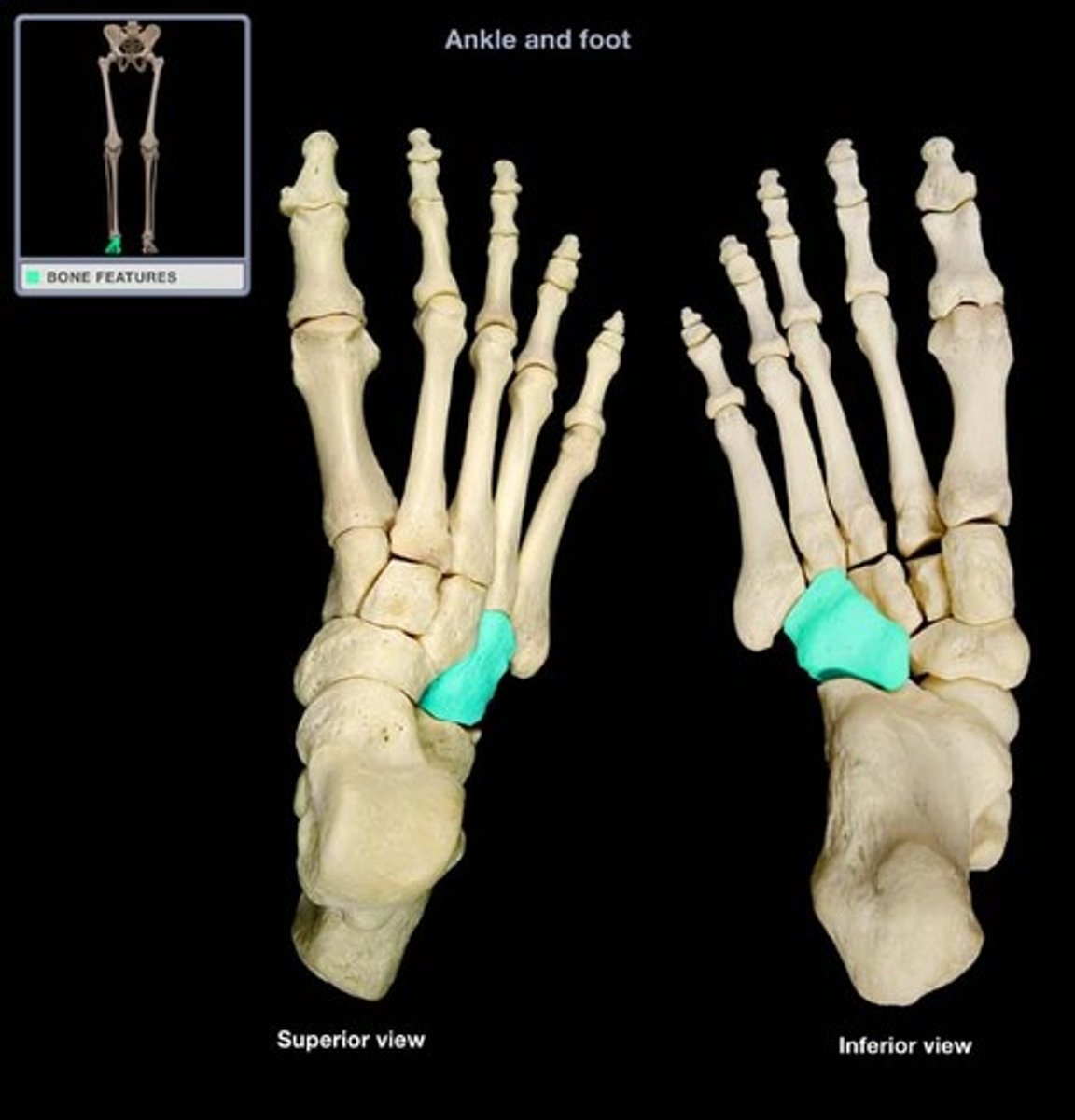
Medial cuneiform
medial bone closest to the metatarsals
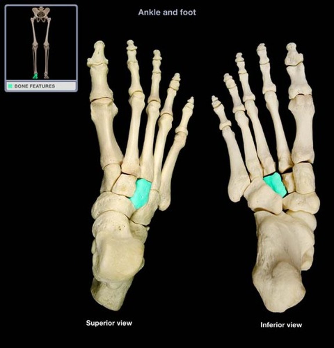
Intermediate cuneiform
Next to the medial cuneiform (middle)
Lateral cuneiform
Most lateral of the cuneiforms, located between the cuboid and the intermediate cuneiform.
Talus
Most superior of the foot bones
Trochlea of talus
Smooth superior portion of the talus that articulates with the tibia.
Cuboid
Cube shaped and most lateral of the bones closest to the metatarsals
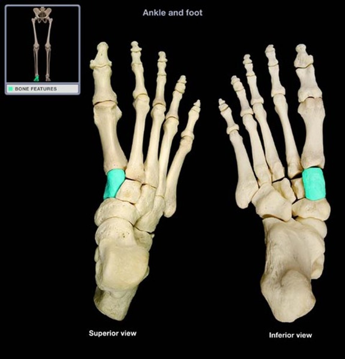
Navicular
Ship shaped bone between the talus and the cuneiforms
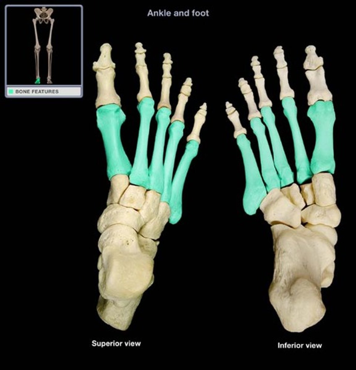
Metatarsals
The long bones in the feet
Phalanges (singular-phalanx)
Bones that form the toes
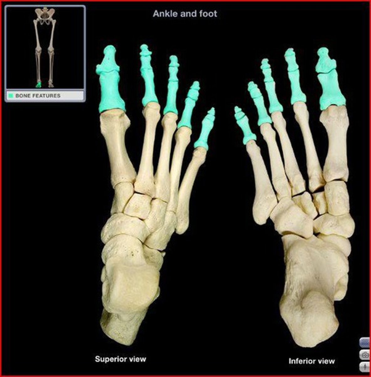
Proximal phalanges
The toe bones closest to the metatarsals
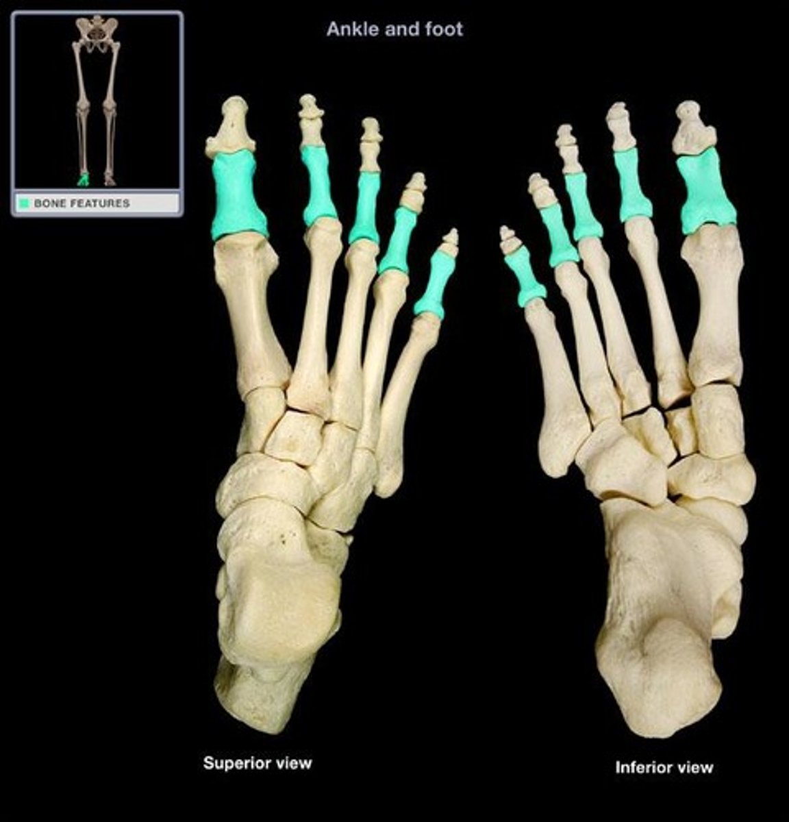
Middle phalanges 2-5
The middle toe bones ( there is not one in the big toe).
Distal phalanges 1-5
The toe bones farthest from the body.

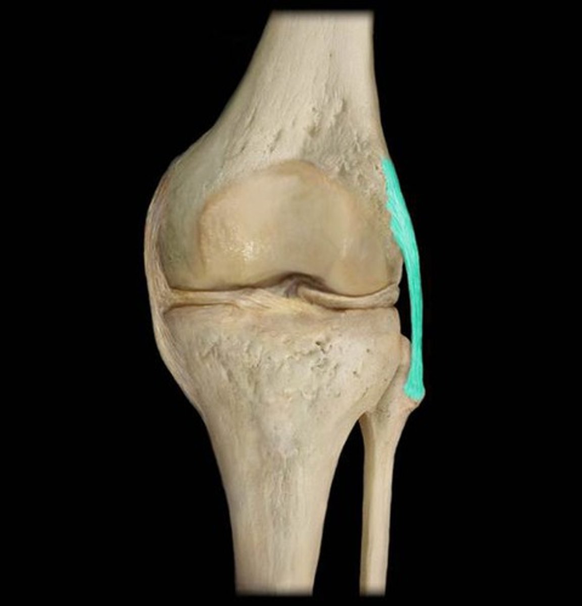
Fibular (lateral) collateral ligament
On lateral side, attaches the femur to the fibula.
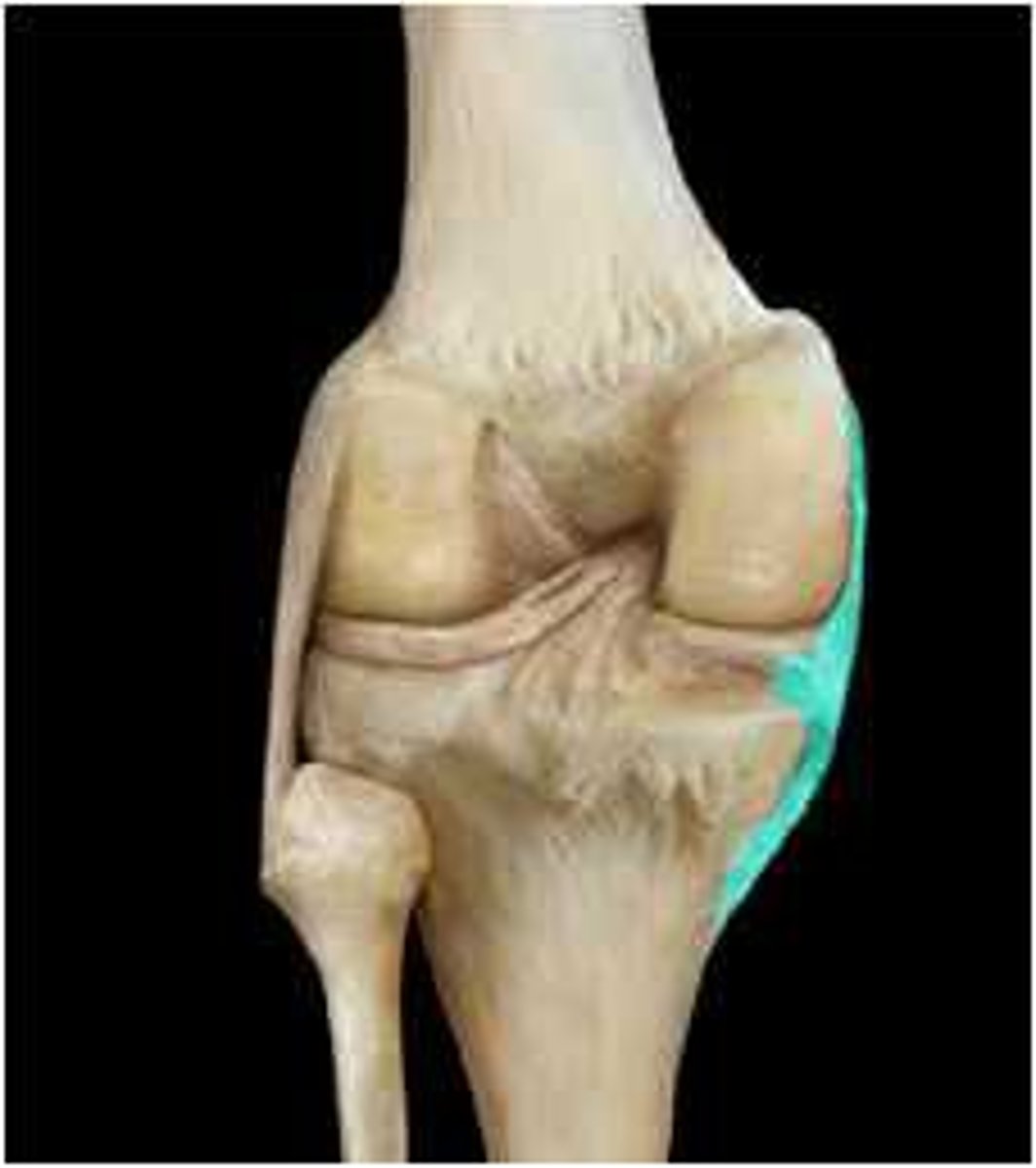
Tibial (medial) collateral ligament
On the medial side, attaches the femur to the tibia
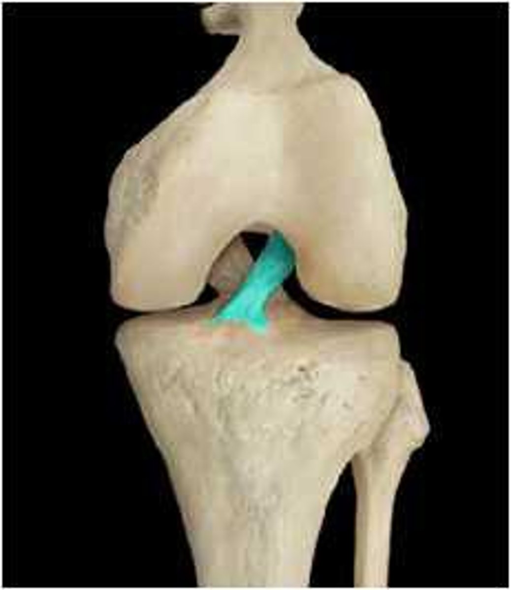
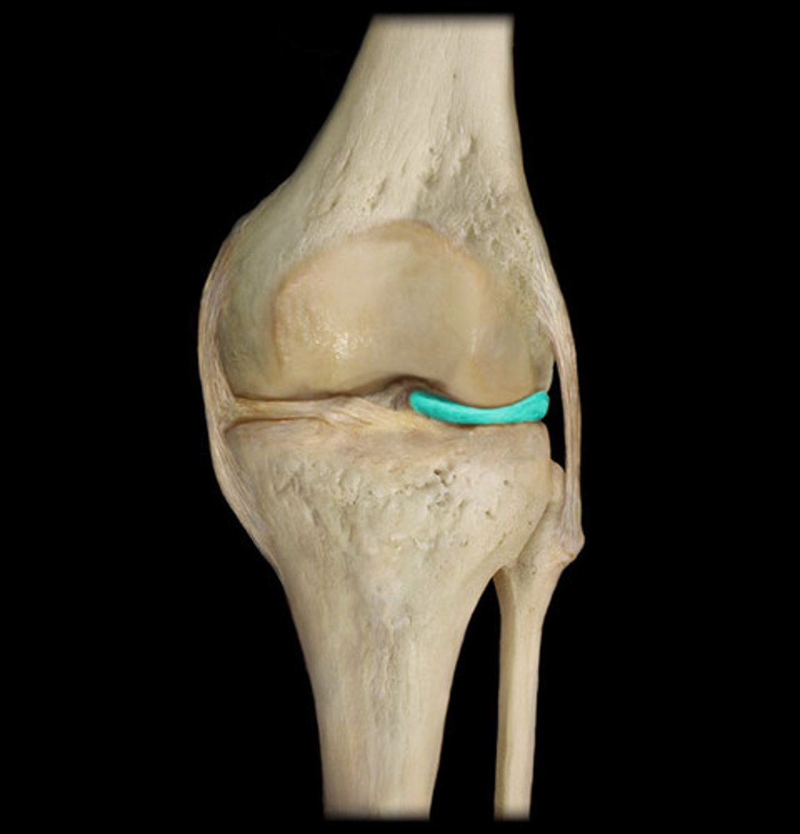
Anterior cruciate ligament
Attaches the femur to the lateral meniscus
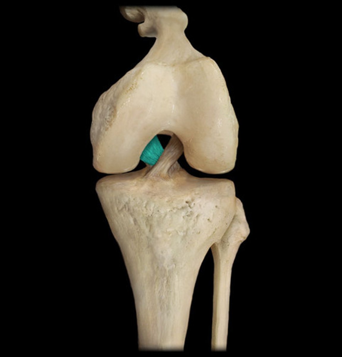
Posterior cruciate ligament
Attaches the femur to the medial meniscus
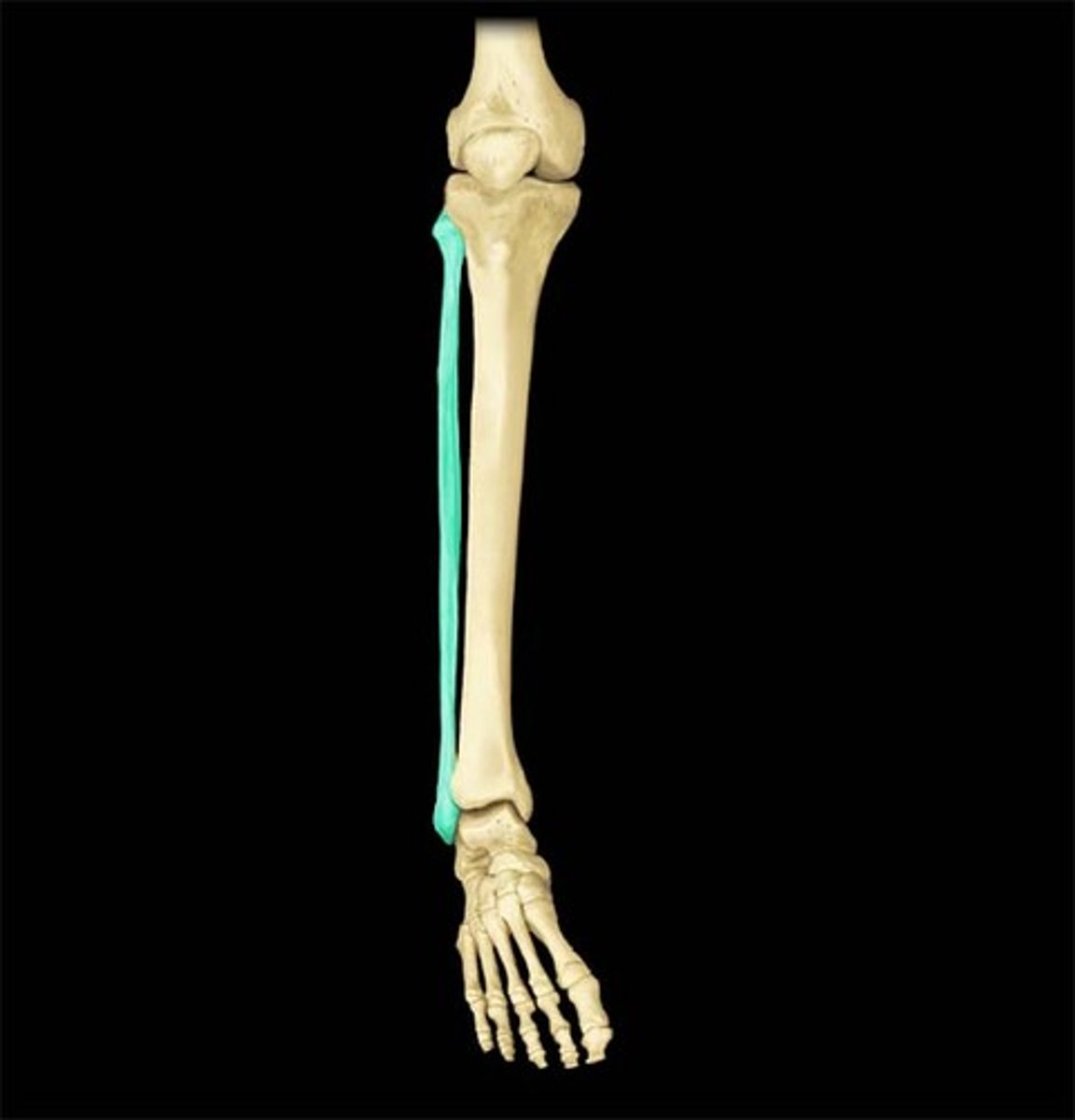
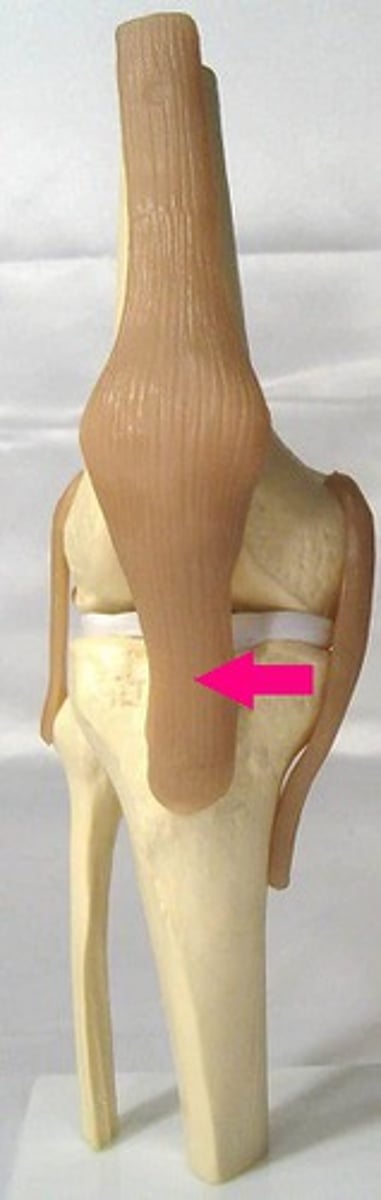
Patellar ligament
Is the lower portion of the ligament that attaches to the patella ( from patella to tibia)
Medial meniscus
A semicircular joint found on the medial side between the femur and the tibia
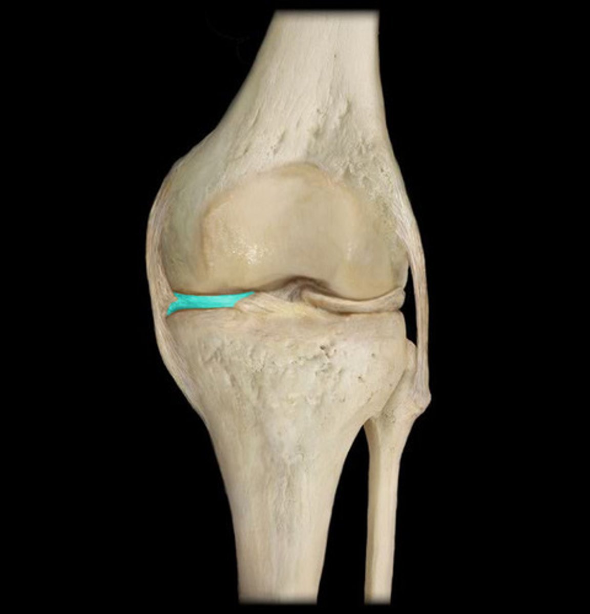
Lateral meniscus
A semicircular joint found on the lateral side between the femur and the tibia
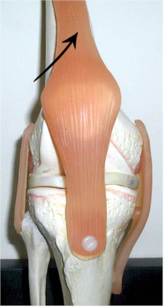
Quadriceps tendon
Tendon that attaches the patella to the femur
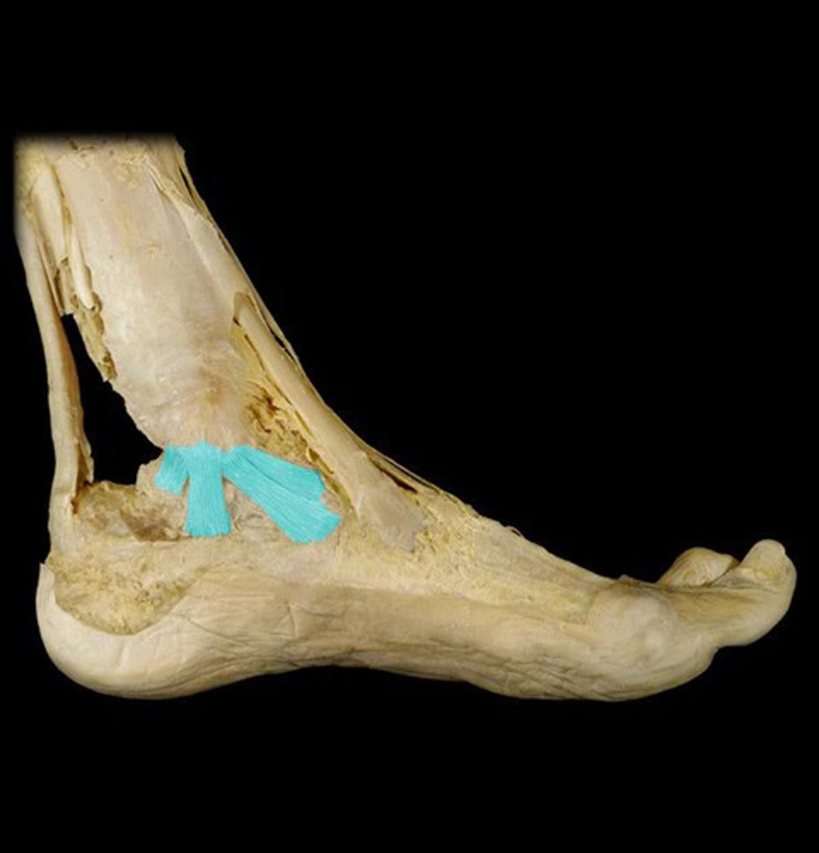
Lateral calcaneofibular ligament
The ligament found on the outside of the ankle that attaches the fibula and the calcaneus
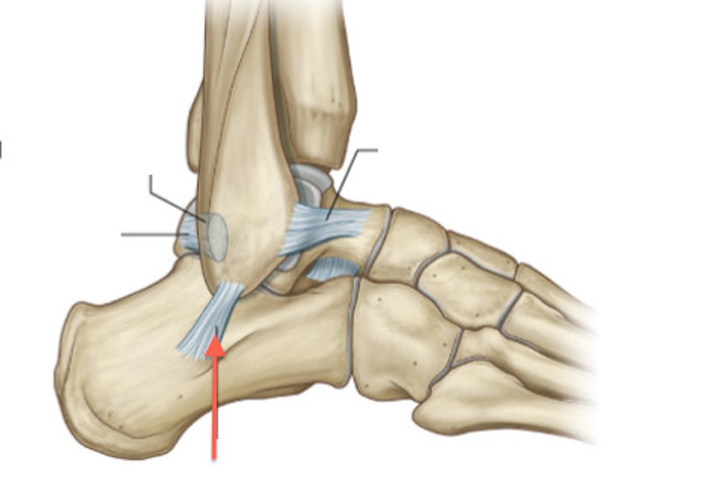
Medial deltoid ligament
Made of three ligaments that attach the tibia to the calcaneus, talus, and navicular bones.
Metatarsophalangeal joints
The joints between the tarsals and metatarsals
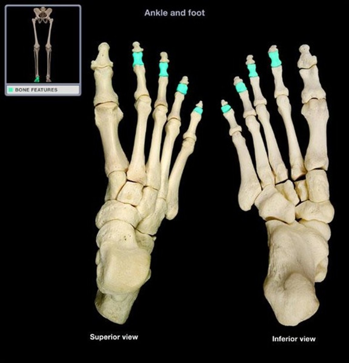
Interphalangeal joint 1
Joint between the proximal and distal phalanges of the big toes only
Proximal interphalangeal joints
Joint between the proximal and middle phalanges

Distal interphalangeal 2-5
Joint between the middle and distal phalanges

Tibialis anterior muscle
Runs from near the patella to the big toe
Extensor hallucis longus muscle
Deep under the extensor digitorum longus muscle
Extensor digitorum longus muscle
From knee to the toes. on the lateral side of tibialis anterior m.
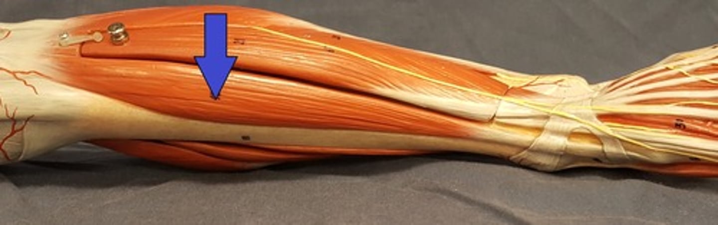
Fibularis (peroneus) brevis muscle
Along the fibula lateral to the extensor digitorum longus muscle.
Fibularis (peroneus) longus muscle
Lateral and superior to fibularis (peroneus) brevis m.
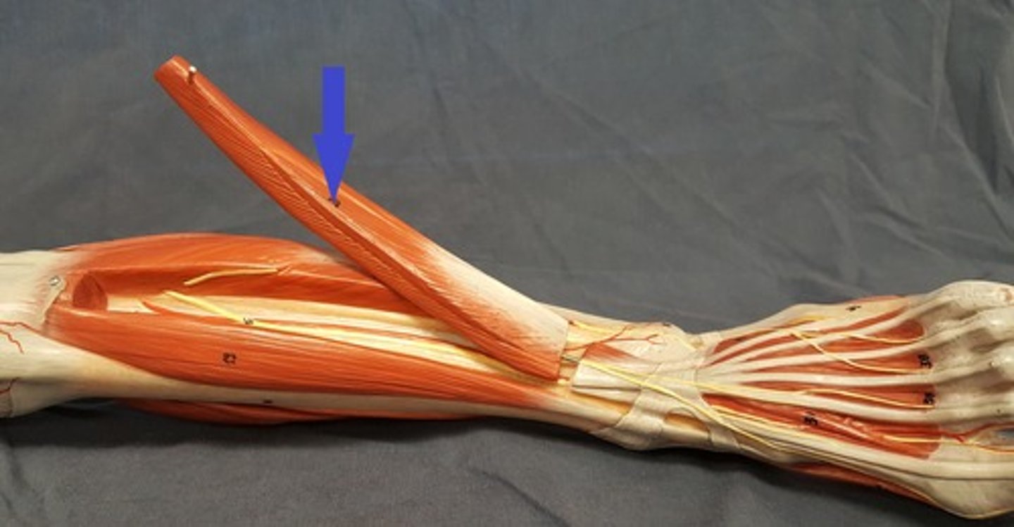
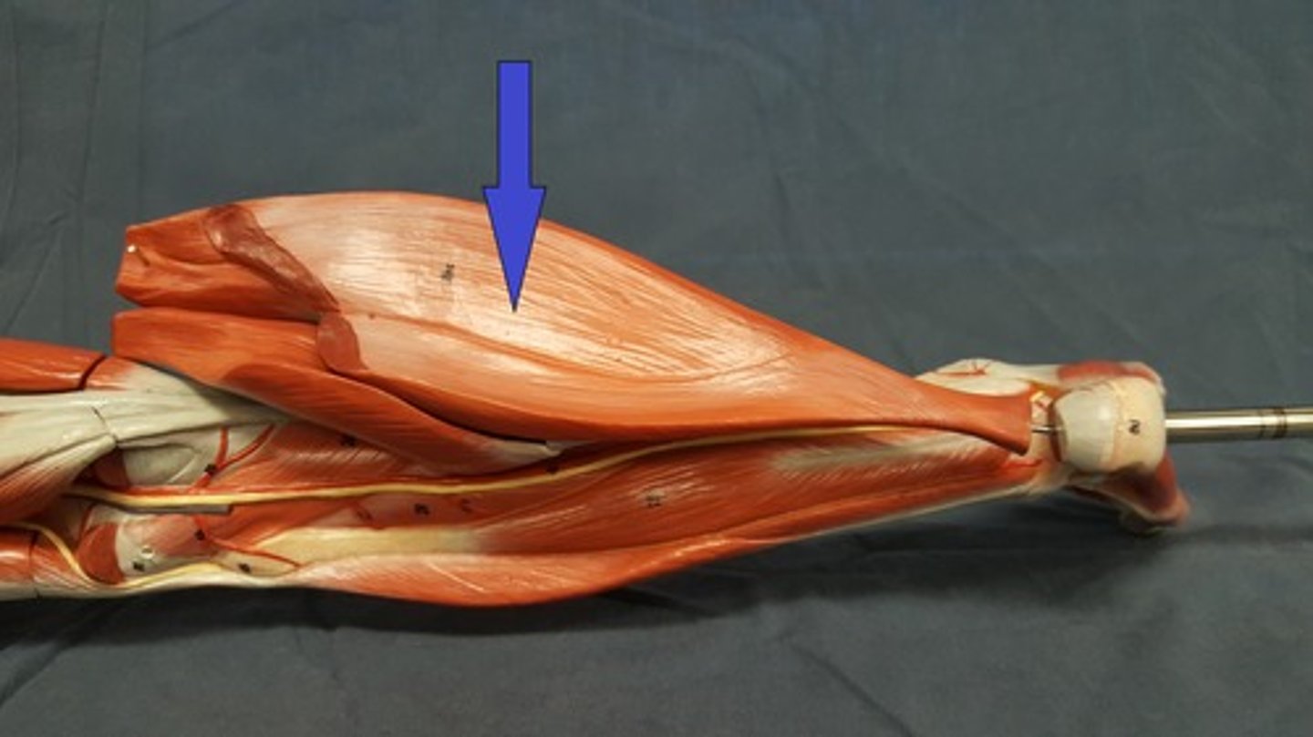
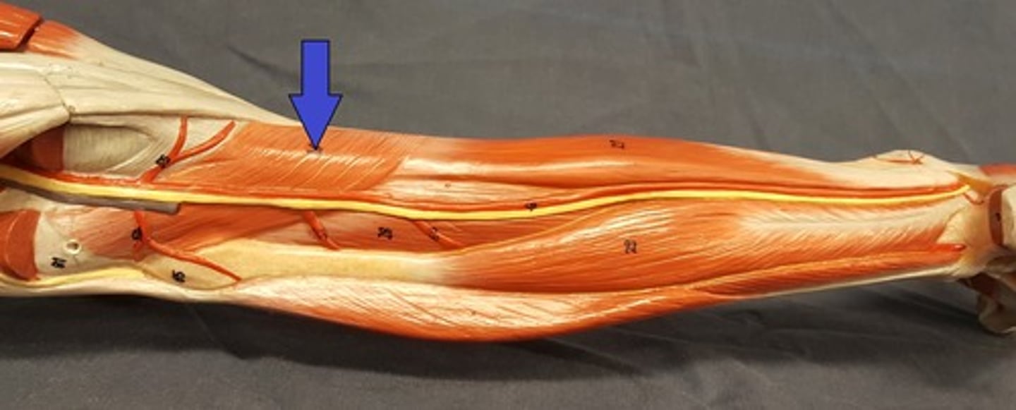
Gastrocnemius muscle
Both sides of the upper calf
Calcaneal (Achilles) tendon
Connects the calf to the calcaneus

Soleus muscle
Deep to the Calcaneal tendon and Gastrocnemius m.
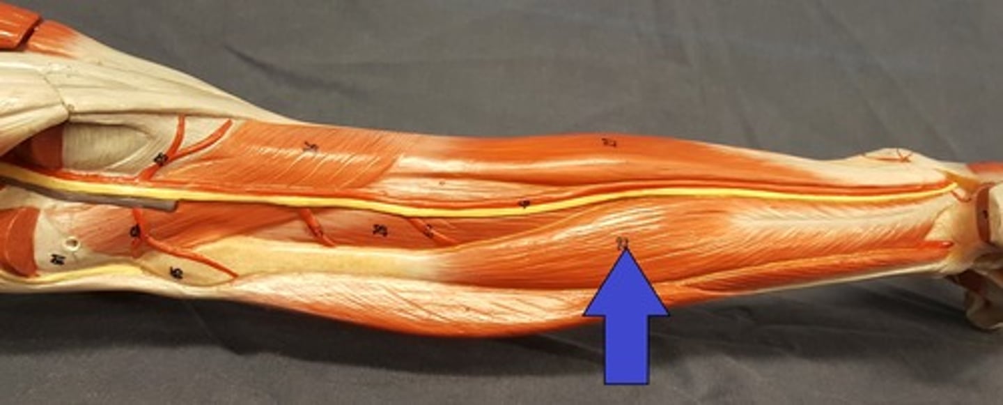
Flexor hallucis longus muscle
Seen after removing the gastrocnemius m. on the lateral side running along the fibula.
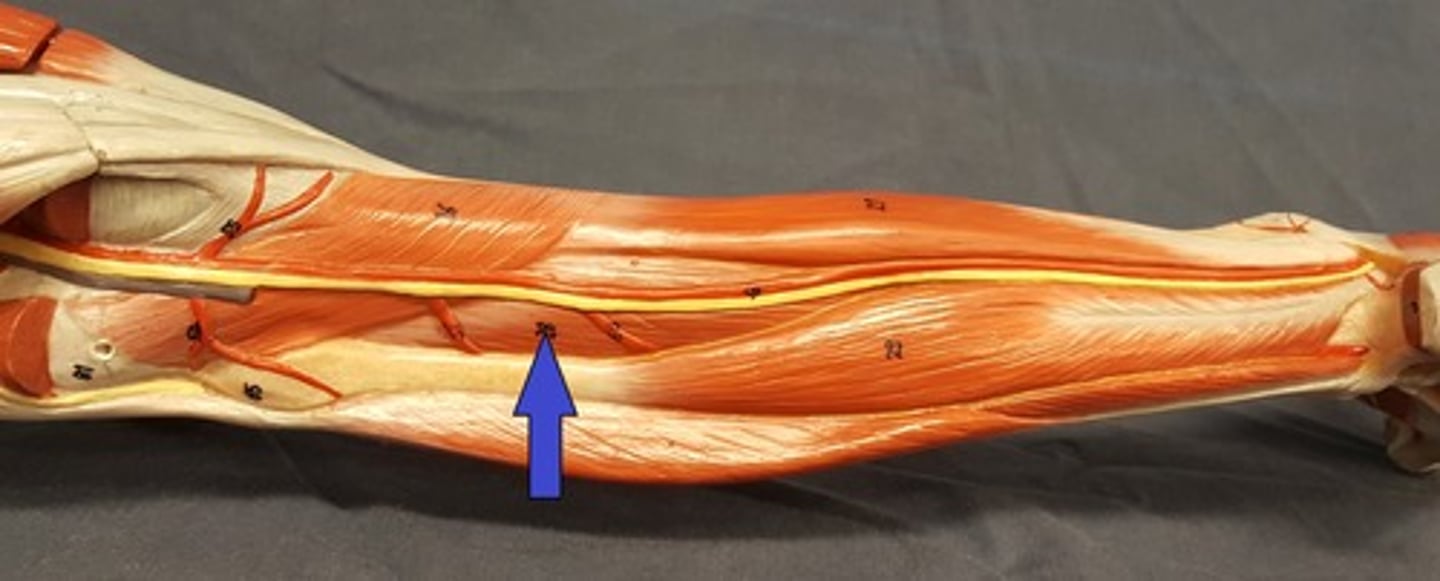
Flexor digitorum longus muscle
Seen after removing the gastrocnemius on the medial side along the tibia.

Tibialis posterior muscle
Seen after removing the gastrocnemius between the flexor hallucis longus m. and the flexor digitorum longus m.
Popliteus muscle
Seen after removing the gastrocnemius m. runs at an angle superior to the flexor hallucis longus m. the posterior tibial m. and the flexor digitorum longus m.
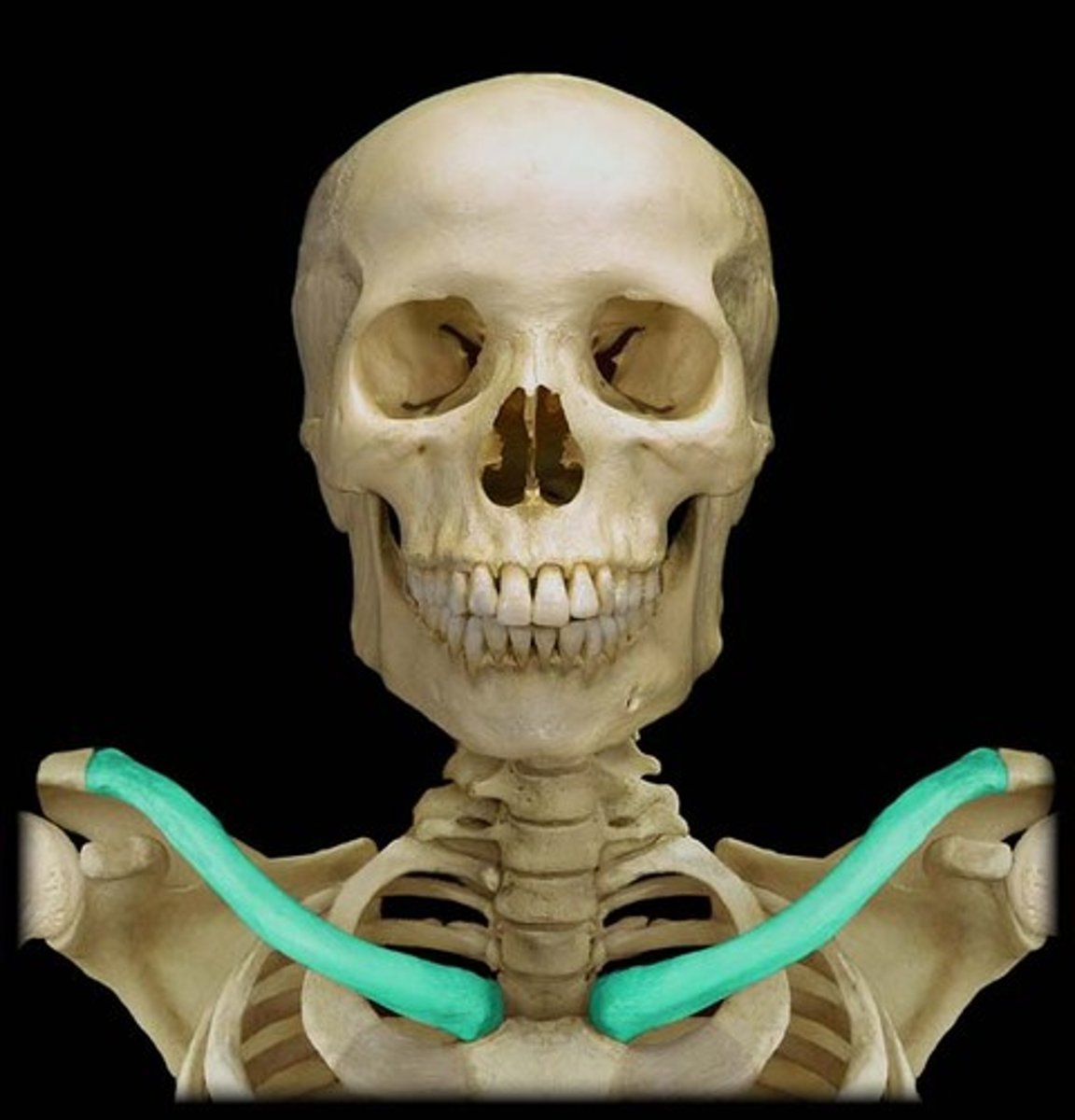
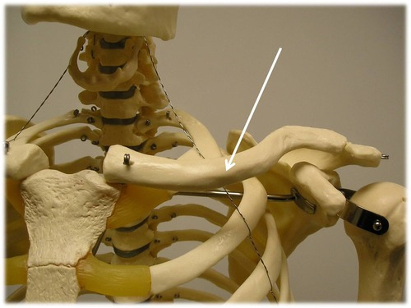
Clavicle
Collar bone
Sternal (medial) end
The (medial) end touching the sternum
(clubbed end)
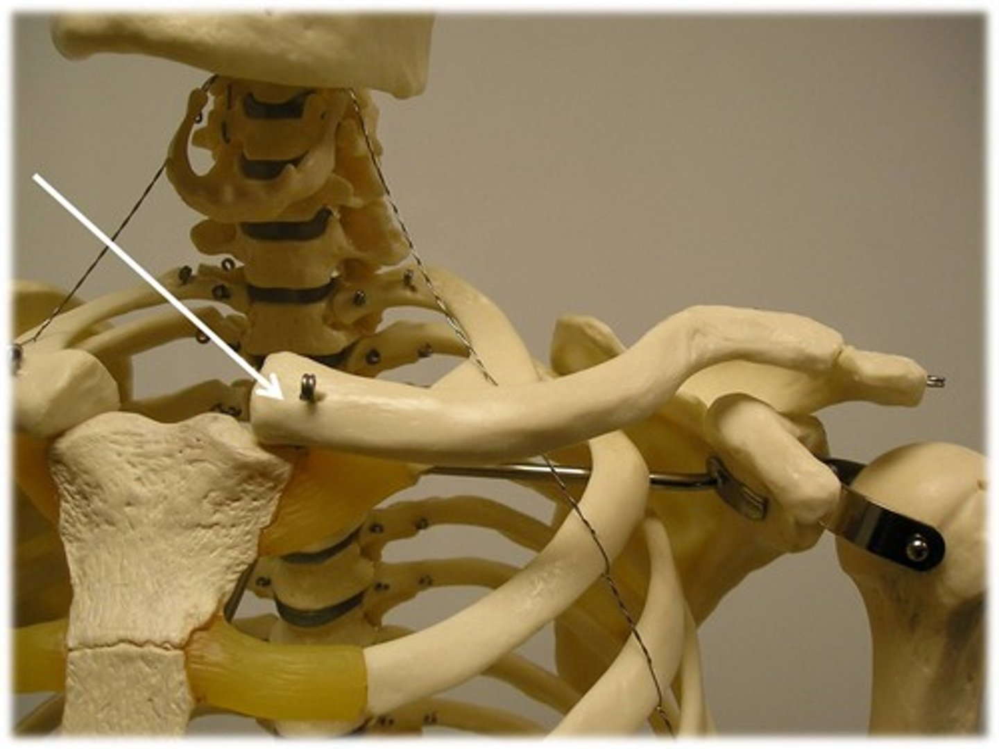
Acromial (lateral) end
The (lateral) end touching the acromial process of scapula (flat end)

Body of clavicle
The middle of clavicle
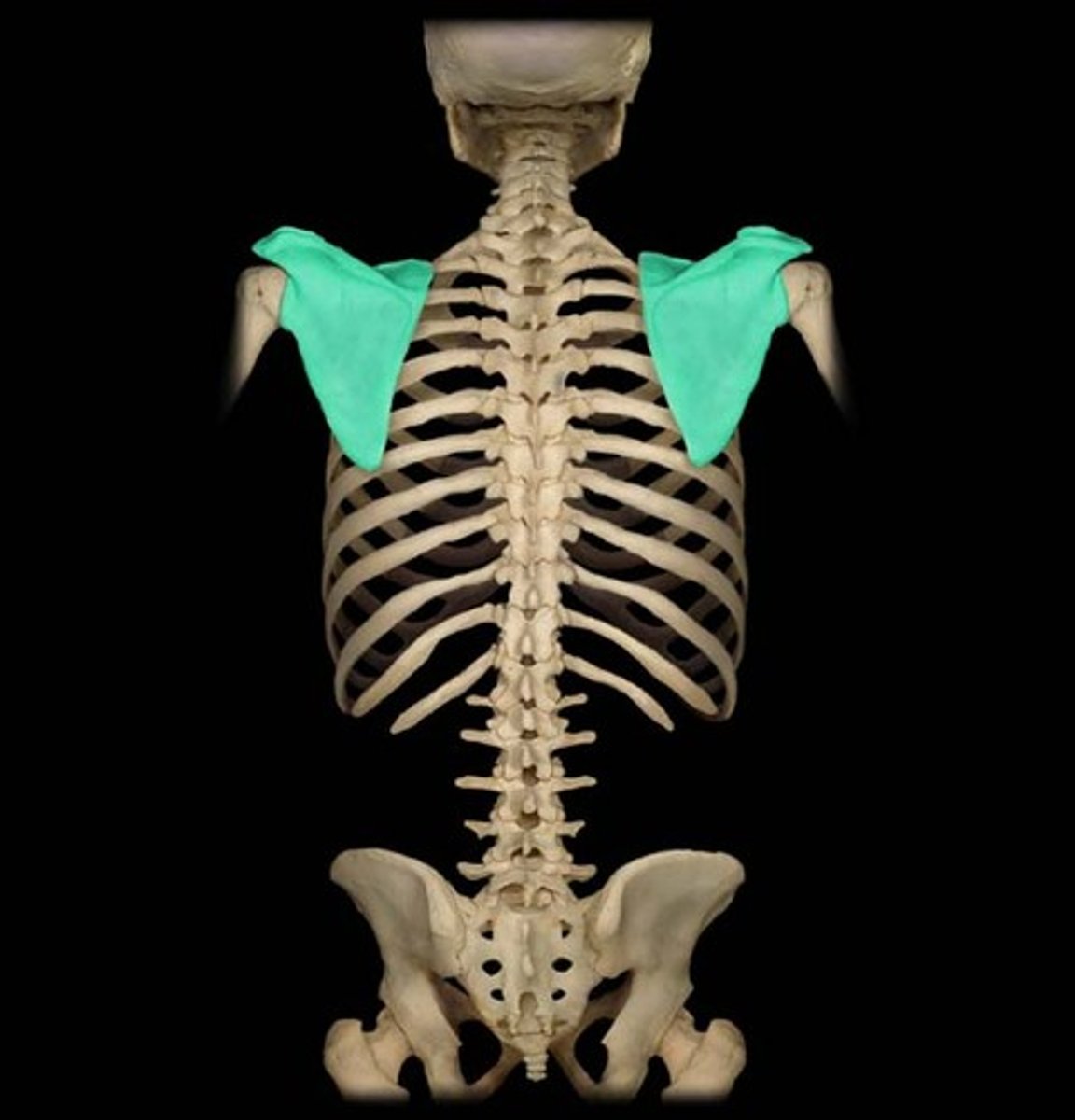
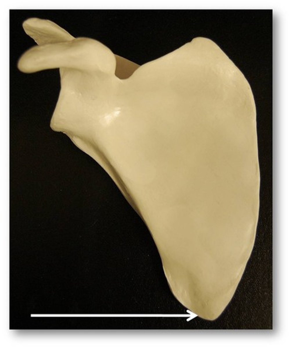
Scapula
Shoulder blade
Acromial process
Superior boney projection on posterior side extending from scapular spine, reaching anteriorly
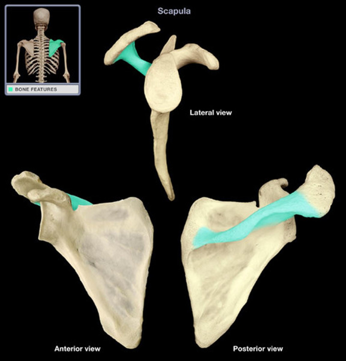
Scapular spine
Boney ridge running laterally on posterior side of scapula
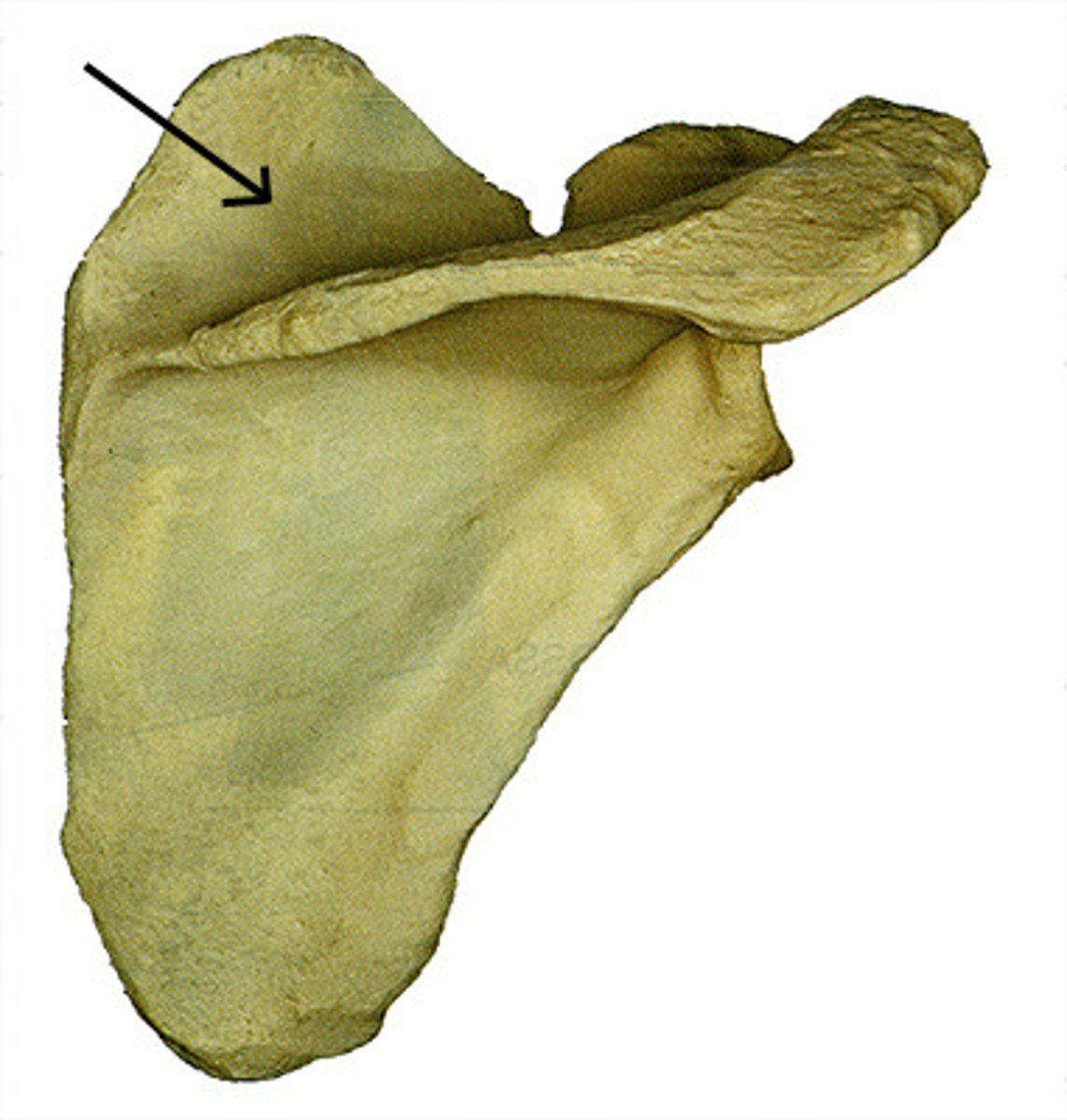
Supraspinous fossa
The "valley" above the scapular spine
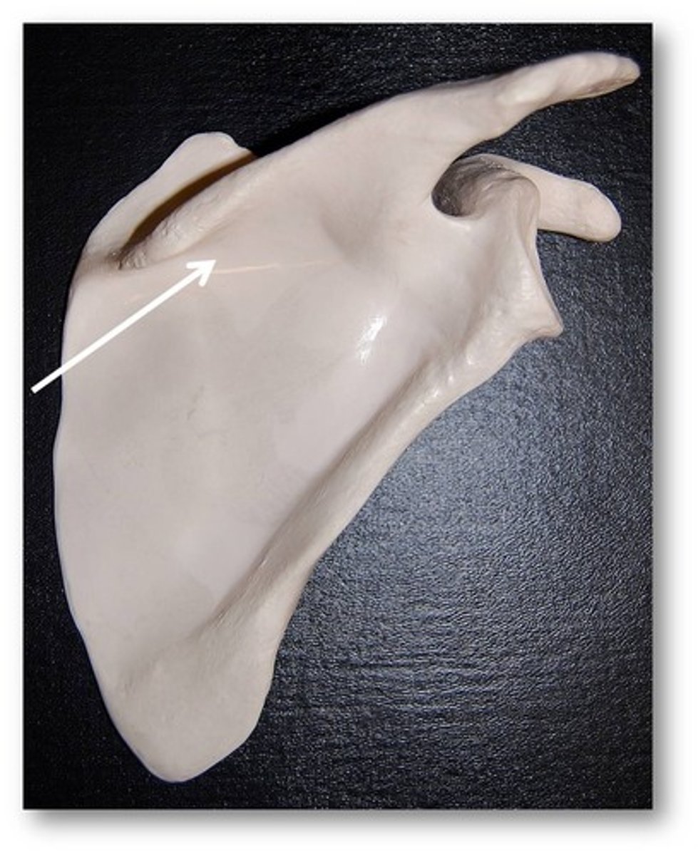
Infraspinous fossa
The "valley" below the scapular spine
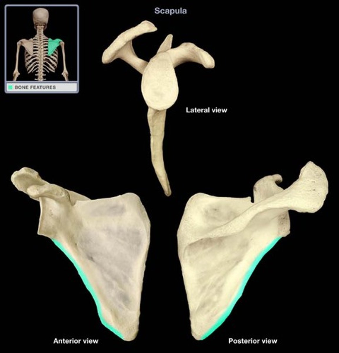
Axillary (lateral) border
Border starting from below glenoid fossa, extending down to inferior angle of scapula
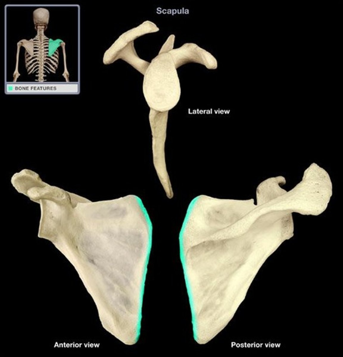
Medial border
Border closest to the spine, extending from superior angle to inferior angle of scapula
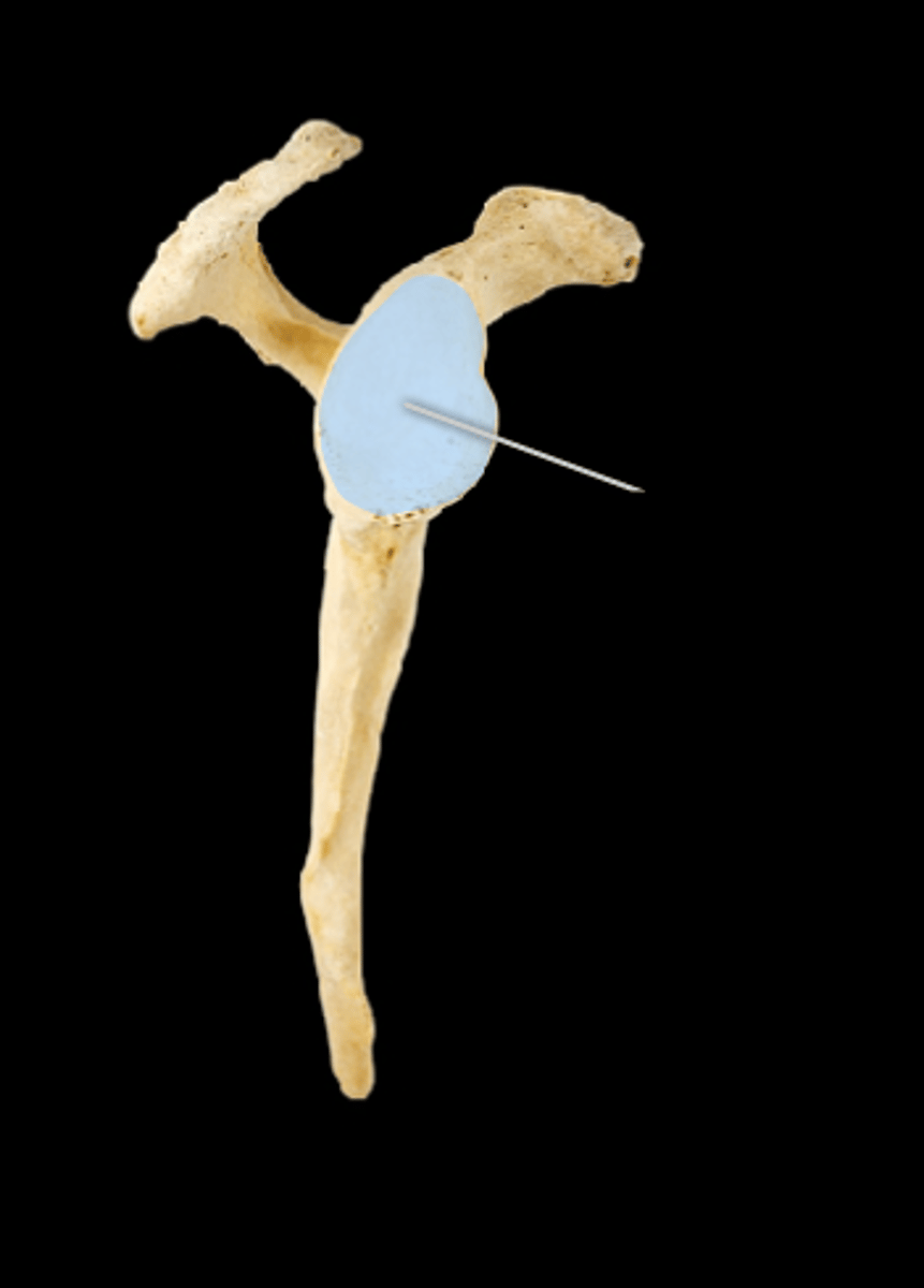
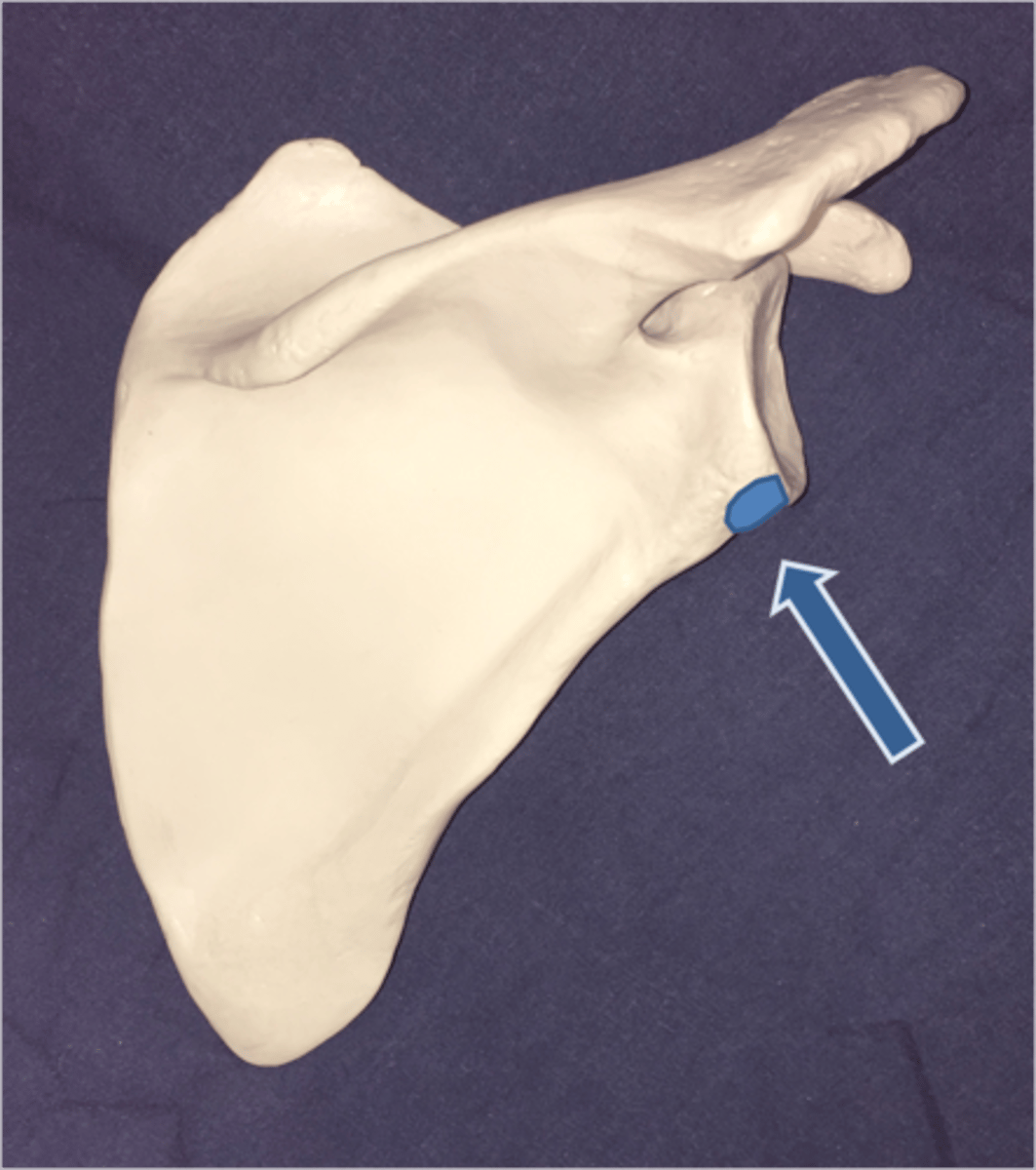
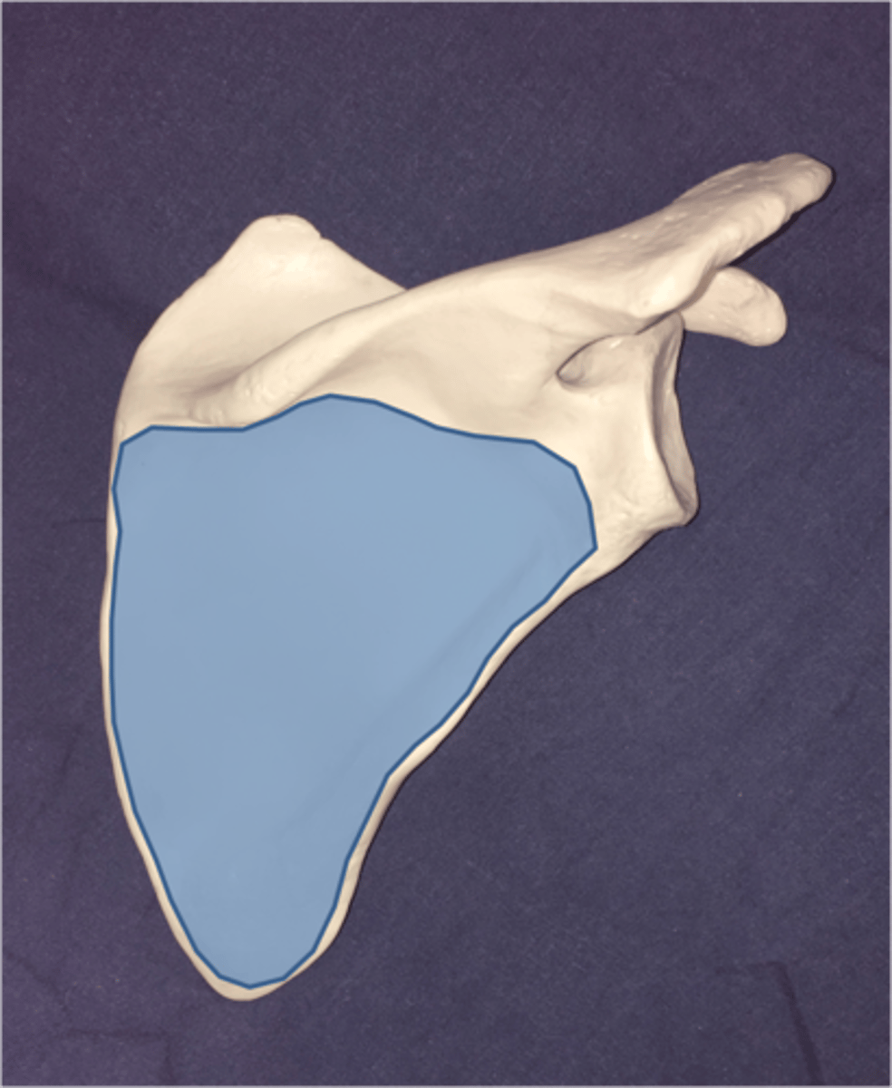
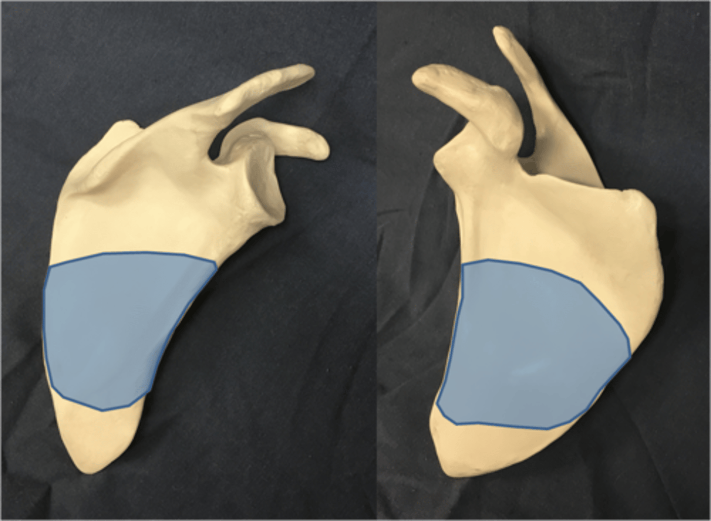
Glenoid fossa (cavity)
Articulation where the head of humerus and scapula meet
Coracoid process
Boney projection inferior to acromion process reaching anteriorly. Greek meaning, "crow's beak"
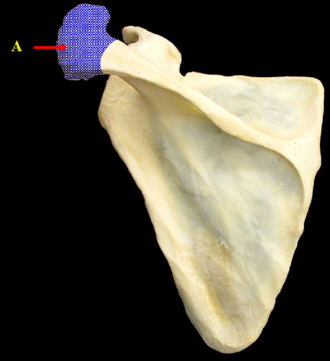
Superior angle
Top angle of scapula on medial side

Inferior angle
Bottom (base) angle of scapula
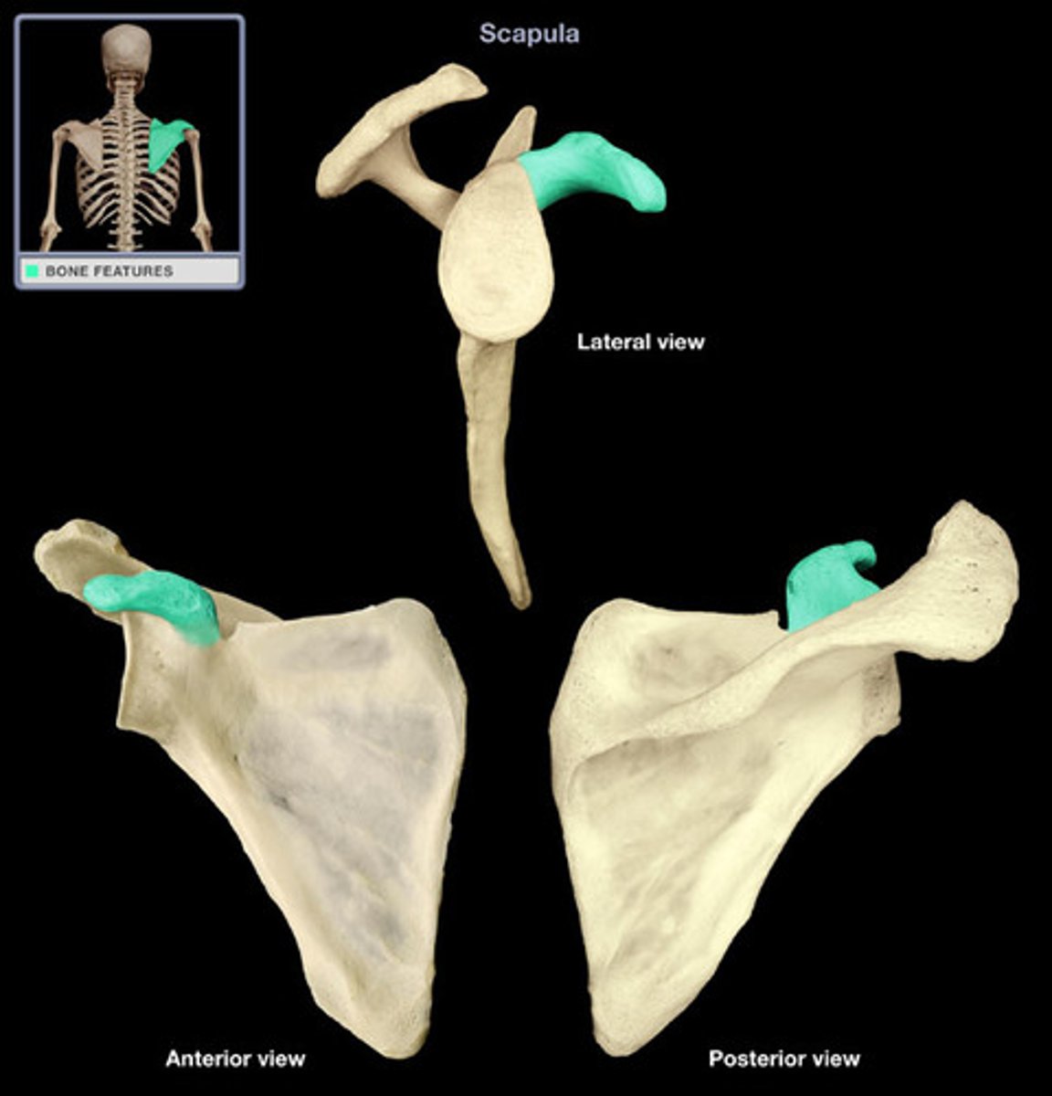
Lateral angle
Lateral side of scapula just below the glenoid fossa
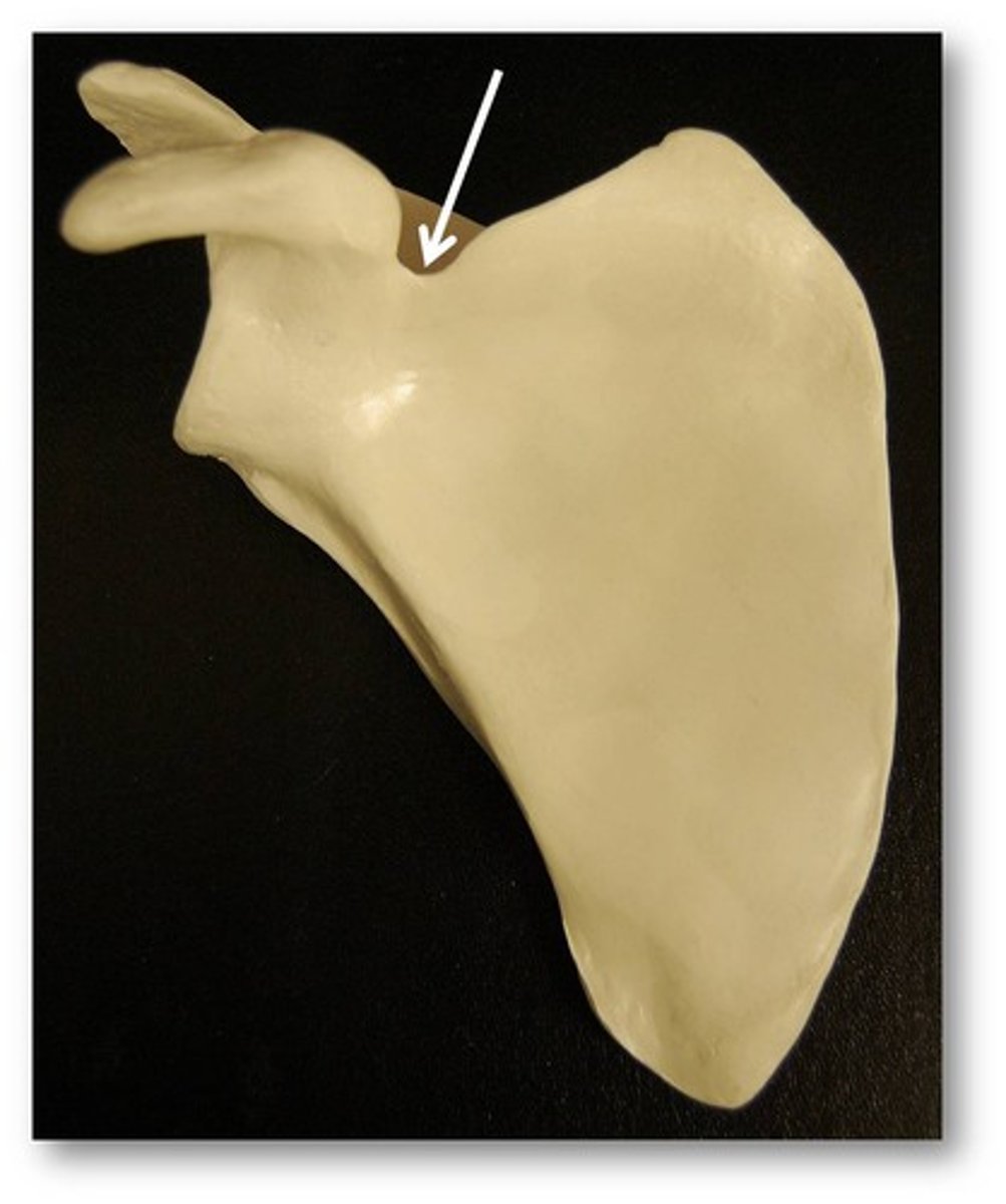
Suprascapular notch
Deep notch between coracoid process and superior angle
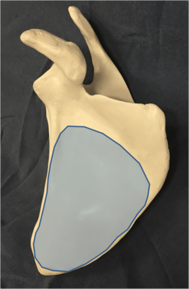
Costal surface
Anterior surface which touches the ribs.
Posterior surface
Area posterior (outer) side below scapular spine
Body of scapula
Main portion of scapula. (Blade)
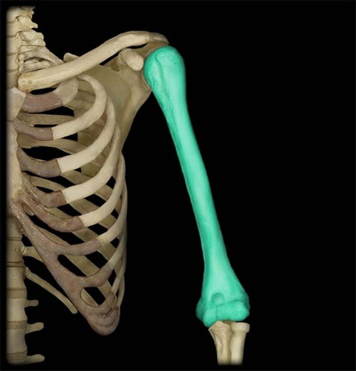
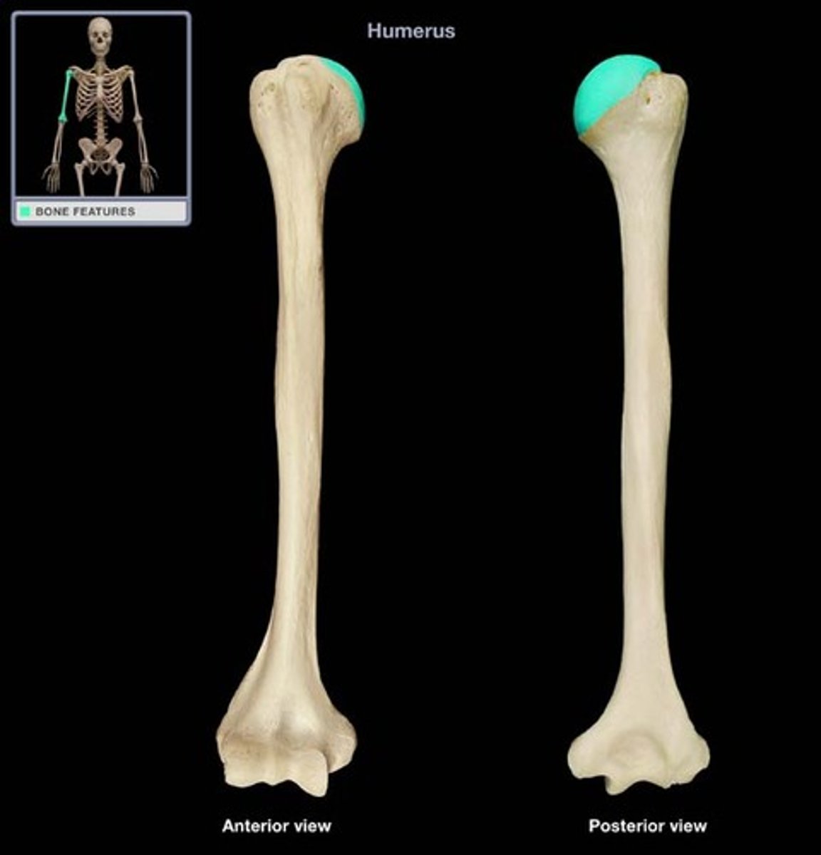
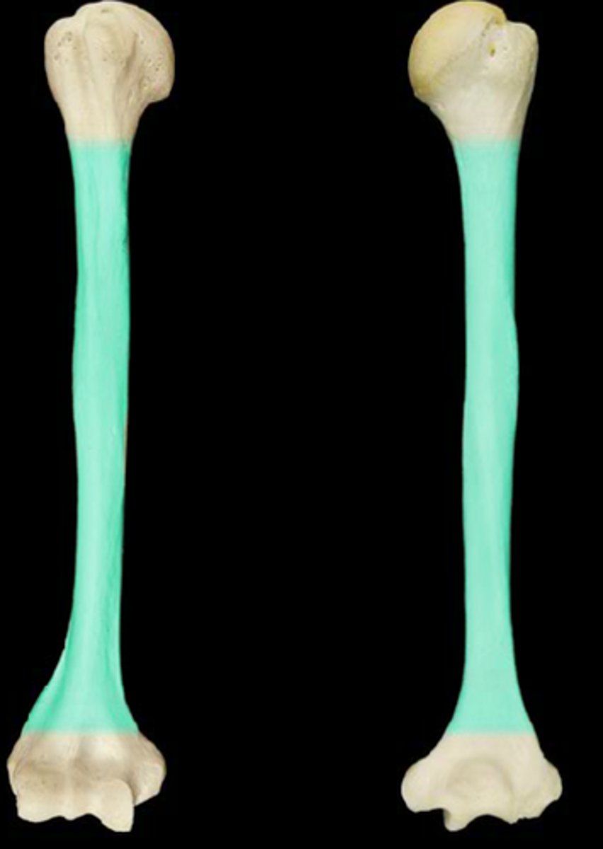
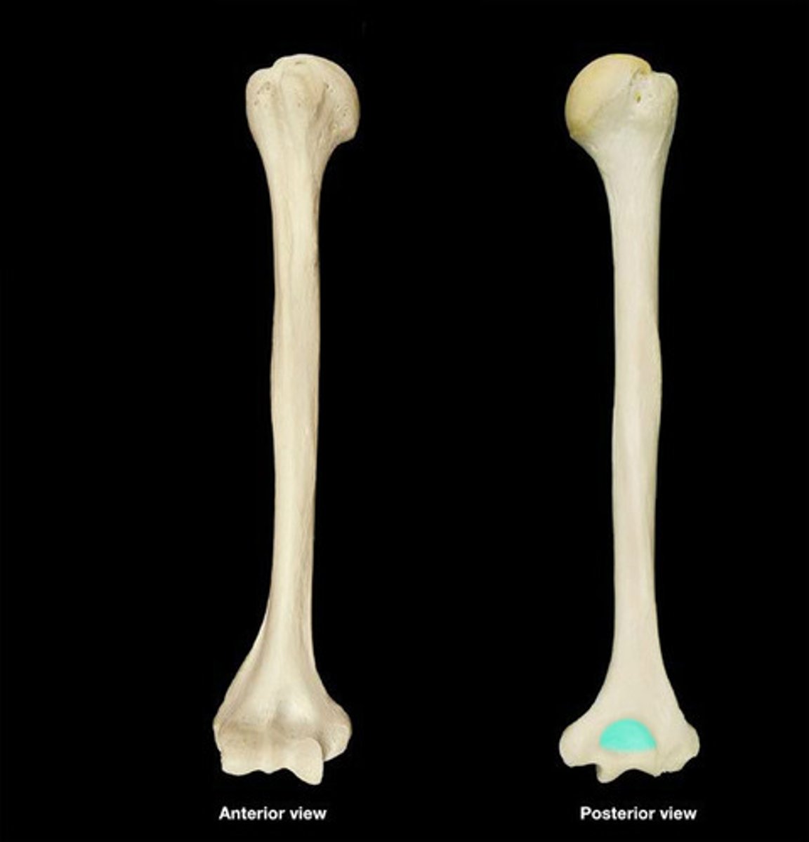
Humerus
Upper arm bone
Head of humerus
The round "ball" which articulates with glenoid fossa of scapula
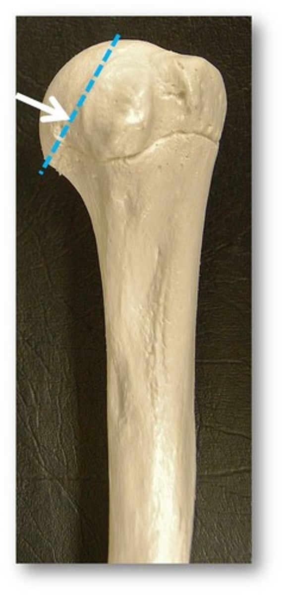
Anatomical neck of humerus
45 degree angled line at base of head of humerus
Surgical neck
The line encompassing below head of humerus
Greater tubercle
Proximal end of humerus. Larger bumpy protrusion on lateral side of humerus, opposite of the head of humerus

Lesser tubercle
Proximal end of humerus. Smaller bumpy protrusion more anterior on humerus,
Deltoid tuberosity
Area where deltoid muscle attaches on lateral side, midway down humerus as it slightly curves outward.
Body (shaft) of humerus
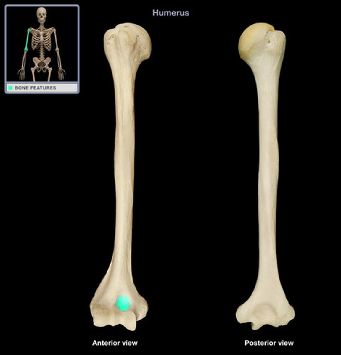
Lateral epicondyle of humerus
Distal end of humerus. The lateral protuberance. Epi=outside. Condyle=knuckle.
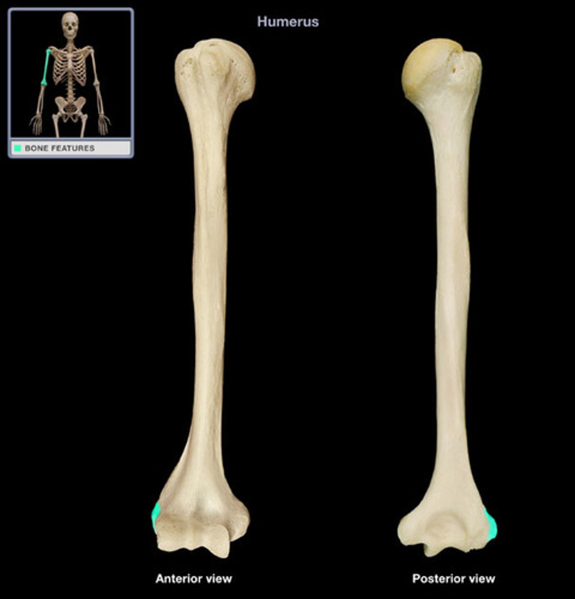
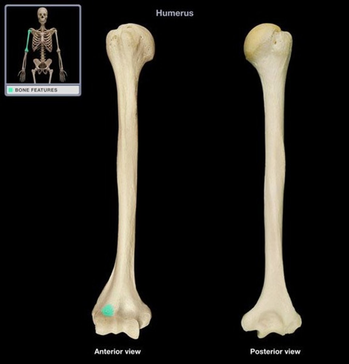
Medial epicondyle of humerus
Distal end of humerus. The medial protuberance. Epi=outside. Condyle=knuckle
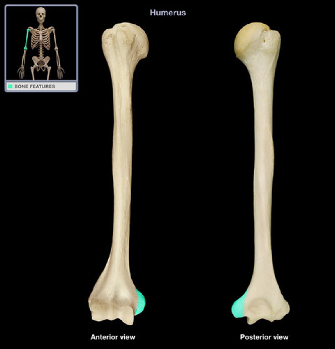
Olecranon fossa
Posterior side, distal end of humerus, large depression
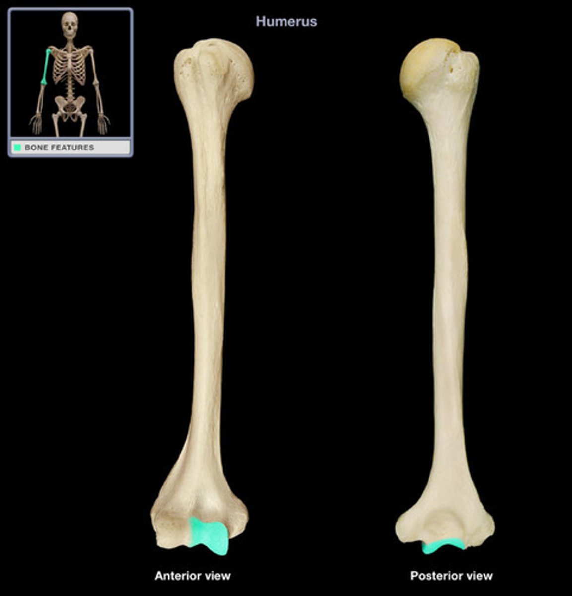
Coronoid fossa
Anterior side, distal end of humerus, medial small depression
Radial fossa
Anterior side, distal end of humerus, lateral small depression
Trochlea of humerus
Anterior side, distal end of humerus, medial smooth structure with a ridge at medial side
Capitulum
Anterior side, distal end of humerus, lateral smooth rounded structure

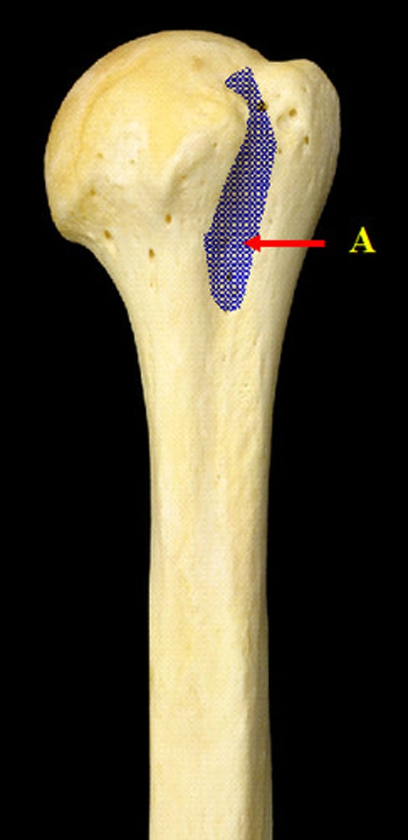
Bicipital groove
Anterior side, proximal end, groove between greater and lesser tubercles
Articular capsule
3

Glenohumeral
1

Coracohumeral ligament
2

Coracoacromial ligament
4

Coracoclavicular ligament
what is 5 and 6 called together
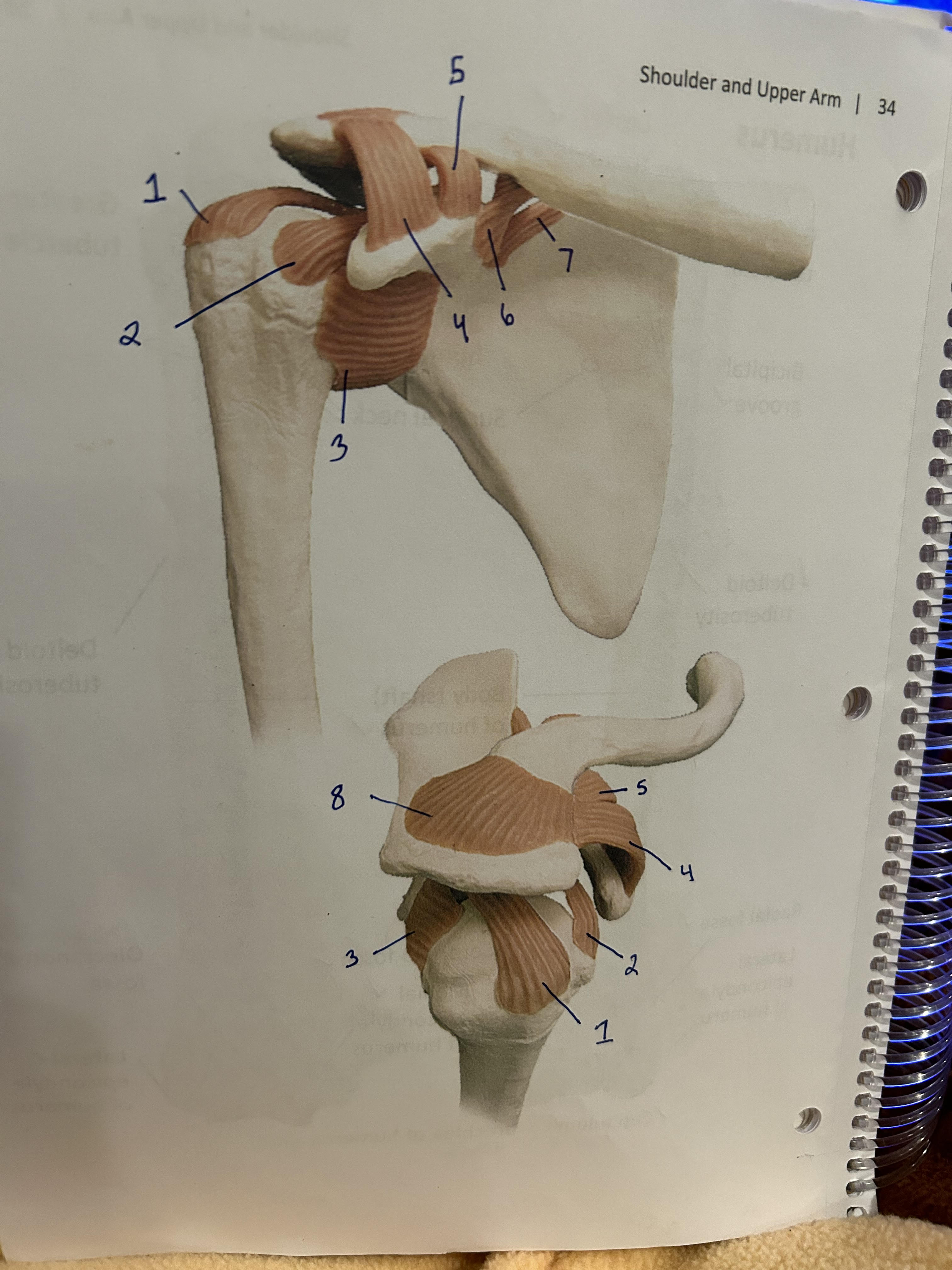
Conoid ligament
6

Trapezoid ligament
5
