BIO 1130: Chapter 5
 Call Kai
 Learn
Learn Practice Test
 Spaced Repetition
 Match
Match Flashcards
1/63

There's no tags or description
Looks like no tags are added yet.
Name | Mastery | Learn | Test | Matching | Spaced | Call with Kai | Chat |
|---|

64 Terms
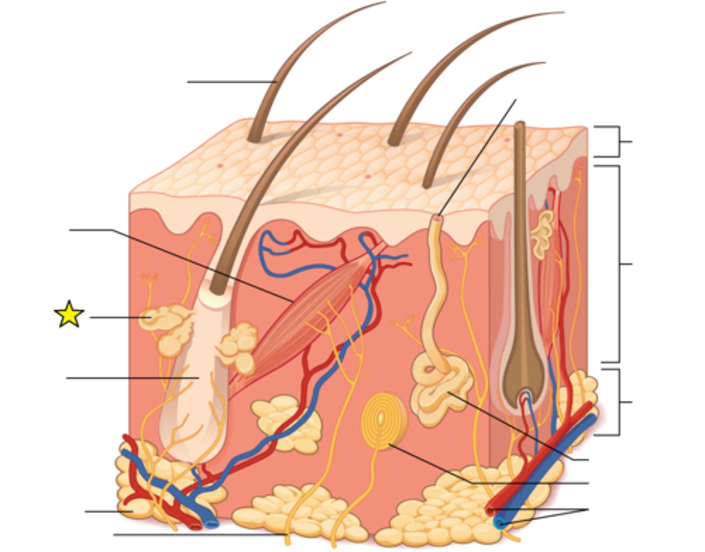
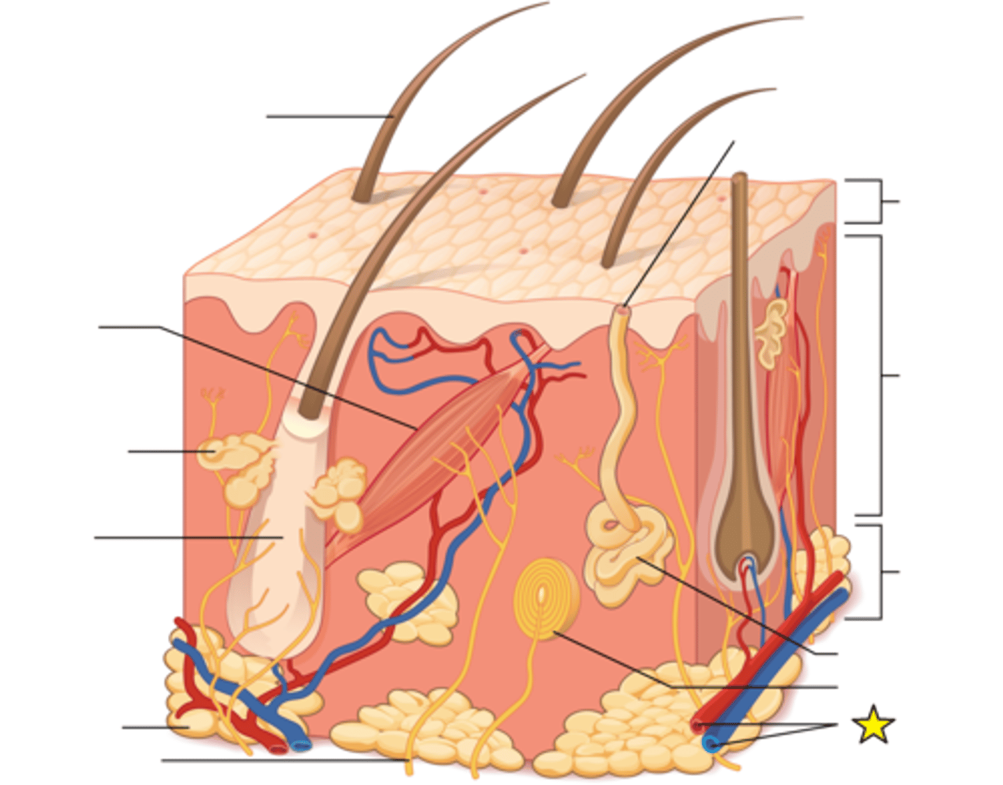
Sebaceous gland
Glands that secrete sebum, associated with hair follicles.
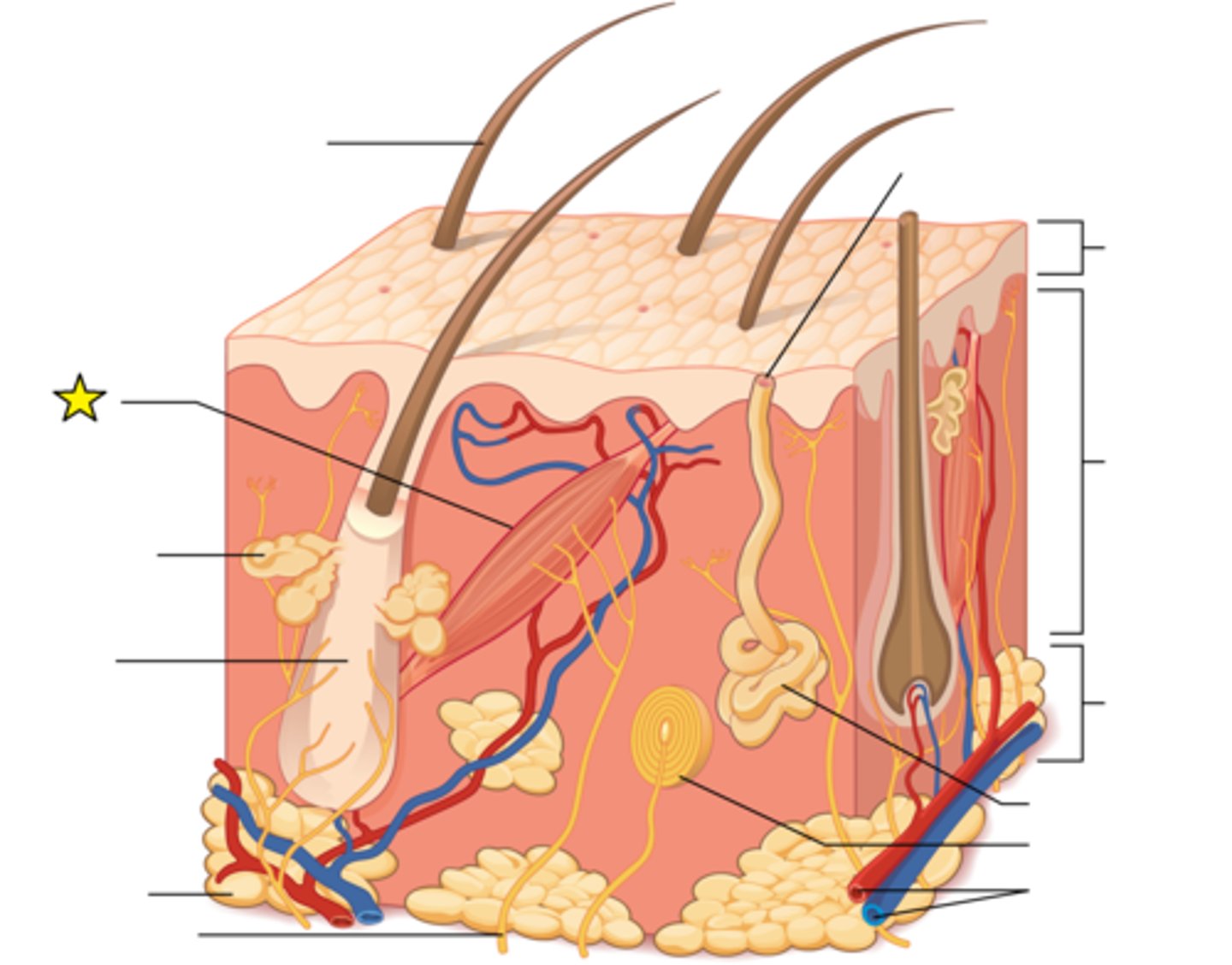
Arrector pili muscle
Attaches to the base of a hair follicle at one end and to dermal tissue on the other end. In order to generate heat when the body is cold, they all contract all at once, causing the hair to "stand up straight" on the skin.
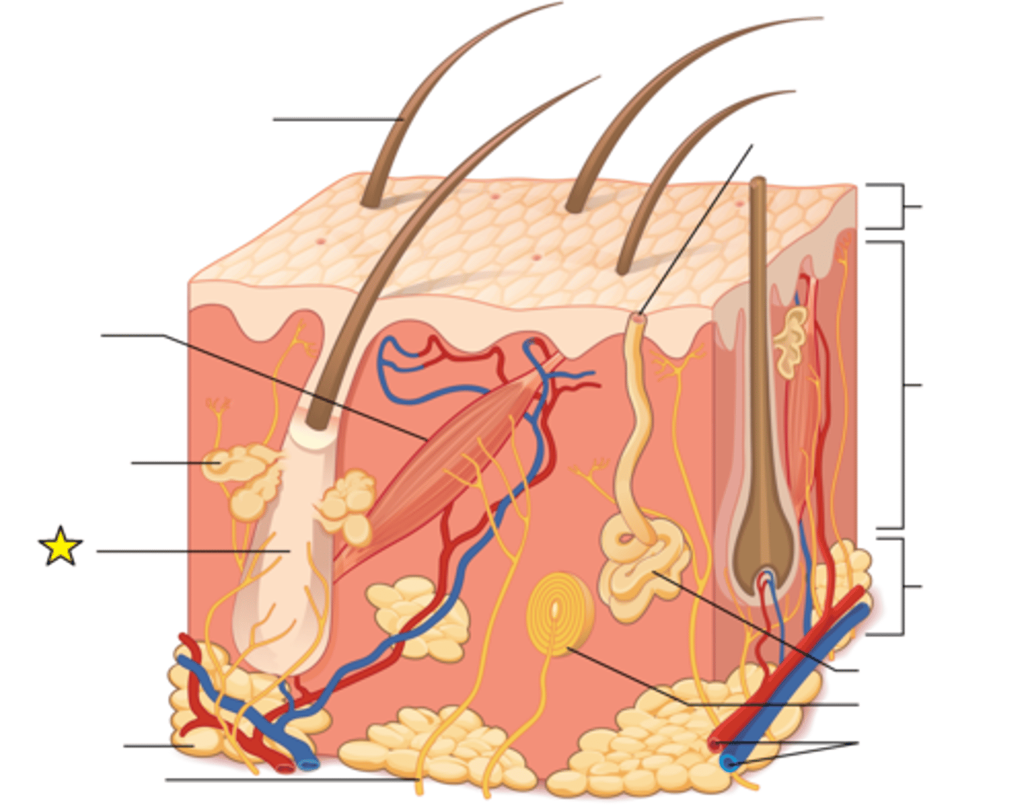
Hair follicle
Regulates hair growth via a complex interaction between hormones, neuropeptides, and immune cells.
Adipose tissue
Mainly fat globules. Many cells contain large lipid deposits. Functions: Insulates the body, protects some organs and serves as a sit of fuel
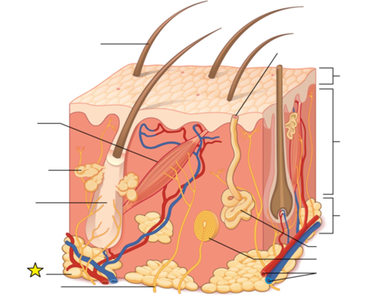
Sensory nerve fiber
Serve to sense and transmit heat, pain, and other noxious sensations. When these are not functioning properly they can produce sensations such as numbness, pins-and-needles, pain, tingling, or burning.
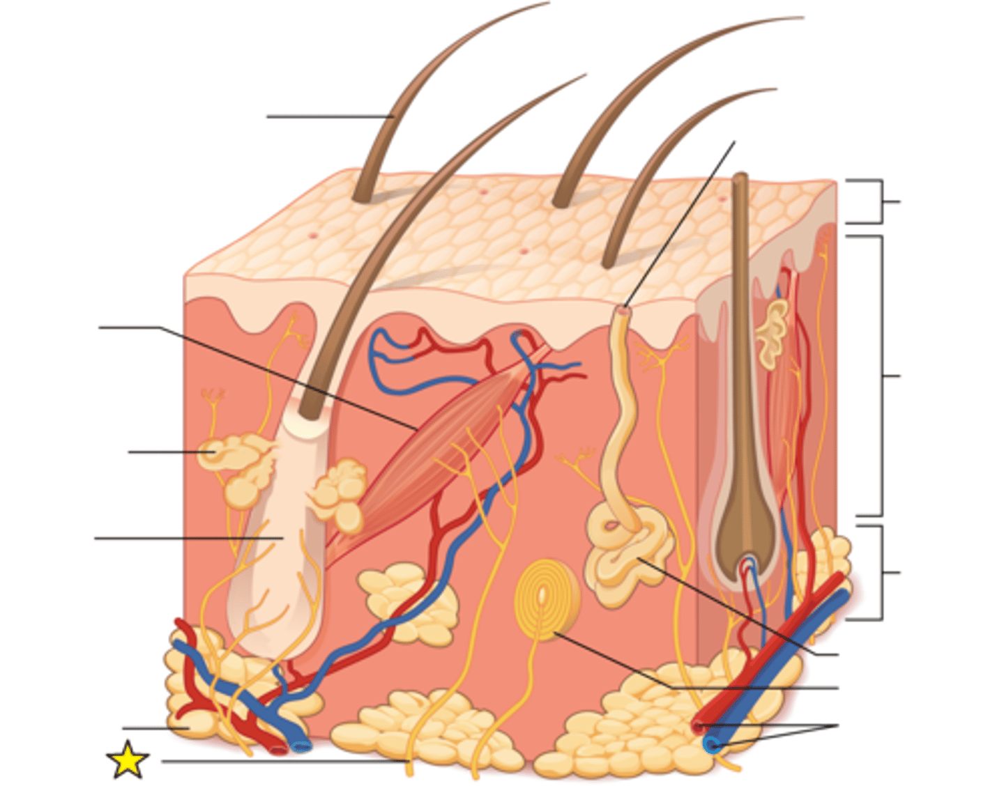
Hair shaft
The part of the hair not anchored to the follicle, and much of this is exposed at the skin's surface.
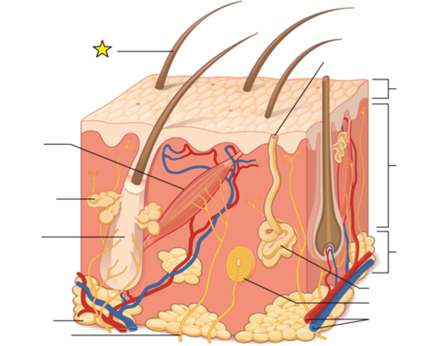
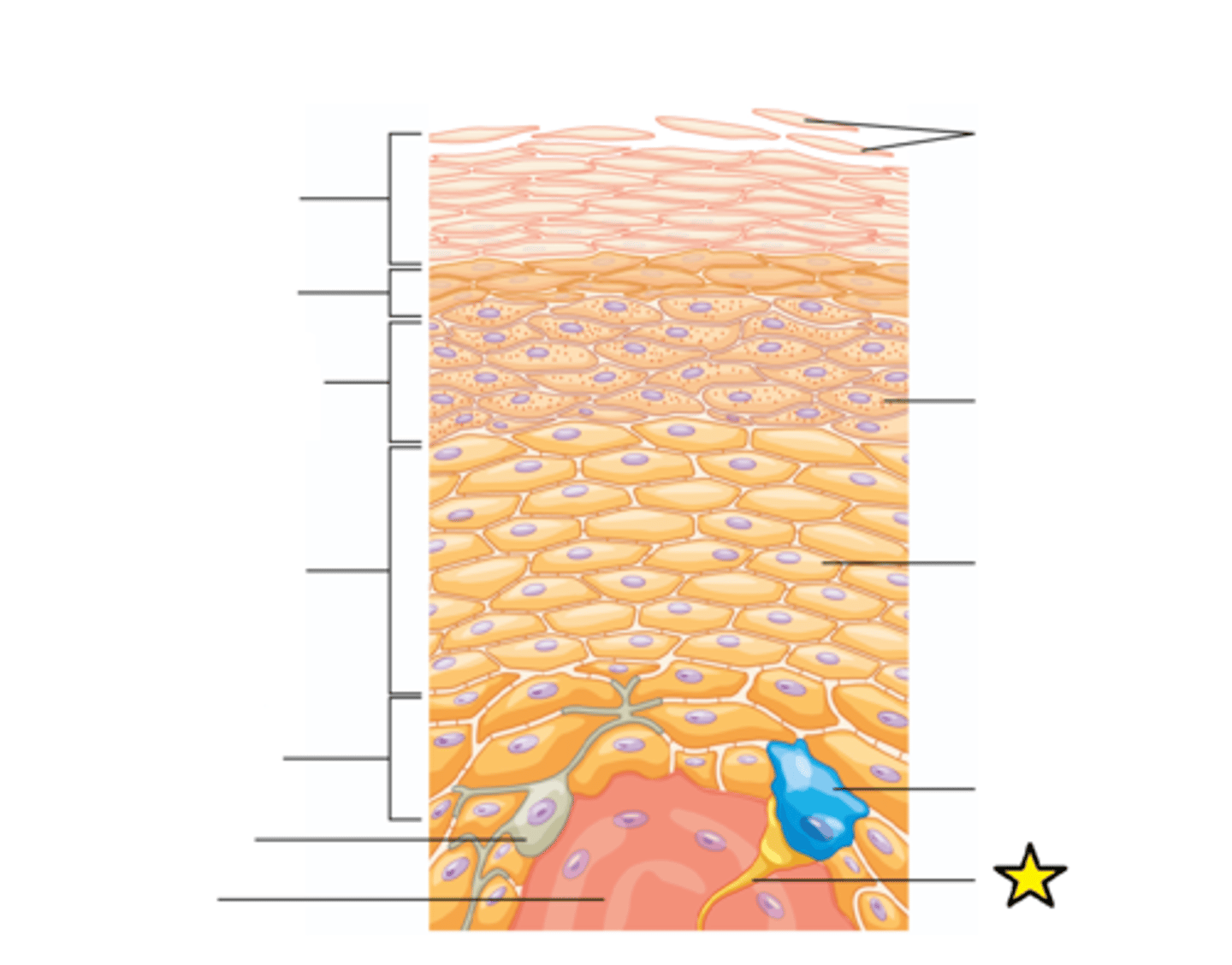
Epidermis
Composed primarily of keratinocytes arranged into layers called strata; Thin skin epidermis has 4 strata layers; Thick skin epidermis has 5 strata layers (palms and soles)
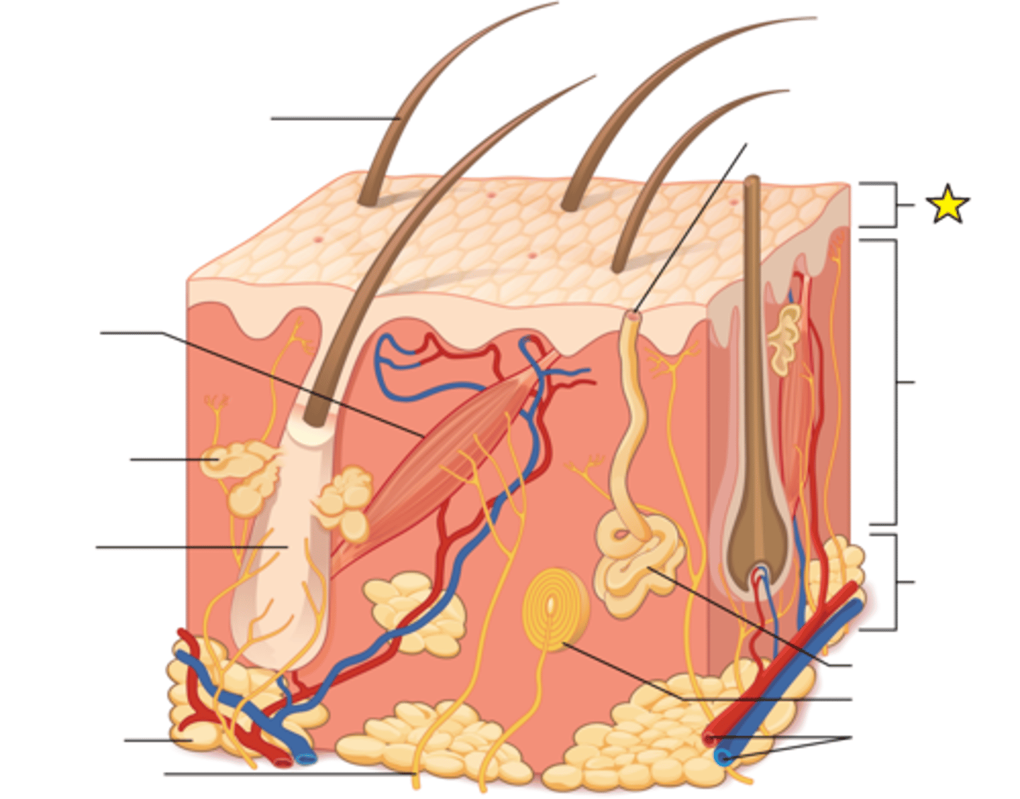
Dermis
The middle layer of skin. Contains hair follicles, collagen, blood vessels (vascular), nerve endings, glands, sensory receptors, and smooth muscle.
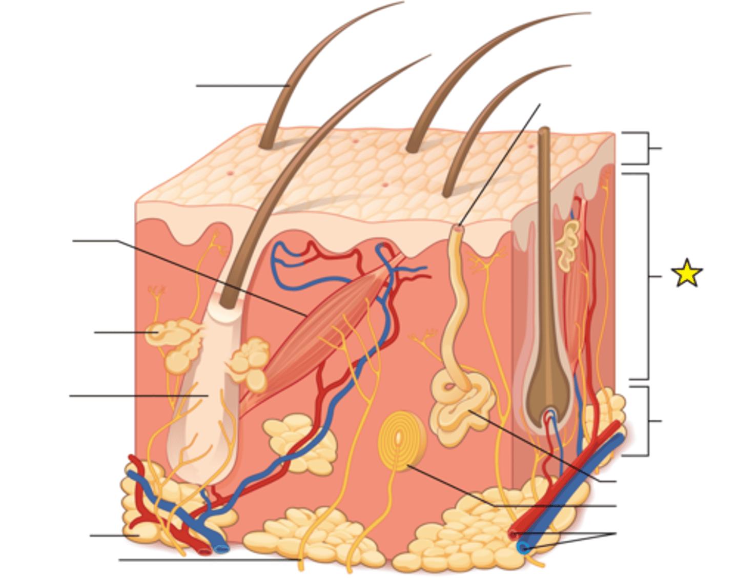
Hypoderms
The innermost (or deepest) and thickest layer of skin; Separates skin from deeper structures; Stabilizes position of skin relative to underlying tissues; Allows independent movement; Dominated by adipose tissue
Important energy storage site.
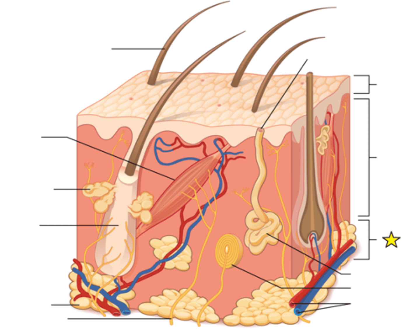
Eccrine sweat gland
Open onto surface of skin; Active when body heats up; helps lower body temperature; Sweat (perspiration) is mostly water, but also excretes wastes
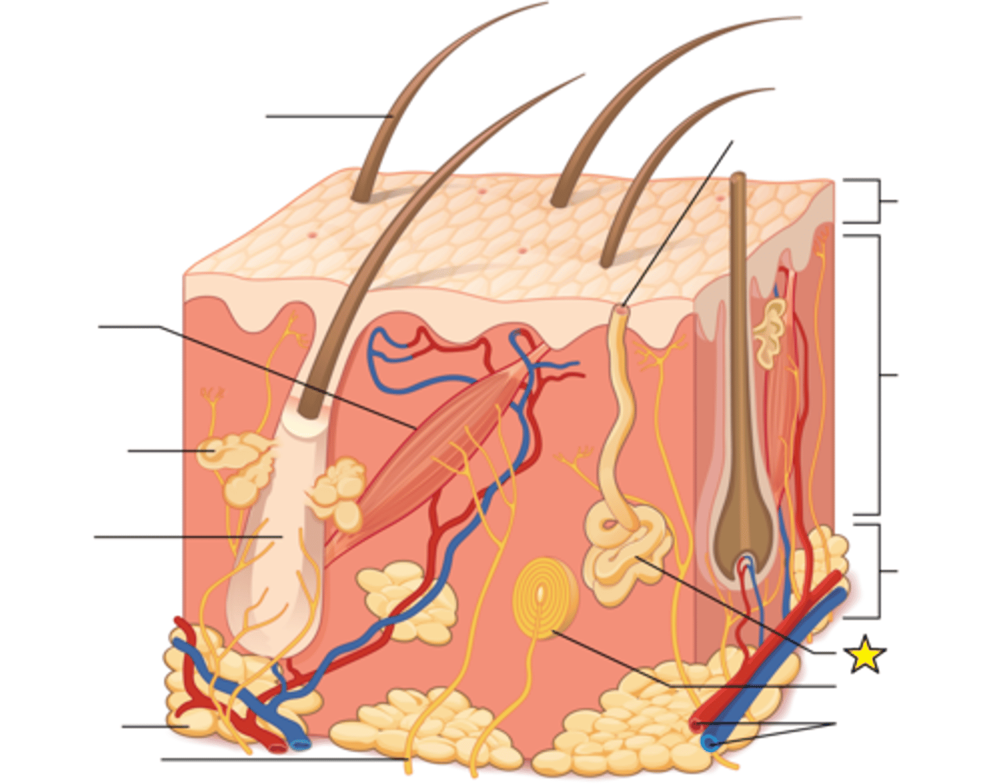
Pacinian corpuscle
Also called Lamellated corpuscles; Detect deep pressure and vibration; Found in both dermal layers and in hypodermis
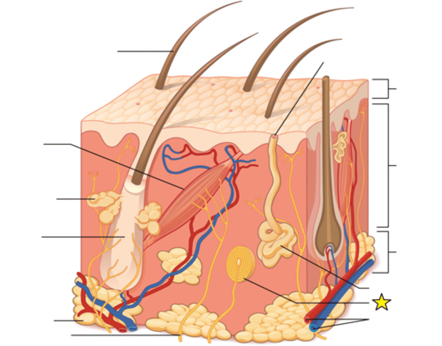
Cutaneous vascular plexus
Responsible for supplying blood to the structures of the dermis, including follicles and glands.
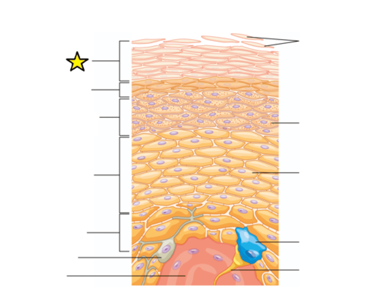
Stratum corneum
The “horny layer”; outermost layer; Cells in surface layer remain about 2 weeks before being shed or washed away; 15–30 layers of dead keratinized cells held together by desmosomes; Water resistant, not waterproof since we lose water through insensible perspiration (unable to see or feel) and sensible perspiration (sweat).
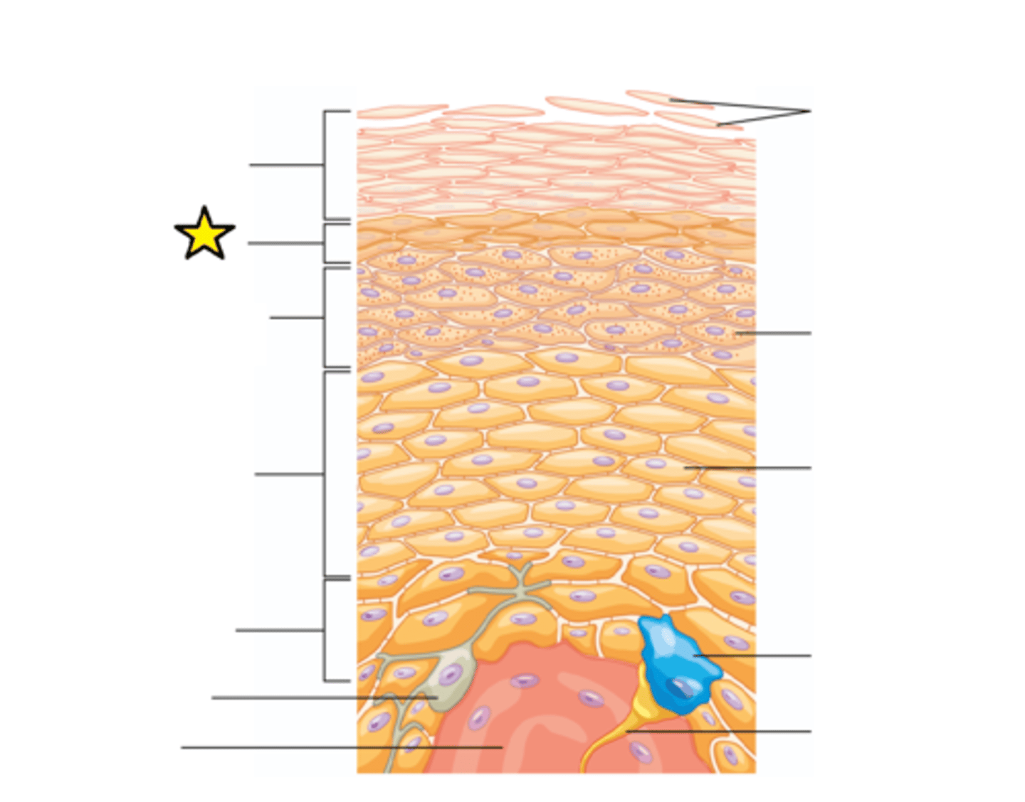
Stratum lucidum
The "clear layer"; Flattened, densely packed dead cells filled with keratin.
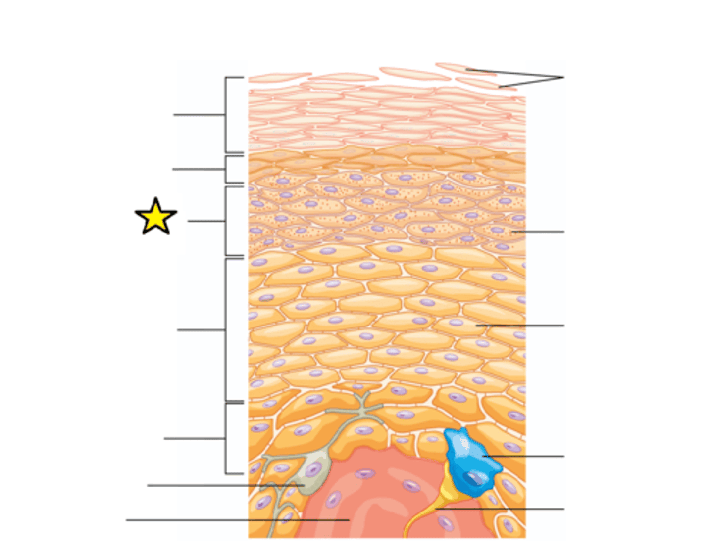
Stratum granulosum
The "grainy layer"; 3-5 layers of keratinocytes that have stopped dividing and started producing keratin; Cells grow thinner and flatter and cell membrane becomes less permeable.
Stratum spinosum
The "spiny layer"; Composed of 8-10 layers of keratinocytes bound together by desmosomes; Contains dendritic (Langerhans) cells defending against microorganisms and superficial skin cancers
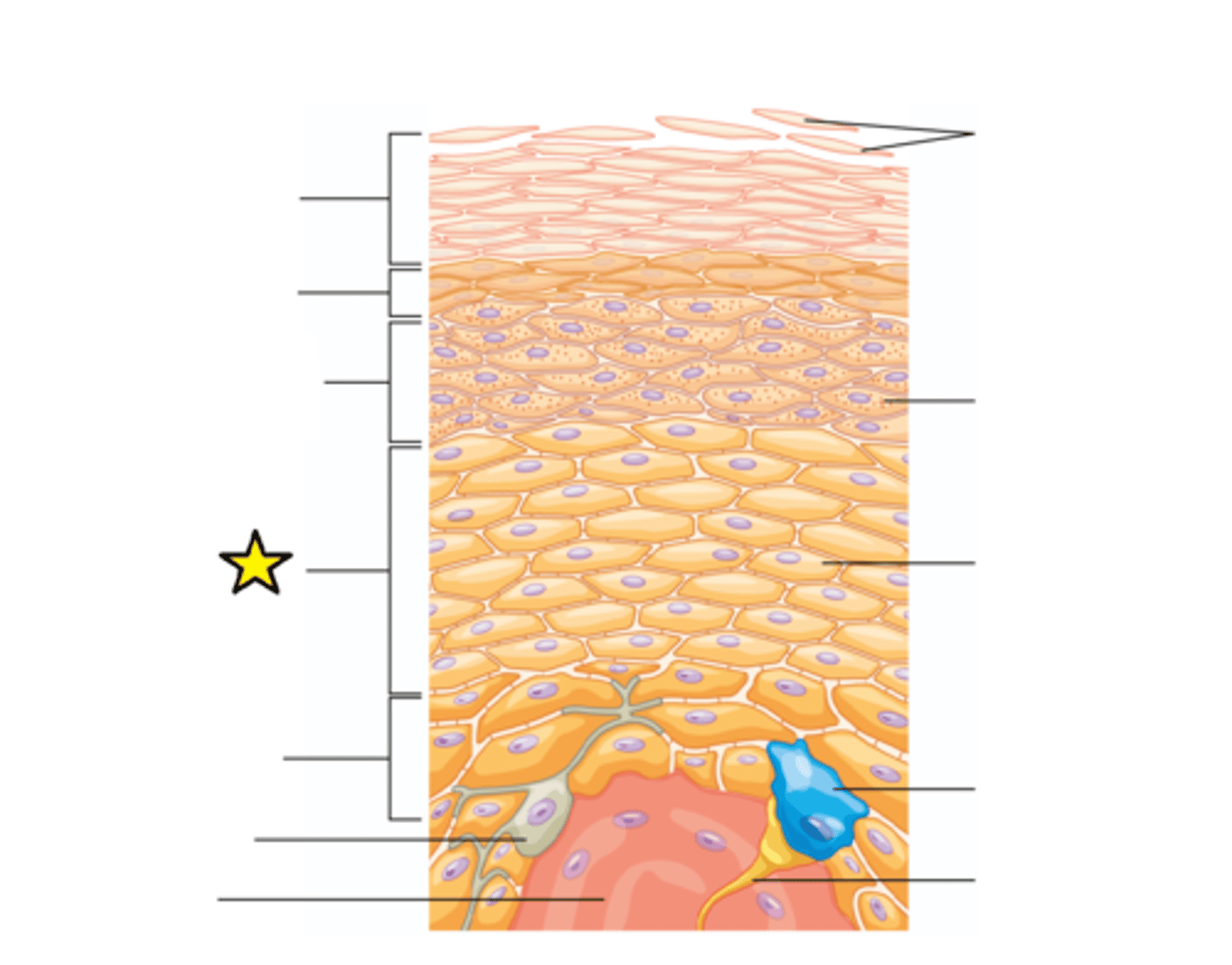
Stratum basale
The "basement layer"; innermost layer; Attached to basement membrane. Most cells here are basal cells, stem cells that divide to replace more superficial keratinocytes. Melanocytes and Merkel cells that respond to touch are also found here.
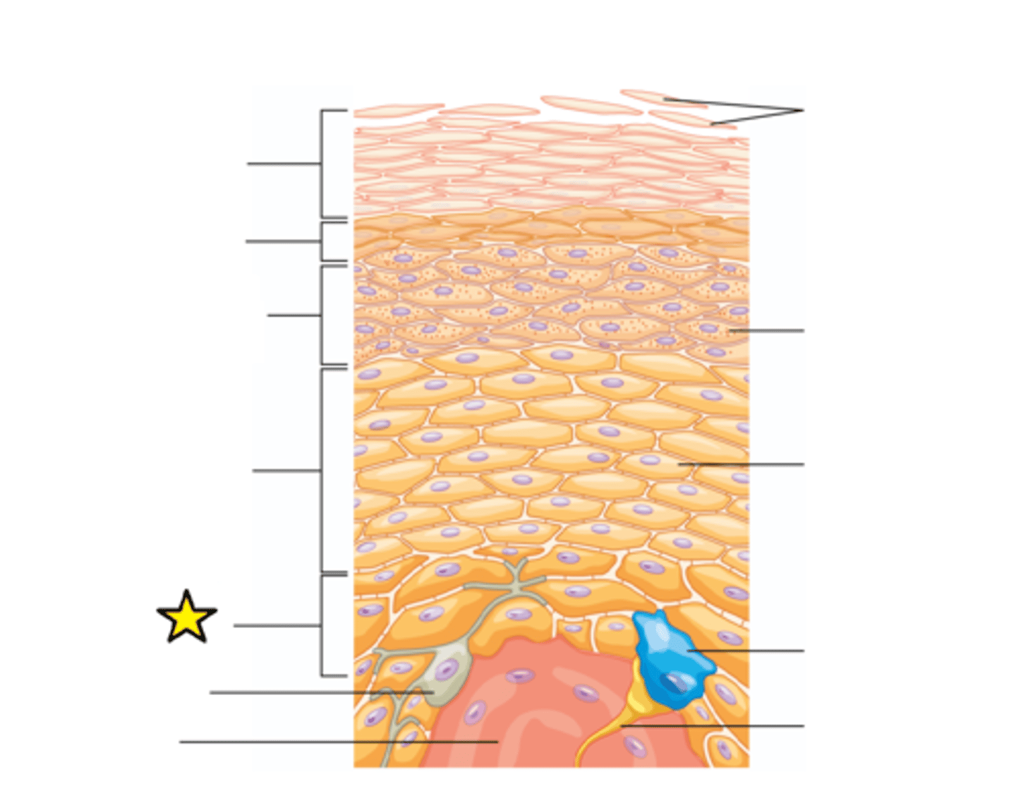
Melanocyte
The relative coloration of the skin depends of the amount of melanin produced by this cell in the stratum basale and taken up by keratinocytes.
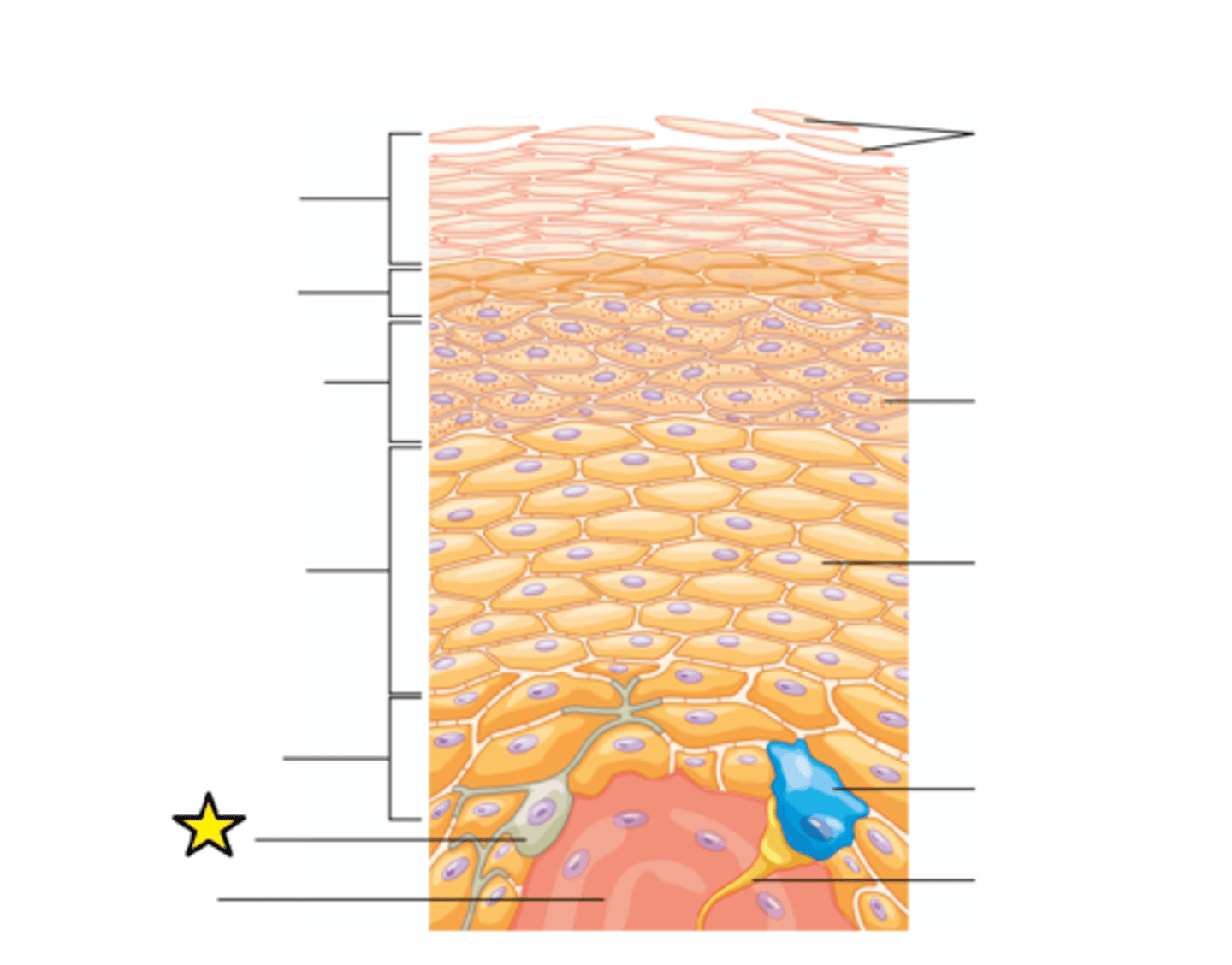
Dermis
The middle layer of skin. Contains hair follicles, collagen, blood vessels (vascular), nerve endings, glands, sensory receptors, and smooth muscle.
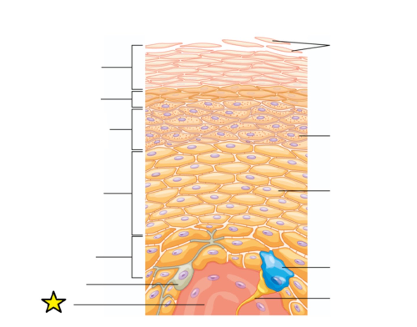
Dead cells filled with keratin
Protein filled cells that are no longer alive.
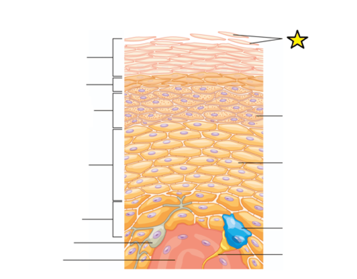
Lamellar granules
Secretory organelles found in type II alveolar cells in the lungs, and in keratinocytes in the skin.
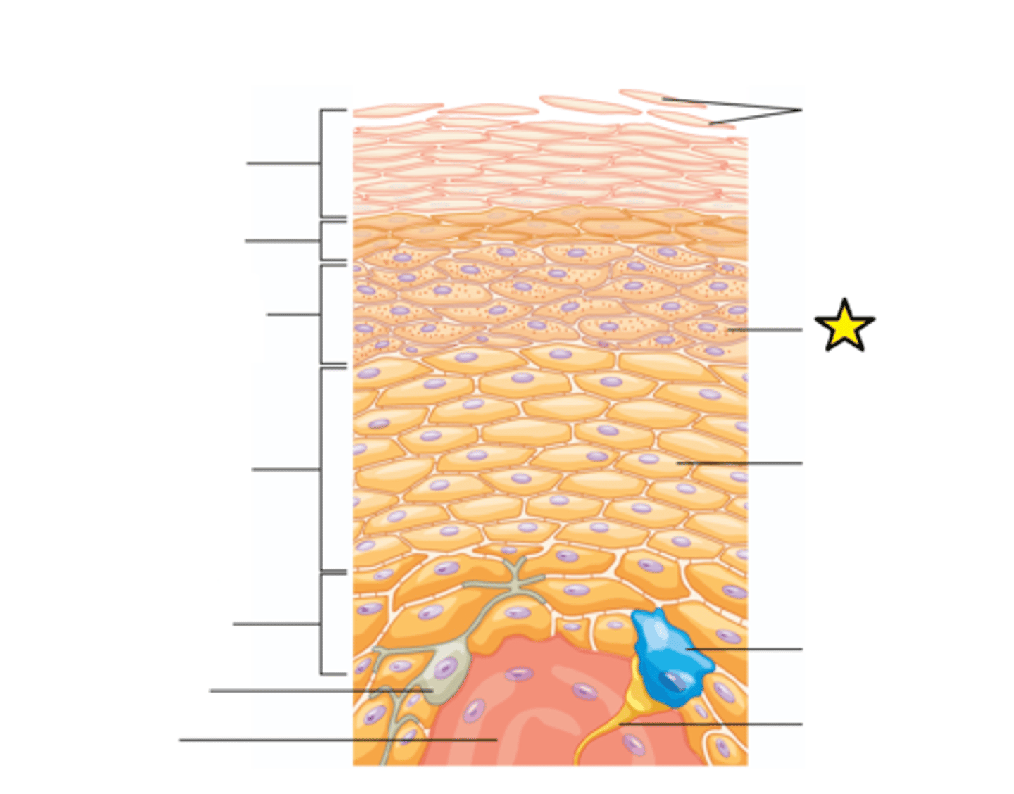
Keratinocyte
Found in the basal layer (stratum basale) of the skin are sometimes referred to as "basal cells"; The predominant cell type in the epidermis.
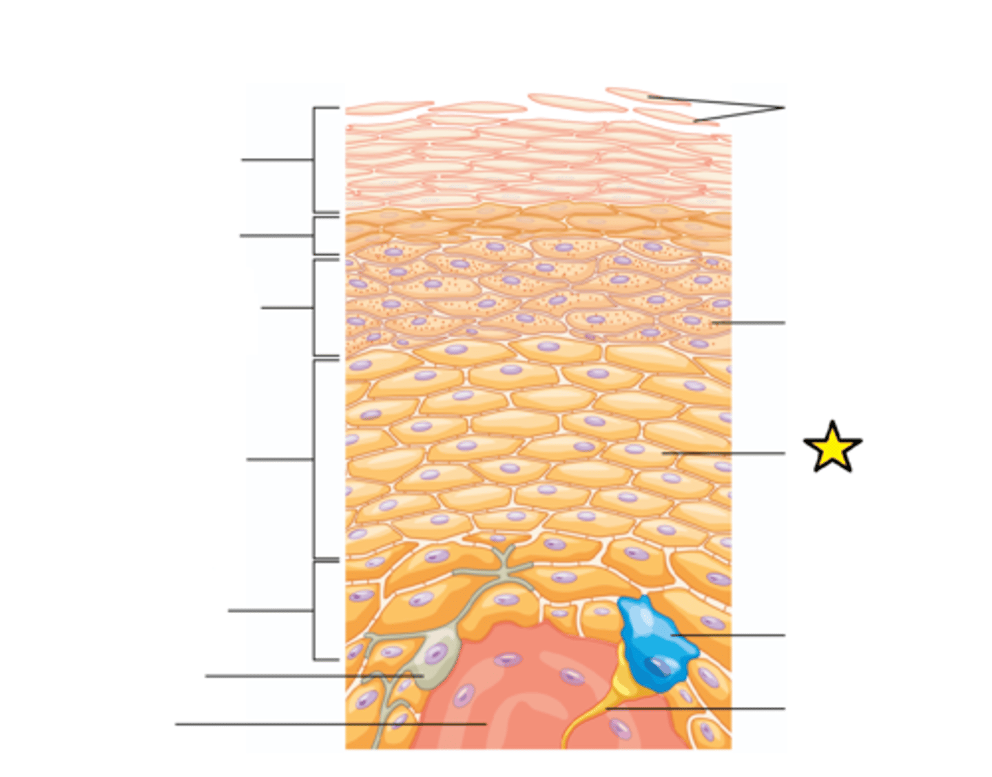
Merkel cells (discs)
Tactile epithelial cells, are oval-shaped mechanoreceptors essential for light touch sensation and found in the skin of vertebrates.
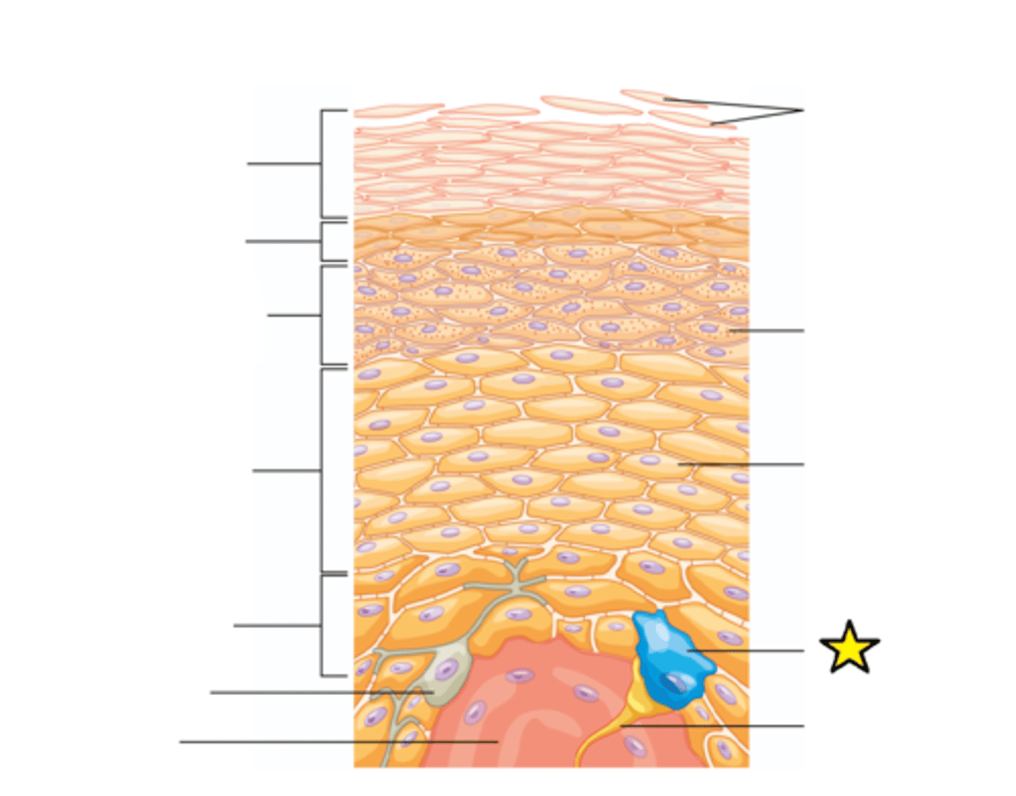
Sensory neuron
A neuron that picks up stimuli from the environment and sends a nerve impulse toward the brain (senses stimuli).
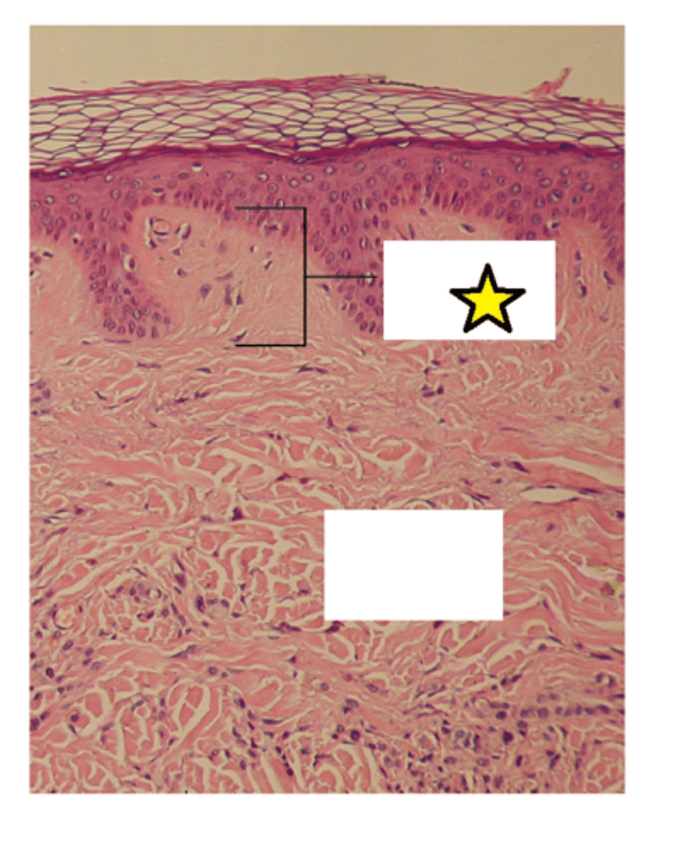
Papillary layer of dermis
Named for dermal papillae in this region; Composed of areolar tissue; Contains capillaries, lymphatic vessels, and sensory neurons
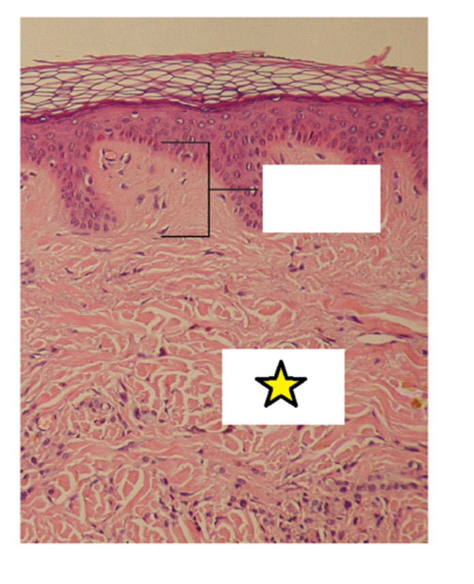
Reticular layer of dermis
The lower layer of the dermis, found under the papillary dermis, composed of dense irregular connective tissue featuring densely-packed collagen fibers. It receives its name from the dense concentration of collagenous, elastic, and reticular fibers that weave throughout it.
Surface
The apical layer of the skin.
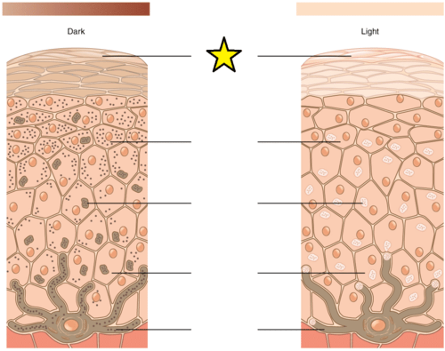
Upper keratinocyte
Closest to the apical surface; Exposure to the UV rays of the sun or a tanning salon causes melanin to be manufactured and built up in in this cell; As sun exposure stimulates these cells to secrete chemicals that stimulate melanocytes; The accumulation of melanin in in these cells results in the darkening of the skin, or a tan.
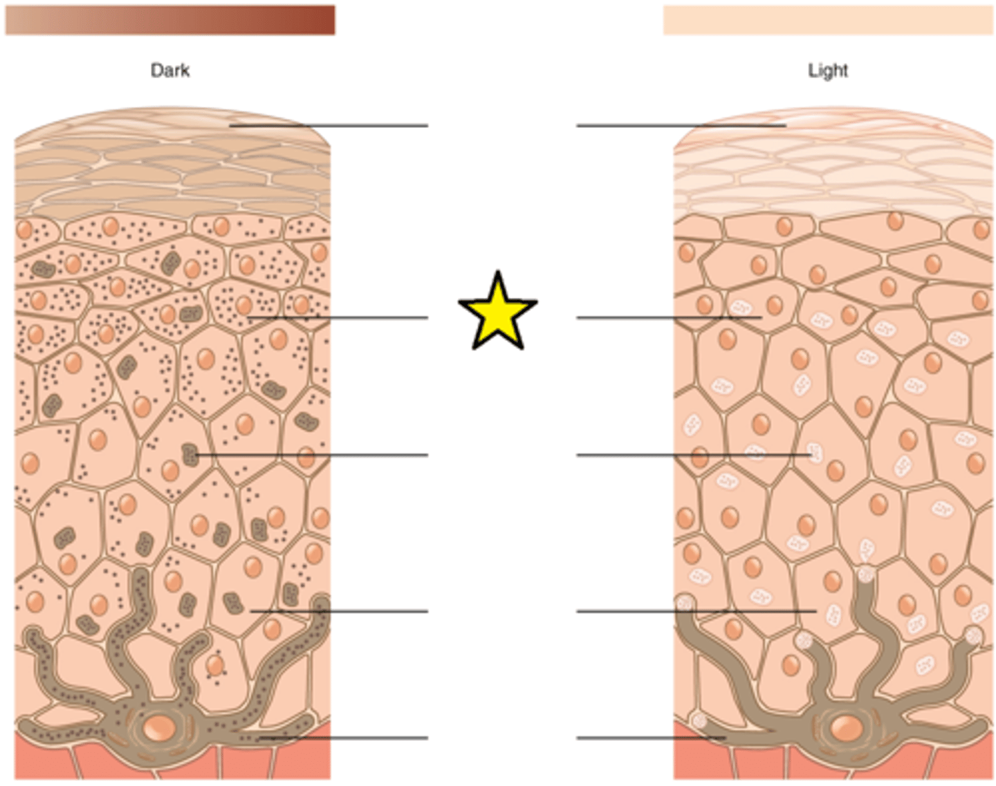
Melanosomes
Temporary structures that are eventually destroyed by fusion with lysosomes; this fact, along with melanin-filled keratinocytes in the stratum corneum sloughing off, makes tanning impermanent.
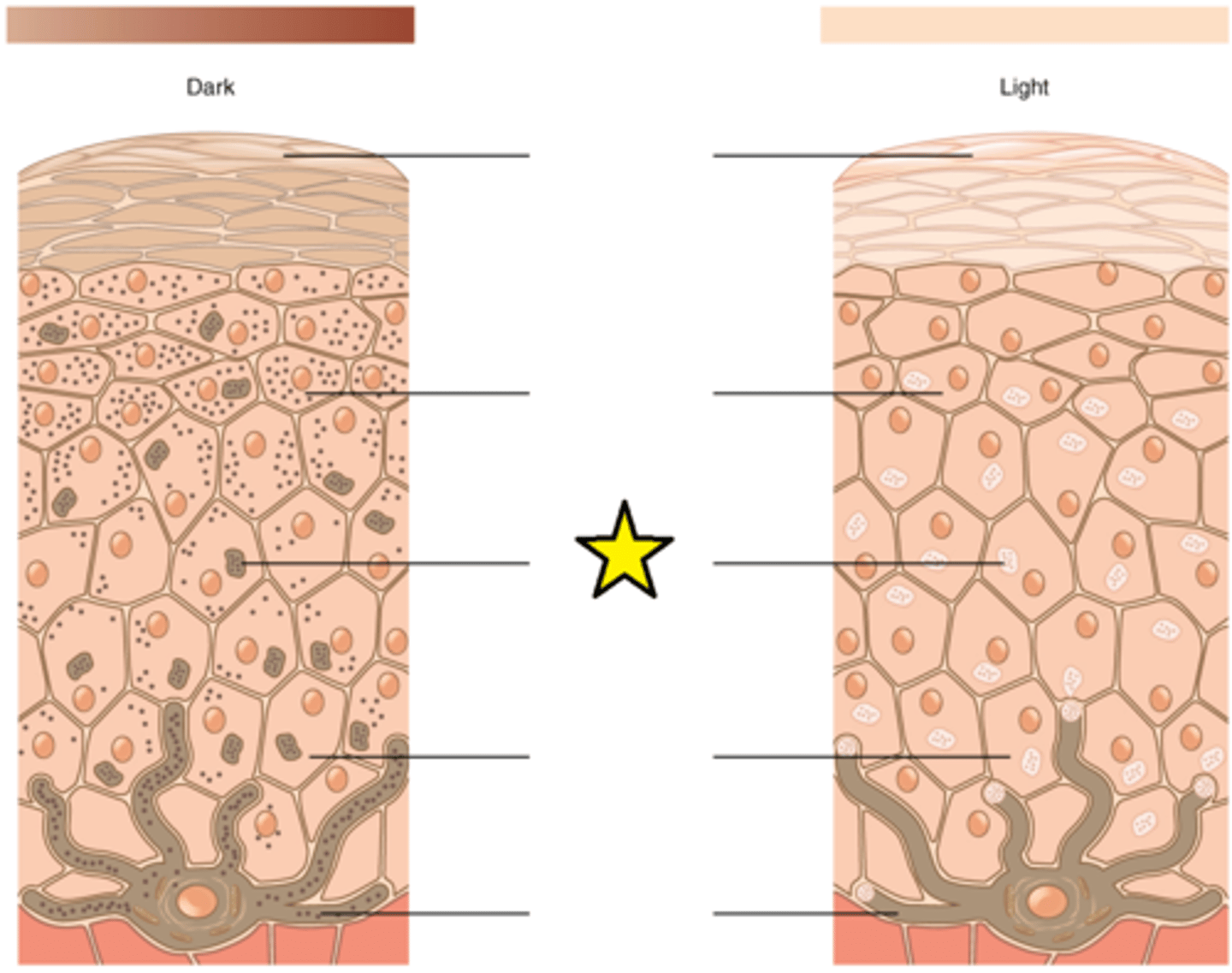
Basal keratinocytes
Closest to the melanocyte; Exposure to the UV rays of the sun or a tanning salon causes melanin to be manufactured and built up in in this cell; As sun exposure stimulates these cells to secrete chemicals that stimulate melanocytes; The accumulation of melanin in in these cells results in the darkening of the skin, or a tan.
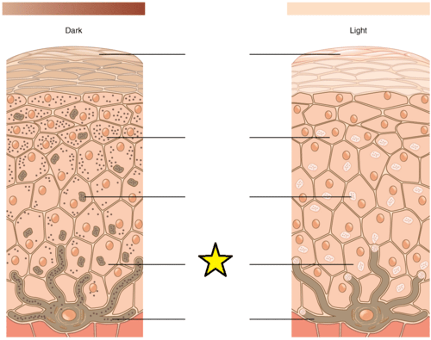
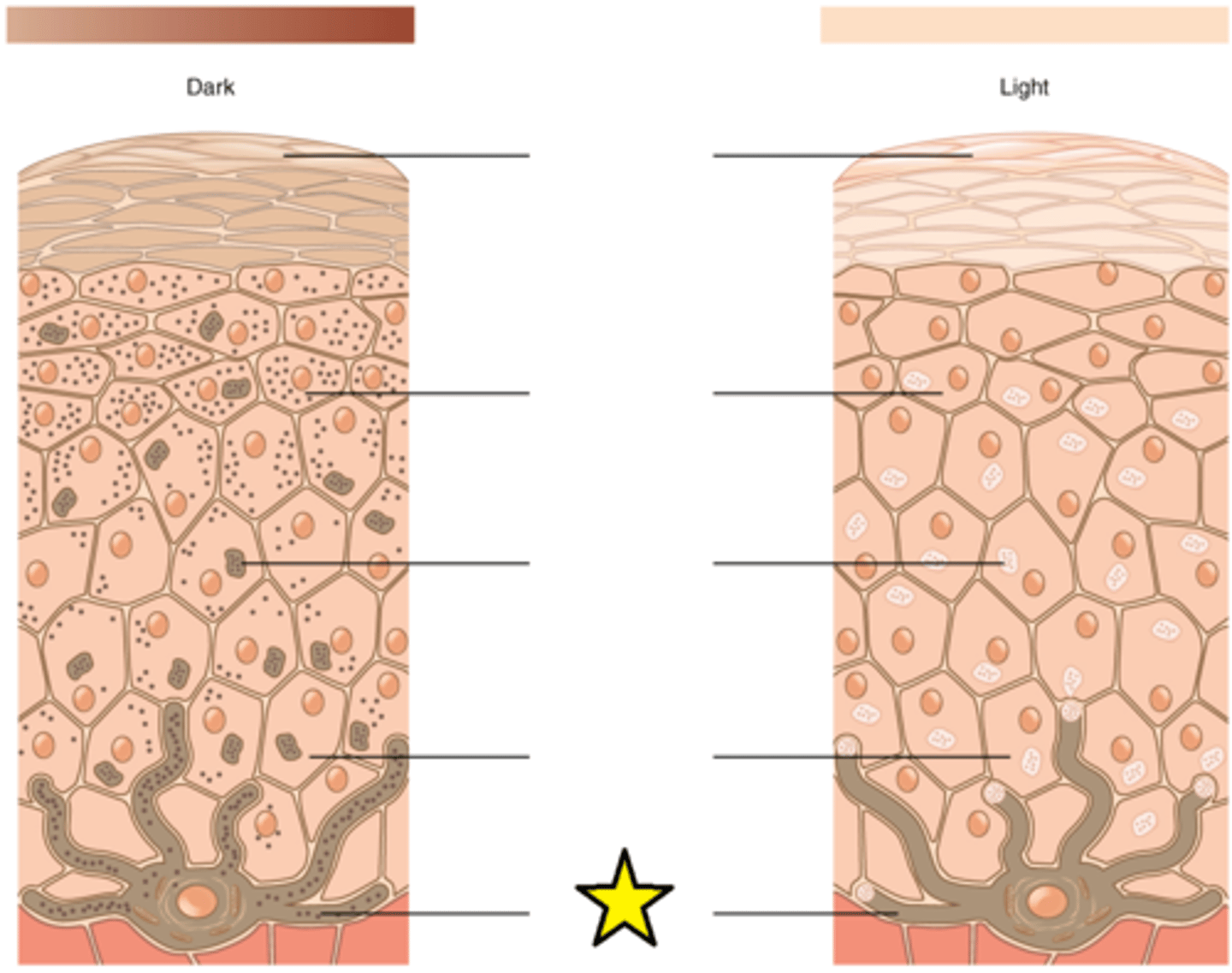
Melanocytes
The relative coloration of the skin depends of the amount of melanin produced by this cell in the stratum basale and taken up by keratinocytes.
Arrector pili muscle
Attaches to the base of a hair follicle at one end and to dermal tissue on the other end. In order to generate heat when the body is cold, they all contract all at once, causing the hair to "stand up straight" on the skin.
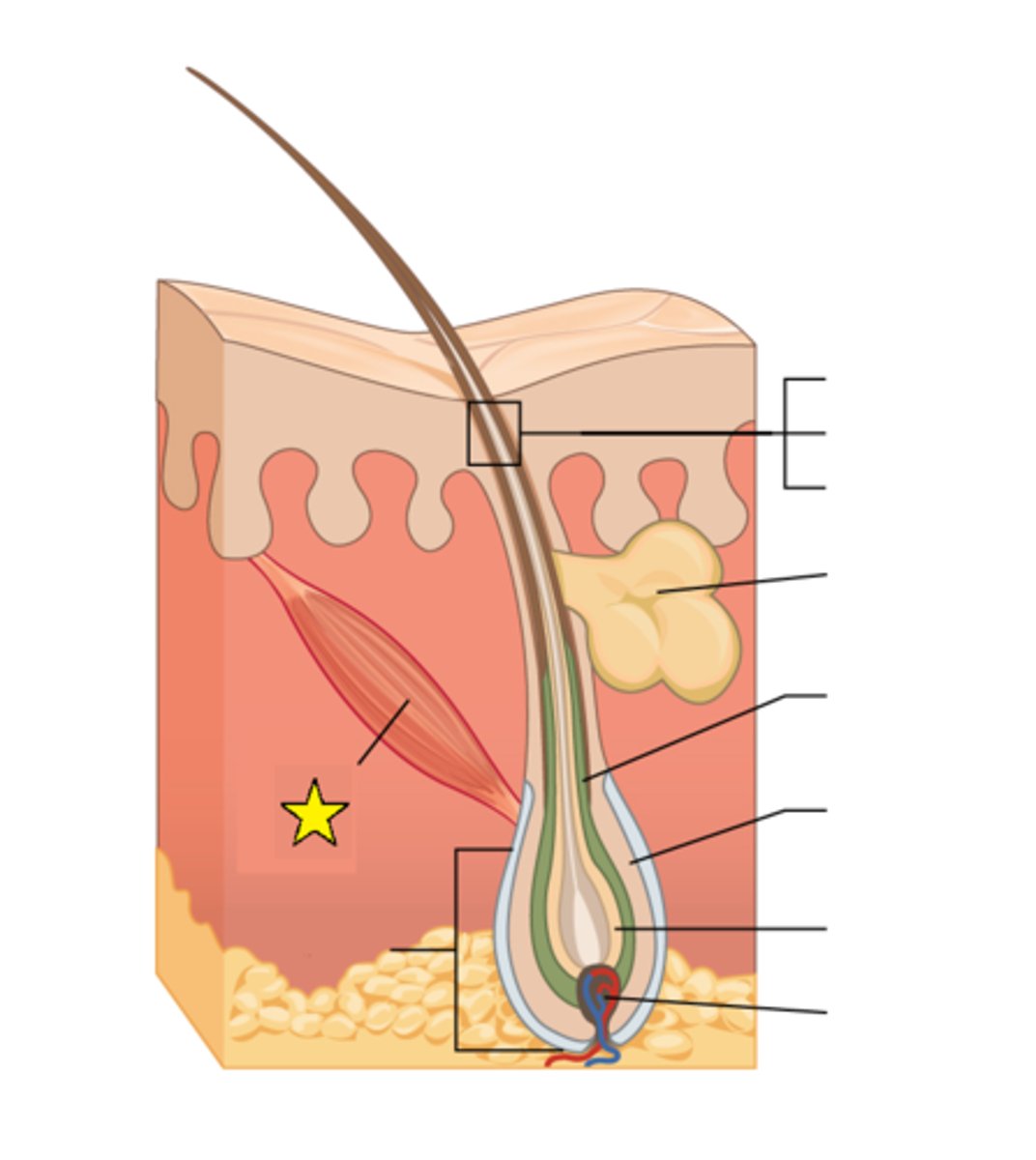
Hair bulb
Forms the base of the hair follicle; In this structure, living cells divide and grow to build the hair shaft. Blood vessels nourish the cells in this region, and deliver hormones that modify hair growth and structure at different times of life.
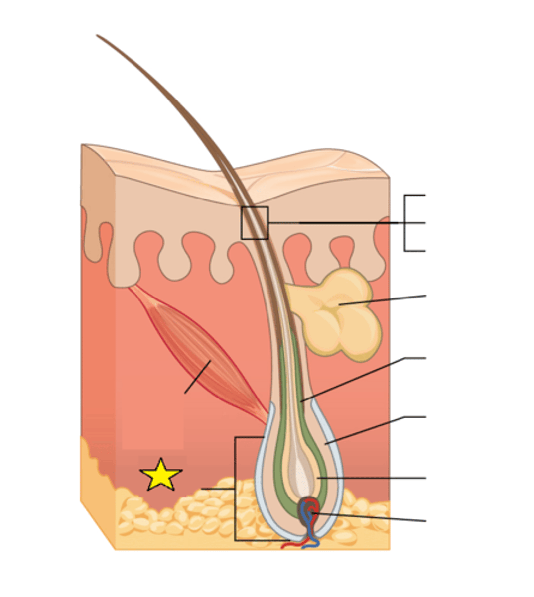
Hair shaft
The part of the hair not anchored to the follicle, and much of this is exposed at the skin's surface.
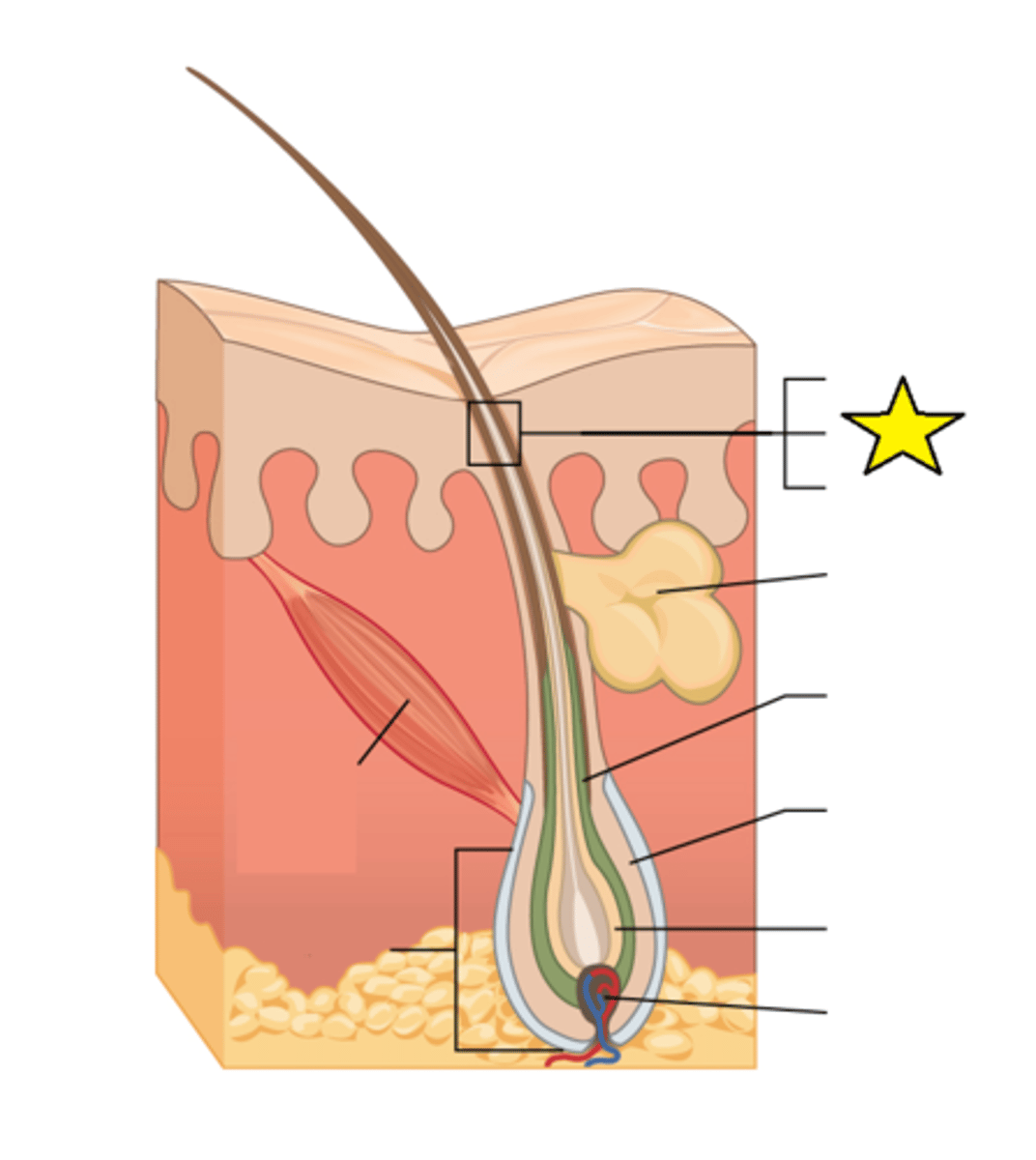
Sebaceous gland
Glands that secrete sebum, associated with hair follicles.
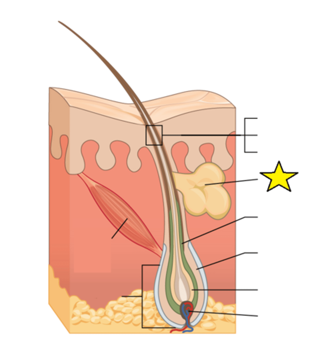
Inner root sheath
Basal cells of the hair matrix in the center differentiate into cells in this region.
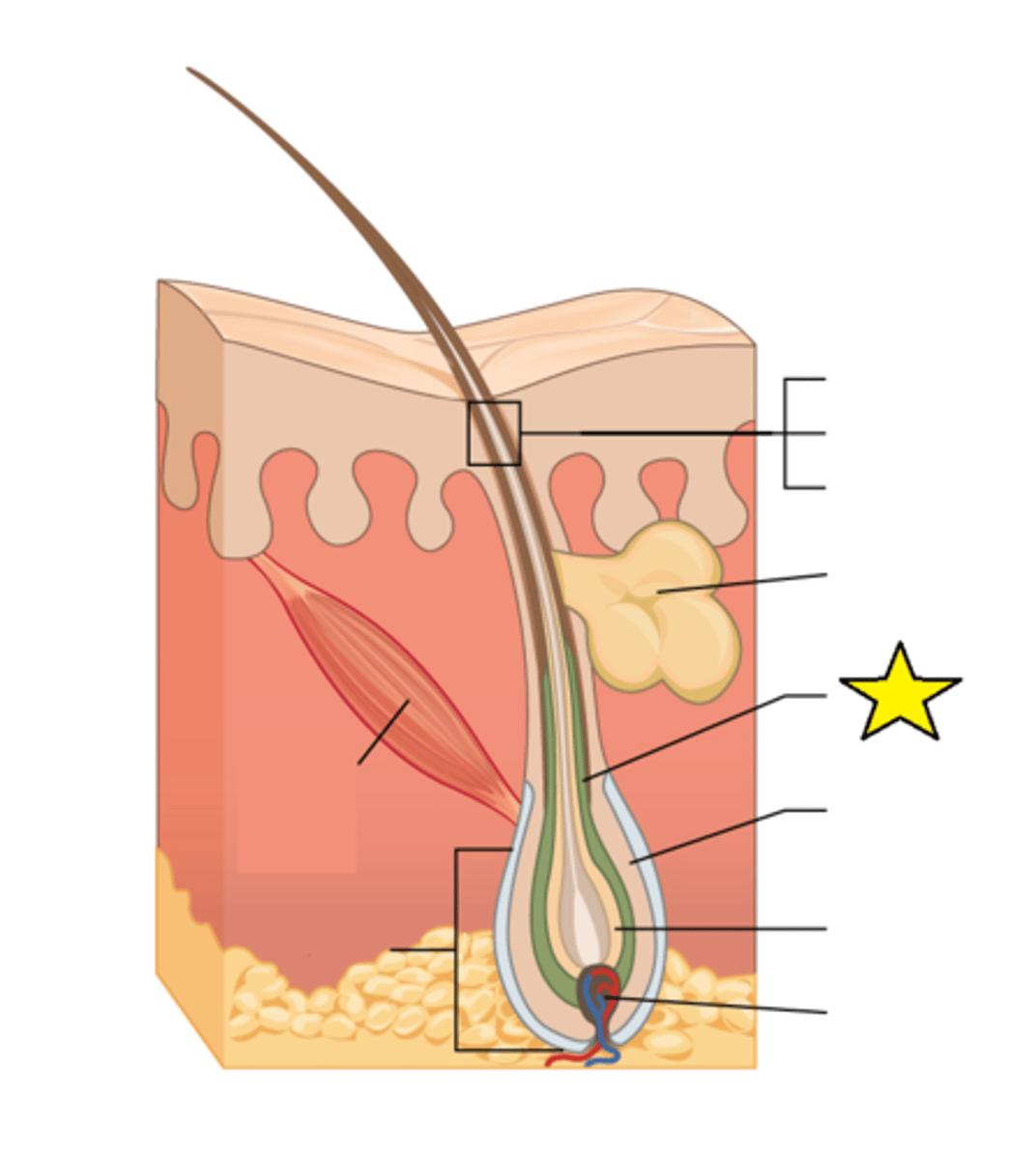
Outer root sheath
Basal cells at the base of the hair root form this structure.
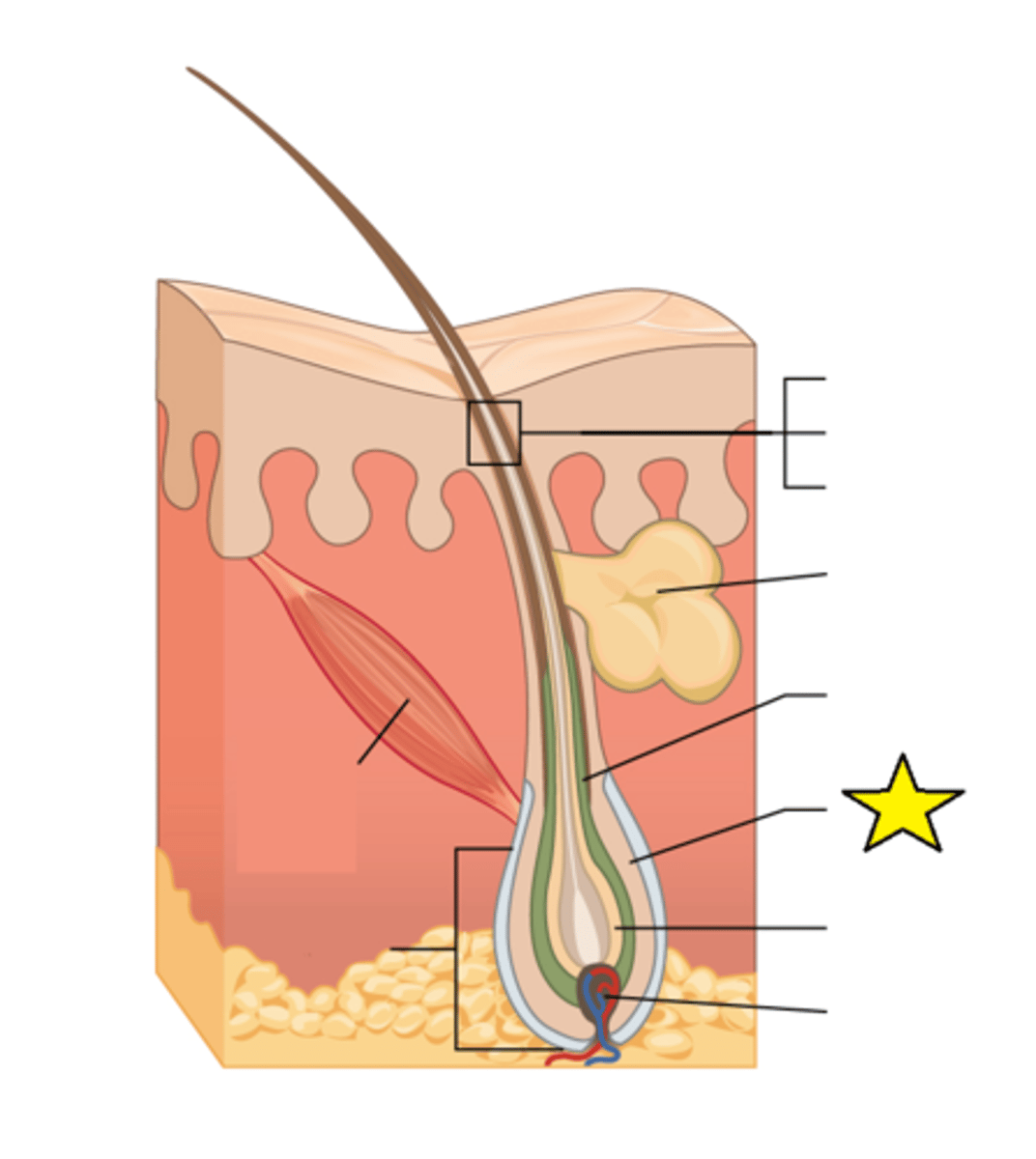
Hair matrix
The basal cells in this region produce a new hair follicle, which pushes the old hair out as the growth cycle repeats itself. Hair typically grows at the rate of 0.3 mm per day.
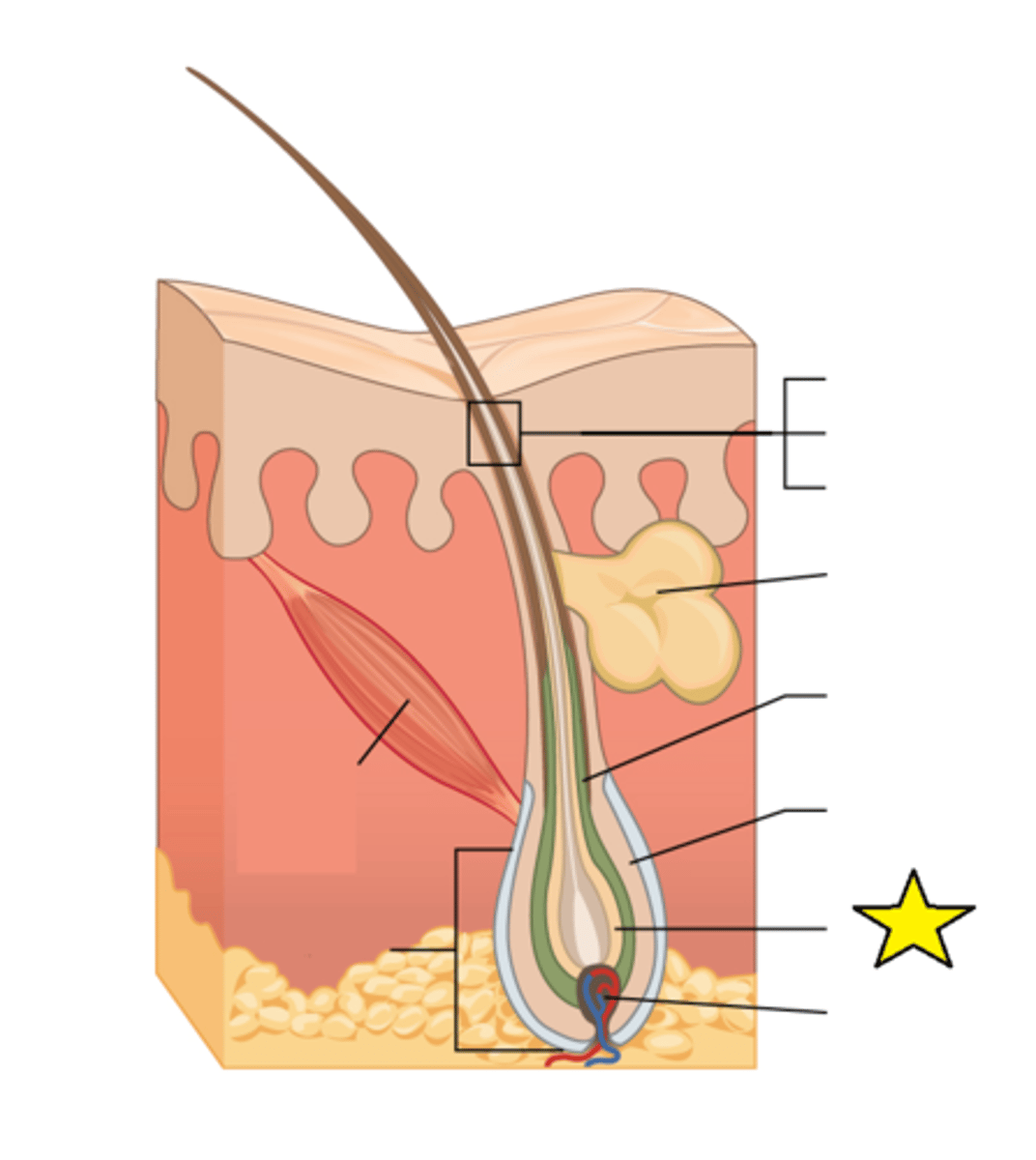
Hair papilla
Made of connective tissue and contains blood capillaries and nerve endings from the dermis; hair gets its color from the pigment melanin, produced by melanocytes in this region.
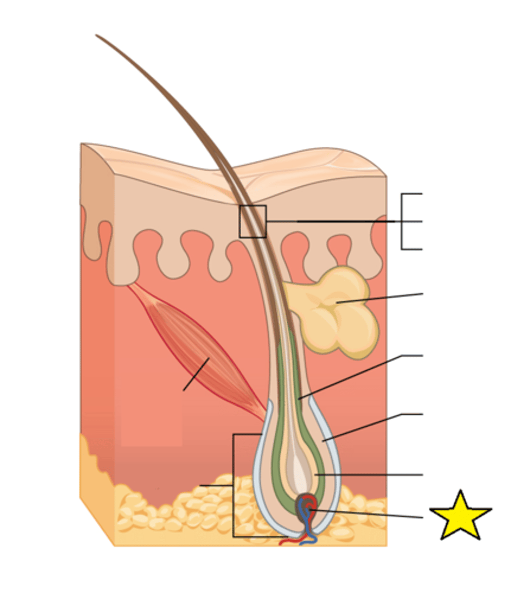
Nail body
Formed on the nail bed, and protects the tips of our fingers and toes as they are the farthest extremities and the parts of the body that experience the maximum mechanical stress; In addition, this structure forms a back-support for picking up small objects with the fingers. It is composed of densely packed dead keratinocytes.
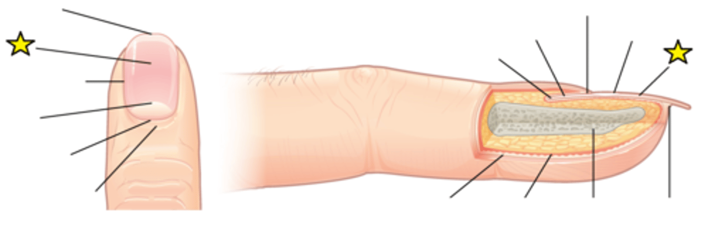
Lunula
The visible portion of the distal nail matrix that extends beyond the proximal nailfold. It is white, half-moon-shaped, appears by week 14 of gestation, has unique histologic features. The lunula has a primary structural role in defining the free edge of the distal nail plate.
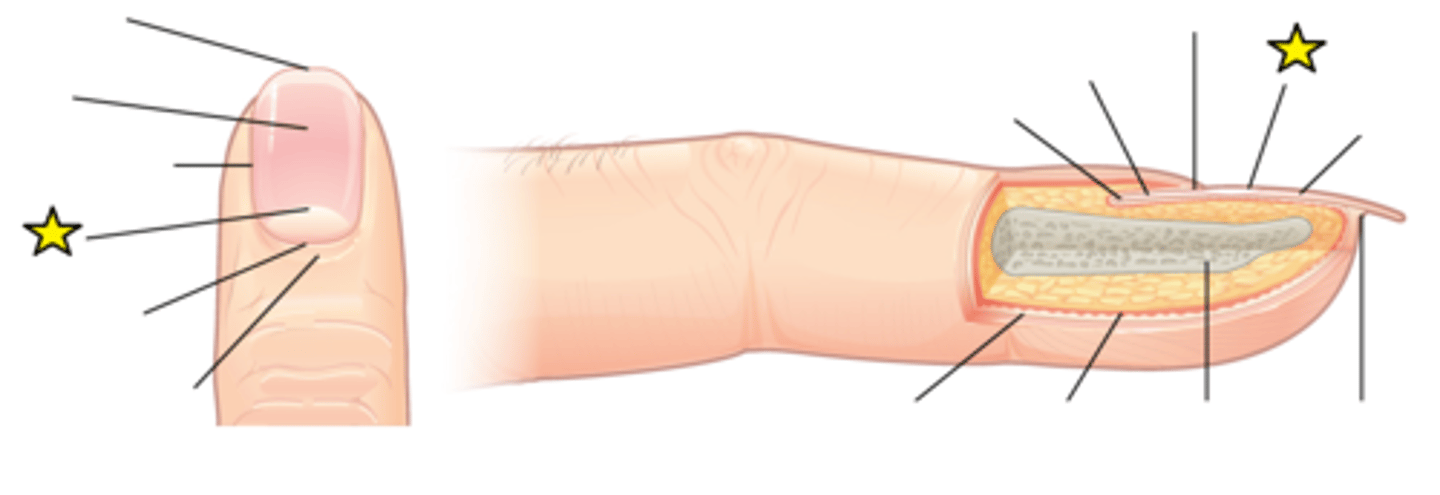
Free edge
The end of the nail plate that is shaped during Manicure & Pedicure.
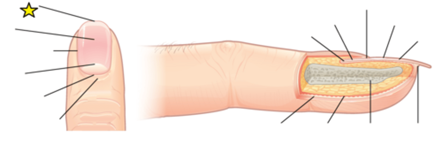
Eponychium
The thickened layer of skin at the base of the fingernails and toenails. It can also be called the medial or proximal nail fold. It differs from the cuticle and comprises live skin cells whilst the cuticle is dead skin cells.
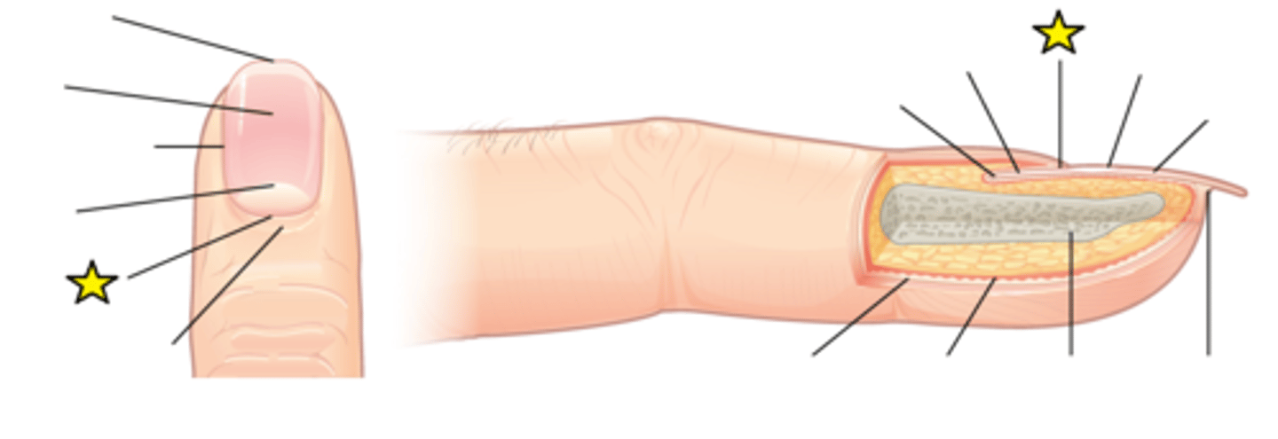
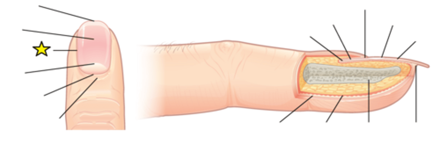
Lateral nail fold
Overlaps the nail on the sides, helping to anchor the nail body.
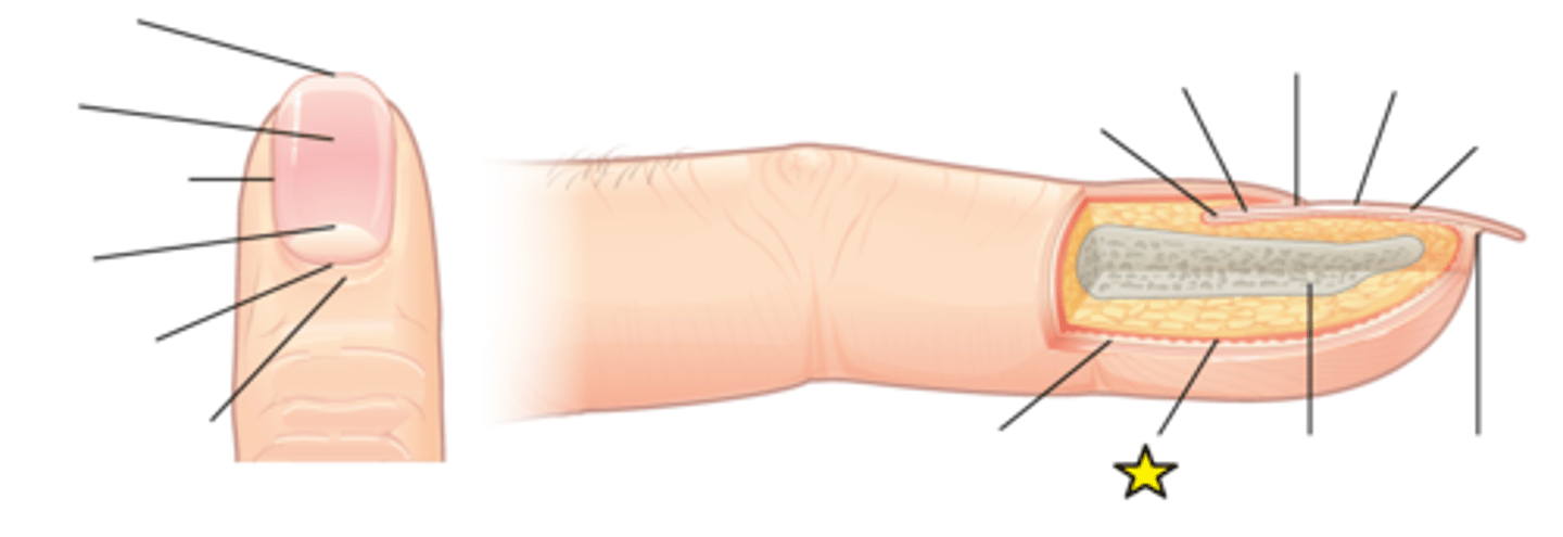
Dermis
The middle layer of skin. Contains hair follicles, collagen, blood vessels (vascular), nerve endings, glands, sensory receptors, and smooth muscle.
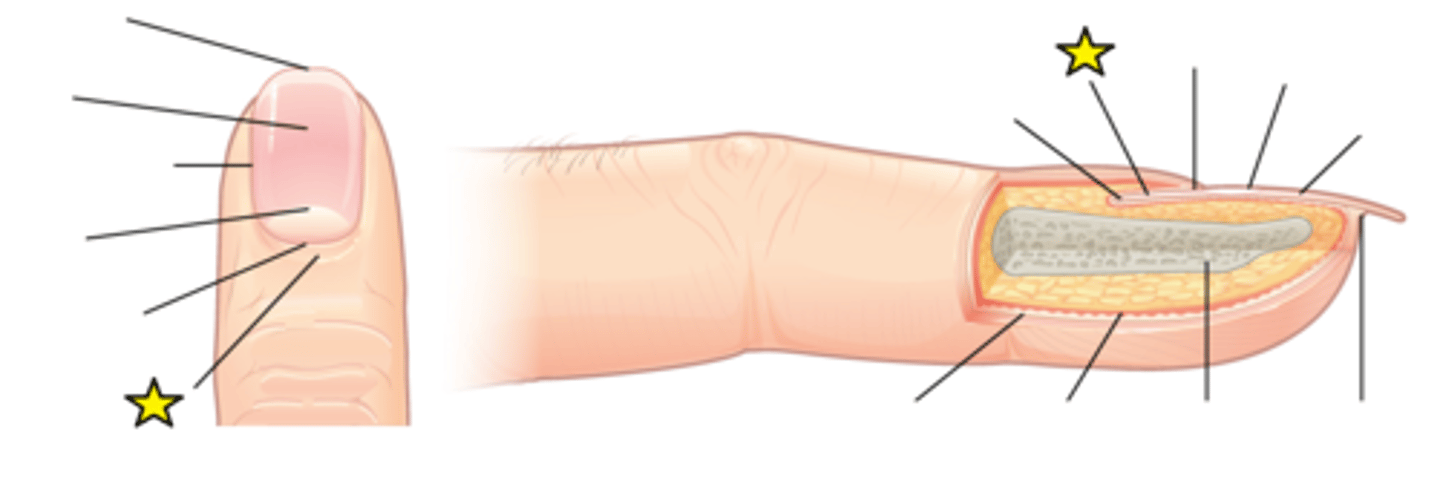
Proximal nail fold
A required guardian seal that prevents germs and bacteria from getting to the nail matrix, where new cells are created.
Hyponychium
A soft tissue seal underneath the extended "free" edge of the nail plate whose purpose is to prevent pathogens from infecting the nail bed.
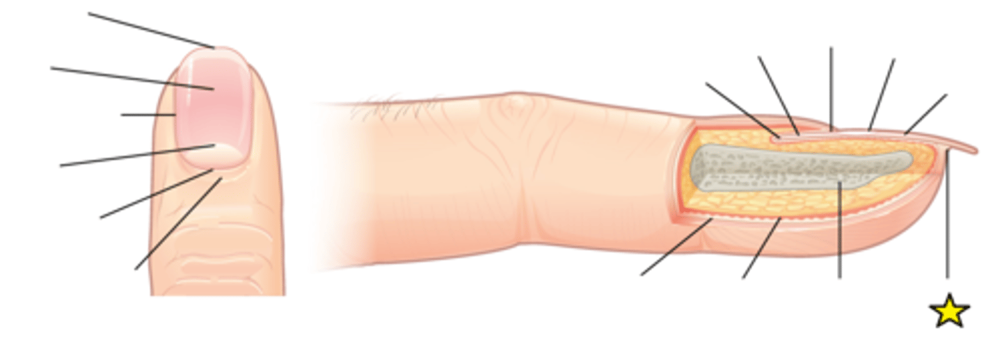
Epidermis
Part of the body has evolved a specialized structure upon which nails can form.
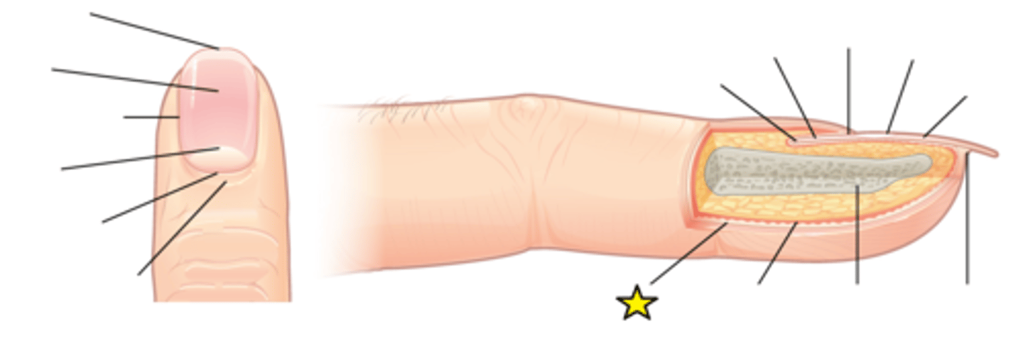
Phalanx
Finger bone
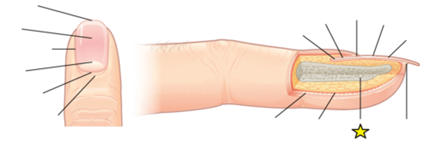
Nail root
Has a matrix of proliferating cells from the stratum basale that enables the nail to grow continuously.
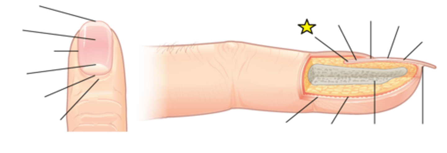
Basal cell carcinoma
Most common form of skin cancer; Originates in stratum basale due to mutations caused by overexposure to UV radiation; Virtually no metastasis and most people survive
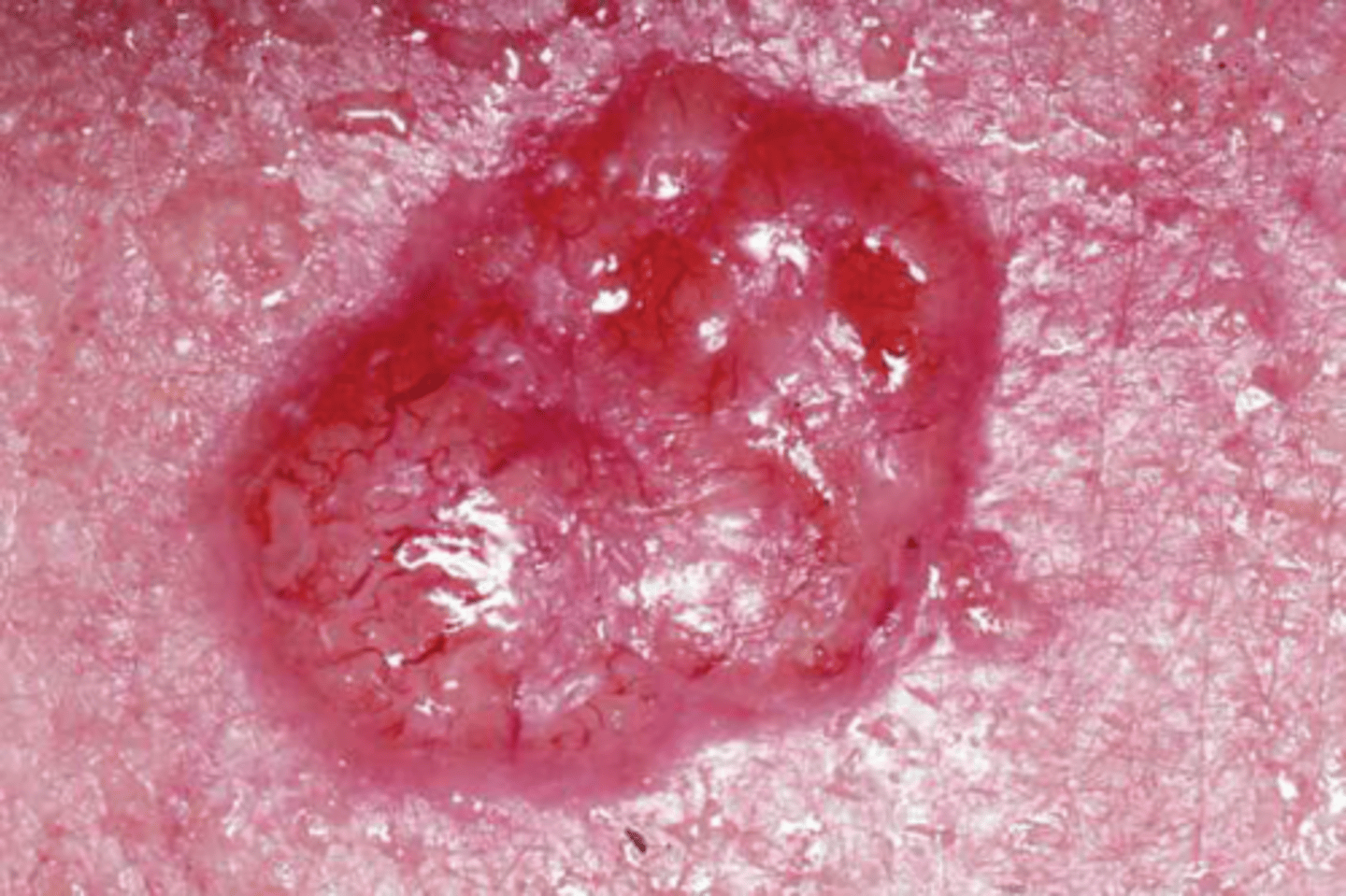
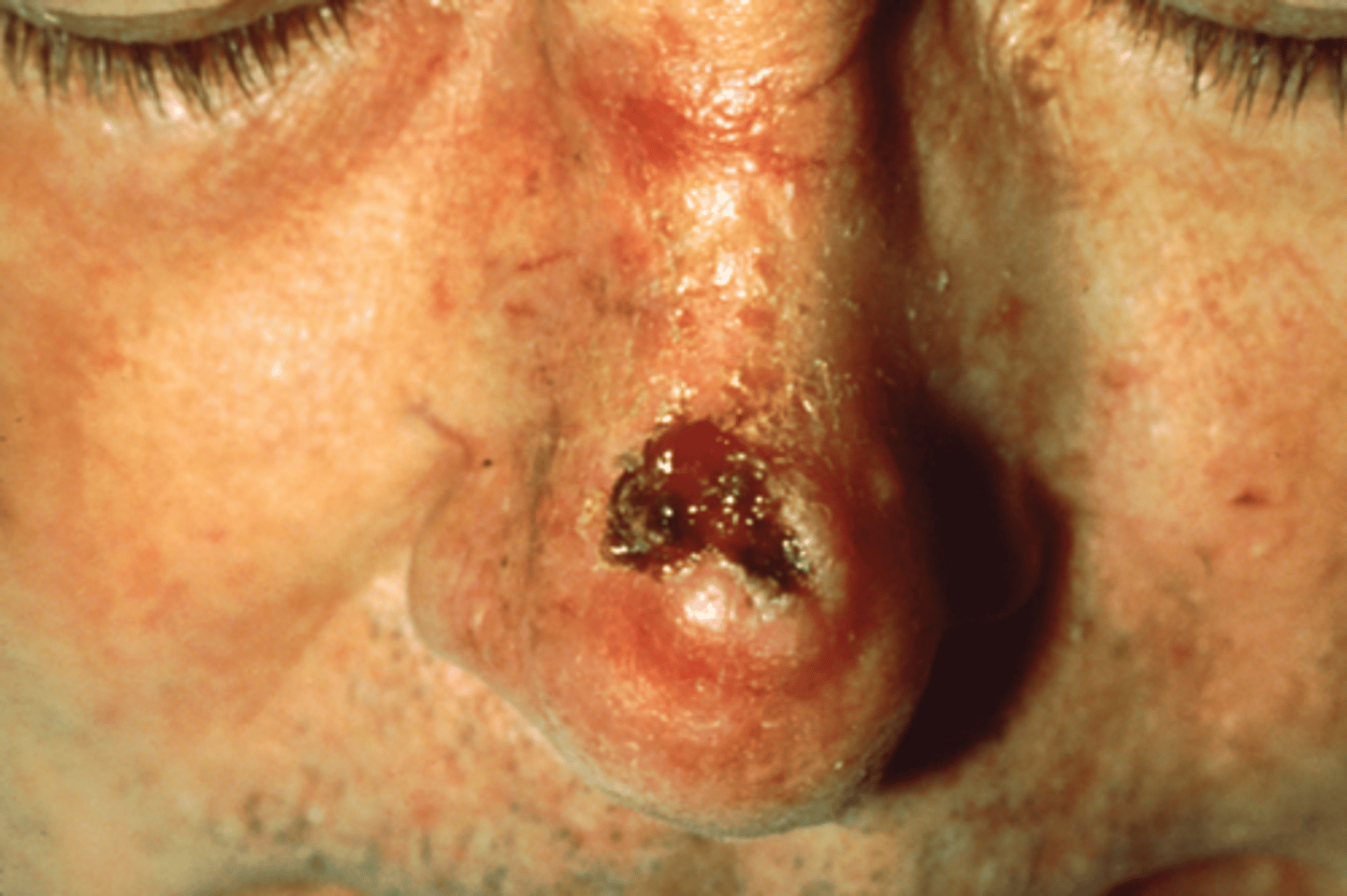
Squamous cell carcinoma
Effects the keratinocytes of the stratum spinosa and presents as lesions; Presents here as a lesion on an individual's nose; Second most common skin cancer
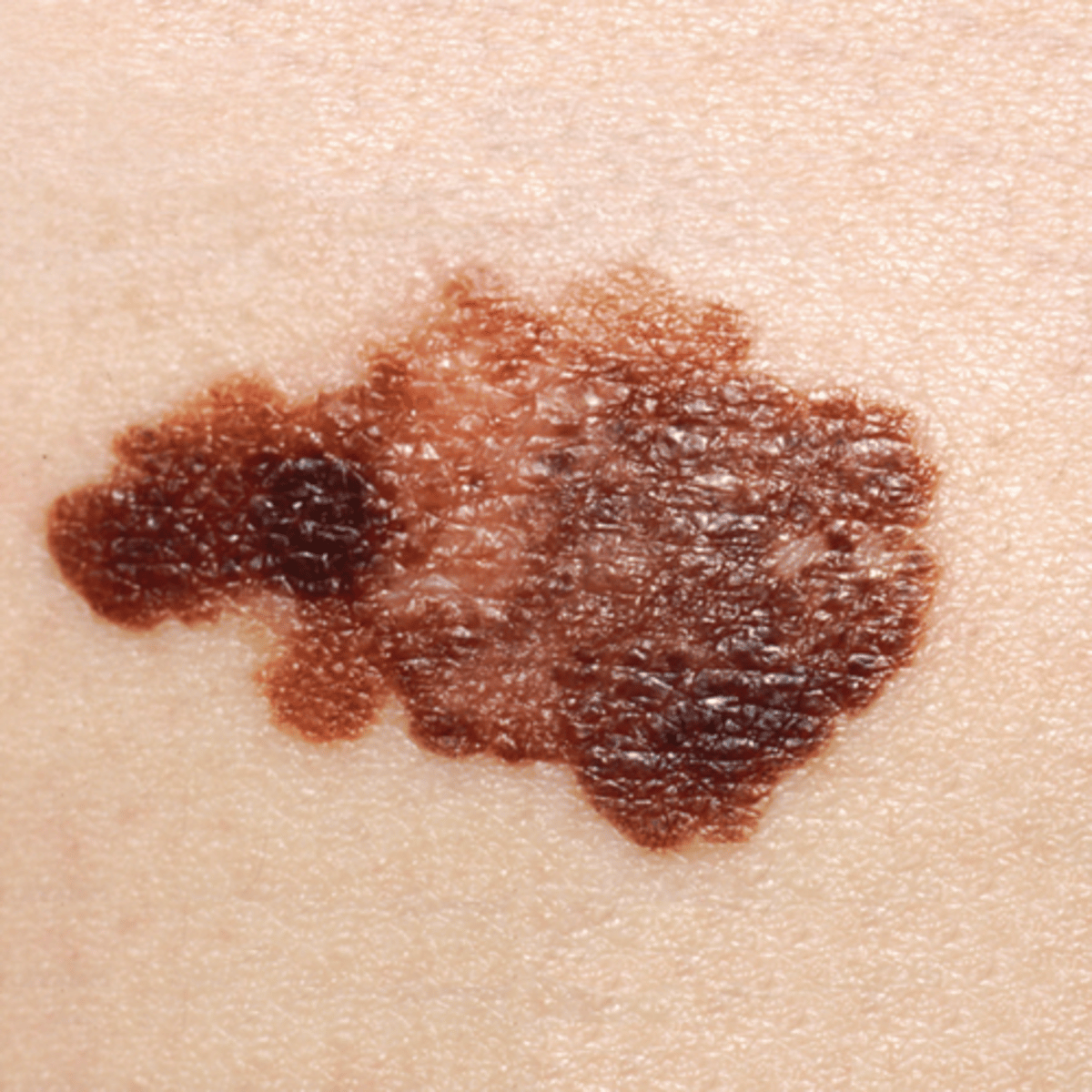
Melanoma
Most serious form of skin cancer; Extremely dangerous
Cancerous melanocytes grow rapidly and metastasize through lymphatic system; If detected early and removed surgically, the 5-year survival rate is 99 percent; If not detected until after metastasis, the 5-year survival rate drops to 14 percent
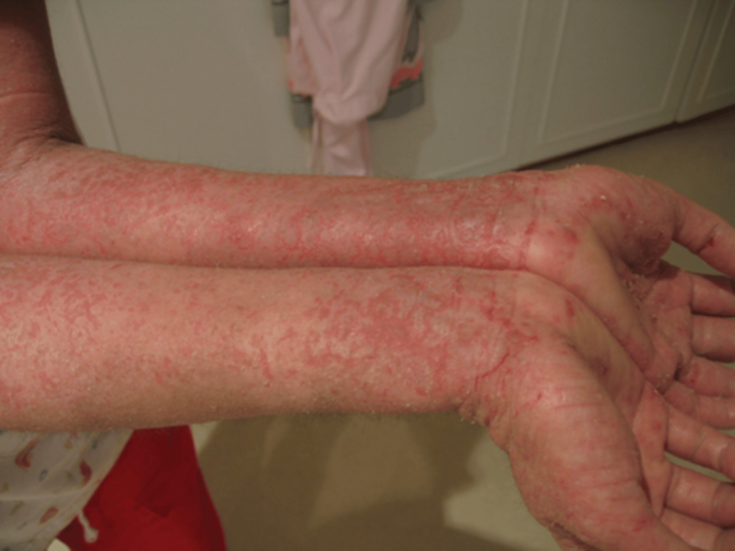
Eczema
A common skin disorder that presents as a red, flaky rash.
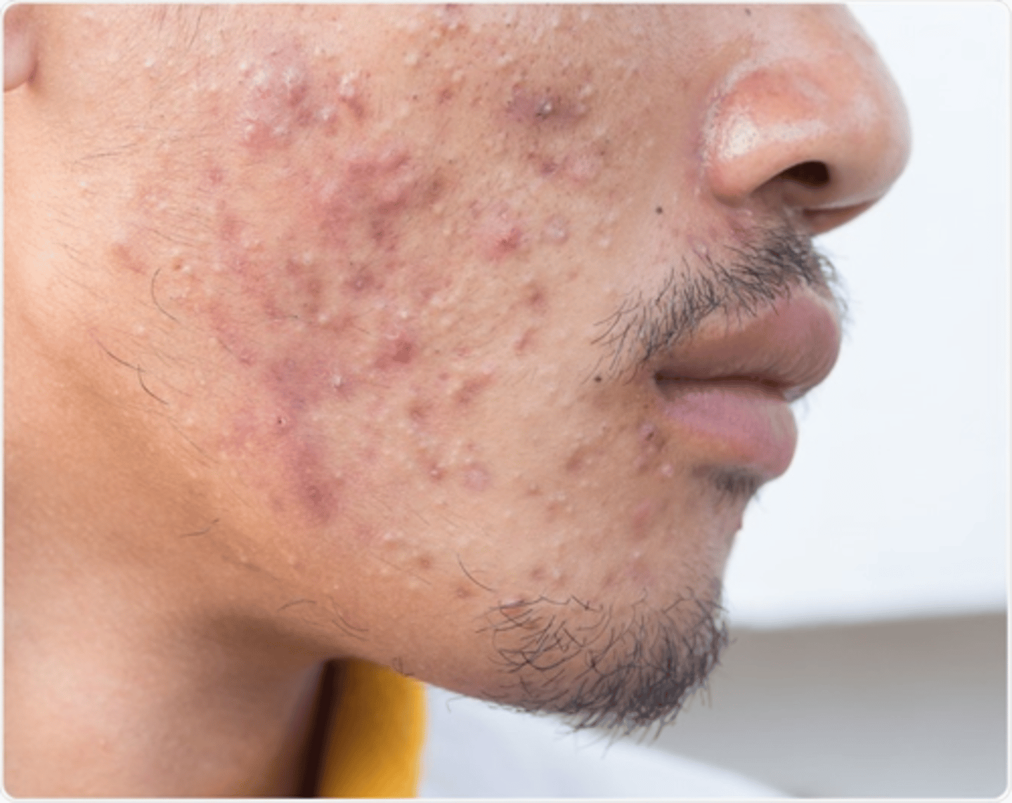
Acne
A result of over-productive sebaceous glands, which leads to formation of blackheads and inflammation of the skin.
First-degree burn
Only epidermis affected; Redness and pain; No blisters or swelling occurs
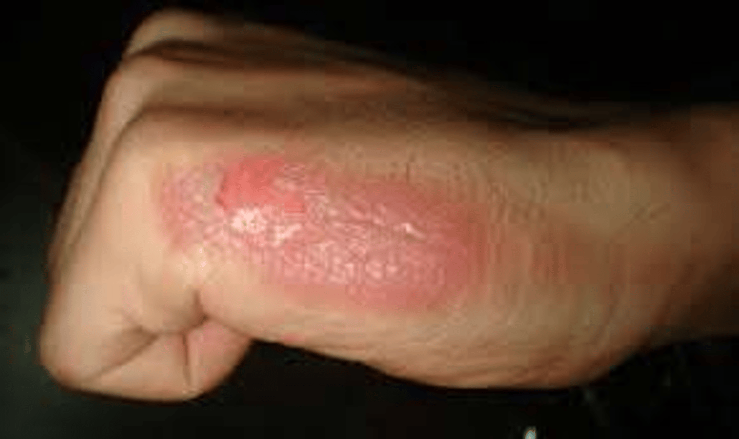
Second-degree burn
Extends through entire epidermis and part of the dermis; Redness, pain, and blistering
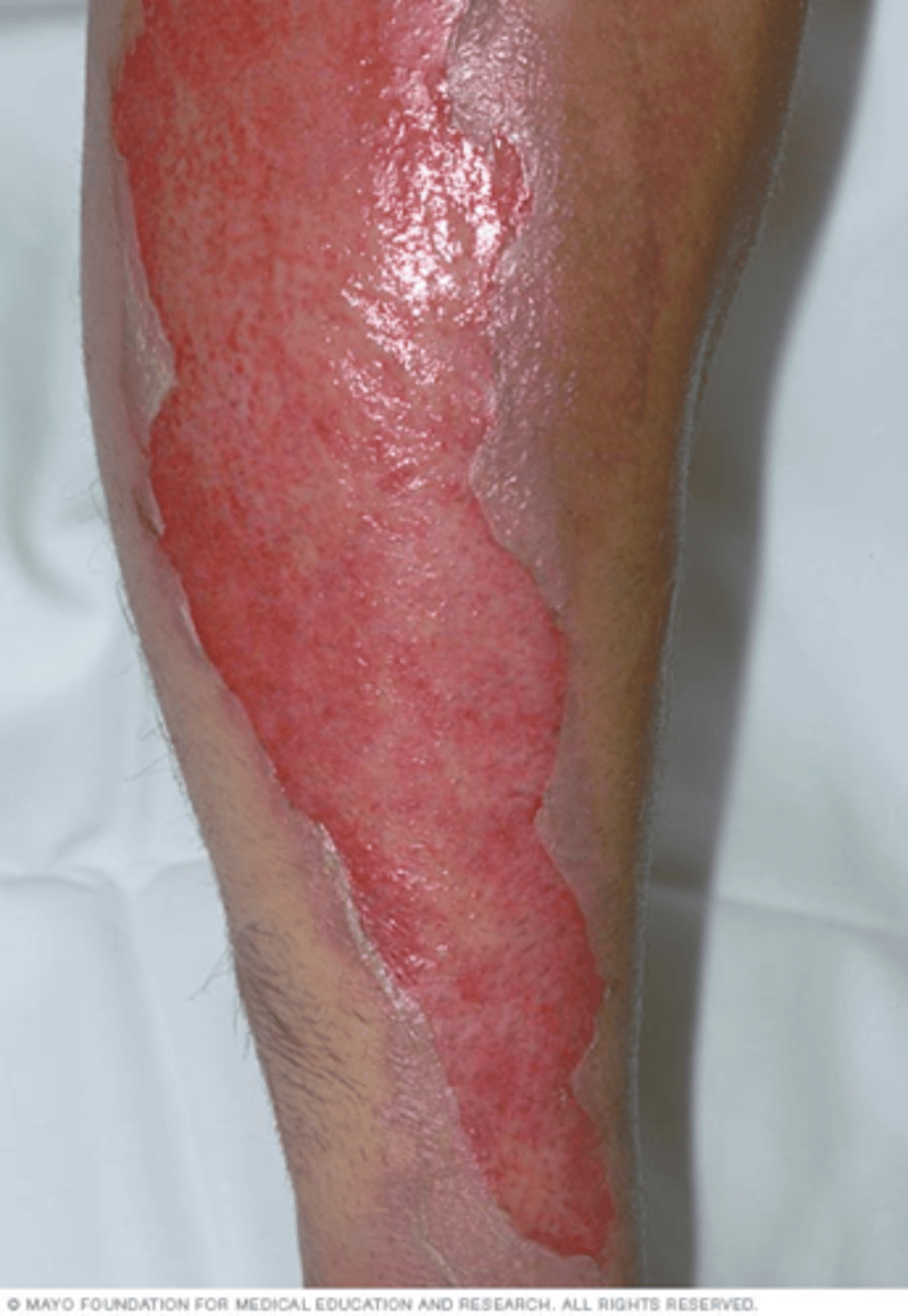
Third-degree burn
Destroys entire thickness of the skin; Surface of wound is leathery and may be brown, tan, black, white, or red; Patient feels no initial pain
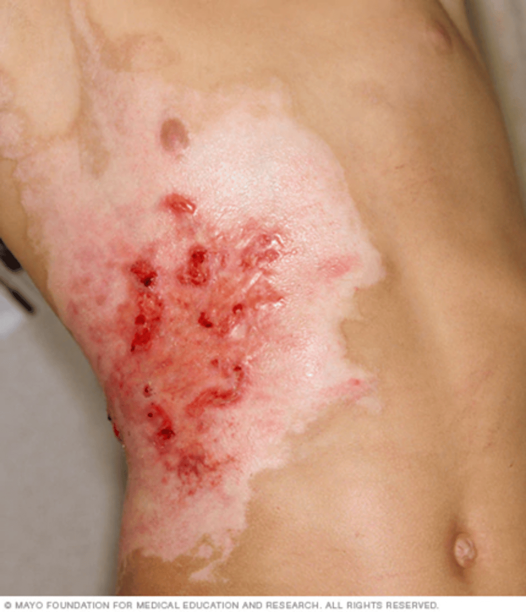
Fourth-degree burn
Involve tissues down to the bone.
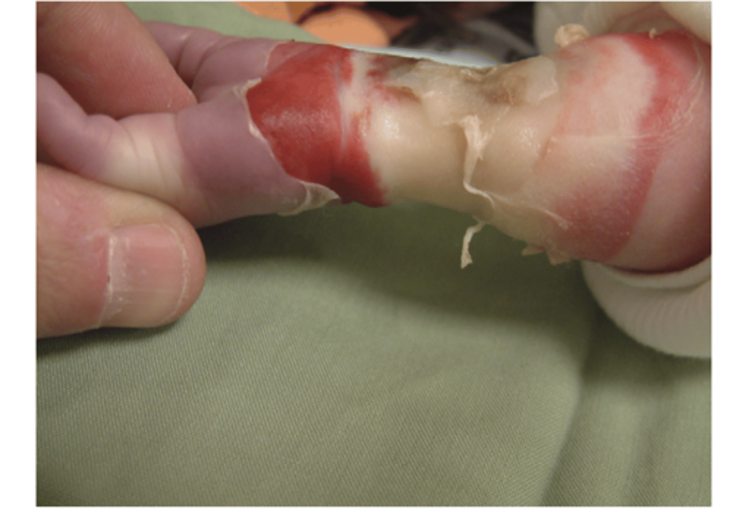
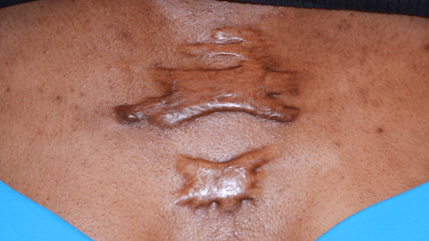
Keloid
Are firm, rubbery lesions or shiny, fibrous nodules, and can vary from pink to the color of the person's skin or red to dark brown in color.
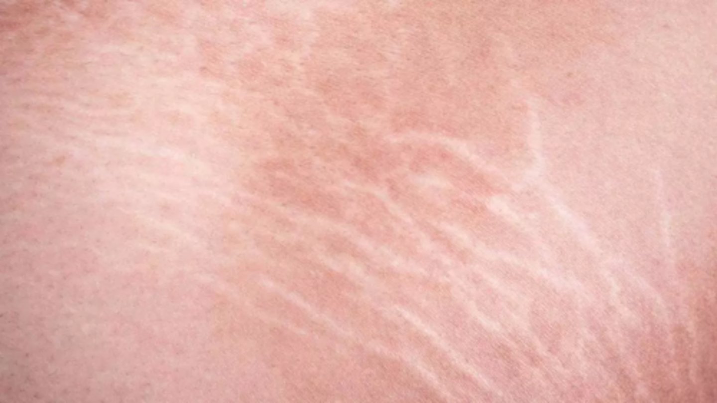
Stretch mark
Also known as Striae or Striae distensae, are a form of scarring on the skin with an off-color hue. Over time they may diminish, but will not disappear completely. Caused by tearing of the dermis during periods of rapid growth of the body, such as during puberty or pregnancy.
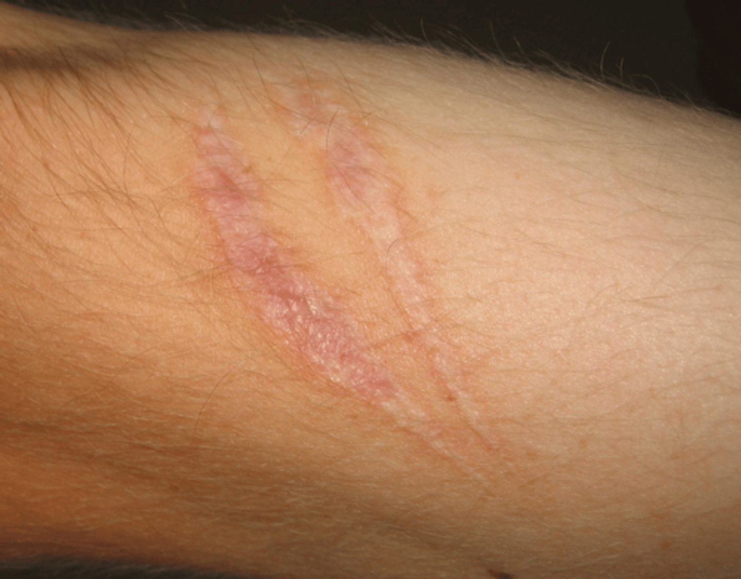
Scar
A mark left on the skin or within body tissue where a wound, burn, or sore has not healed completely and fibrous connective tissue has developed.
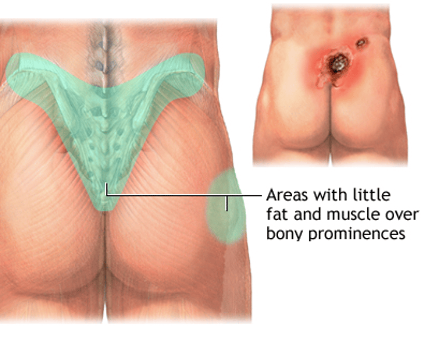
Bed sore
Also called pressure ulcers and decubitus ulcers; Are injuries to skin and underlying tissue resulting from prolonged pressure on the skin. Often develop on skin that covers bony areas of the body, such as the heels, ankles, hips and tailbone.
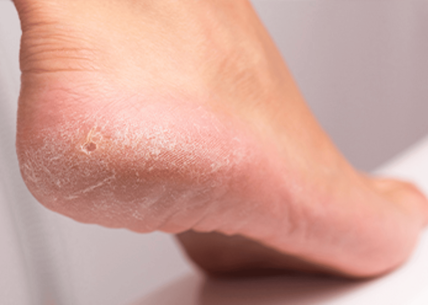
Callus
A thickened and hardened part of the skin or soft tissue, especially in an area that has been subjected to friction.