Sect 4 PPII - Non-Protein Nitrogen Compounds
 Call Kai
 Learn
Learn Practice Test
 Spaced Repetition
 Match
Match Flashcards
 Knowt Play
1/69

There's no tags or description
Looks like no tags are added yet.
Name | Mastery | Learn | Test | Matching | Spaced | Call with Kai | Chat |
|---|
No analytics yet
Send a link to your students to track their progress
70 Terms
Non-protein nitrogen compounds
the breakdown of proteins and nucleic acids results in the formation of:
Creatinine, Urea, Ammonia, Uric Acid
Non-protein compounds:
Creatine
Synthesized in the liver, pancreas, and kidneys
Transported through circulatory system to muscle, brain and other organs where it is converted to phosphocreatine and acts as energy source (like ATP)
arginine, glycine, methionine
Creatine is synthesized in the liver from:
**hint: amino acids
Creatinine
Produced in the muscle (amt in blood is proportional to pts lean muscle mass)
Produced as a waste product of creatine and phosphocreatine
NOT reabsorbed by kidneys; 100% excreted through the urine
serum and plasma
Specimen requirement for Creatinine methods
Fluoride and Ammonium heparin anticoagulant tubes
For Creatinine Measurement, what tube should NOT be used for specimens measured by the enzymatic rxn -- False neg
severe exercise and a high meat diet
For Creatinine Measurement, what can cause false pos
Jaffe Reaction (Chemcial)
Creatininase, Creatinase, Creatinine deaminase (Enzymatic)
What are the 2 methods used to measure Creatinine?
Jaffe reaction
Chemical method for Creatinine measurement
Creatinine reacts with picric acid in alkaline environment to generate an orange-red product
Most common reaction in the lab
picric acid
Chemical used in the Jaffe reaction
alkalkine environment
pH used in the Jaffee reaction
not specific for creatinine
Is the Jaffe reaction specific for creatinine?
interference with Jaffe reaction
False pos: elevated levels of bilirubin and hemolysis; protein, glucose, ascorbic acid, acetone, acetoacetate, pyruvate, guanidine and cephalosporins
Creatininase, Creatinase, Creatinine deaminase
3 enzymes utilized in the Enzymatic method for Creatinine measurement
Enzymatic method for creatinine measurement
Rxns are coupled with enzymatic reactions that can be measured spectrophotometrically
NADH to NAD+ and H2O2 to H2O → measuring colorless product
interference with Enzymatic method for Creatinine measurement
Less susceptible to interference from keto acids & cephalosporin antibiotics
False neg: fluoride and ammonia heparin anticoagulants inhibit enzyme activity
Inulin
gold standard for clearance tests
not typically used though because it is NOT endogenous (must be administered to paitent)
24hr urine and blood specimen
Specimen requirements for Creatinine clearance
Creatinine Clearance Formula
CC = (mL/min)
UC/PC = mg/dL
volume = mL
time = minutes
ASA/PSA = square meters
If you don't have PSA, remove PSA and ASA from formula

Schwarz formular
alternate method used to estimate the creatinine clearance in children
(k x Ht)/Creat
Cockcroft and Gault formula
alternate method to estimate creatinine clearance that adjusts for age difference (only used for 18 and older)
Cystatin C
Produced by all nucleated cells of the body at a constant rate
- releases when cell is broken down
- found in all body fluids EXCEPT urine → ALL is reabsorbed by proximal tubules
Unaffected by: muscle mass, sex, age, diet, ethnicity
Cystatin C increases
What happens to Cystatin C as GFR decreases?
**hint: all should be reabsorbed by proximal tubules
(compromised kidney = decreased reabsorption)
Blood Urea Nitrogen (BUN)
Urea is reported as what in the United States?
BUN
Breakdown of proteins
Synthesized in the liver and transported through blood to kidney, where it is filtered through the glomerulus, where almost HALF is reabsorbed back into the blood by passing though nephron tubule

protein intake and protein breakdown
The rate of urea synthesis depends on (2):
**hint: Amino acids → Ammonia + CO2 → Urea + water
75%
BUN accounts for more than _____% of non-protein nitrogen that is excreted from the body
**hint: the most abundant waste product in the urine
increase in BUN
- Disorders with excessive breakdown of proteins (febrile illnesses, dehydration, corticosteroid/tetracycline therapy, internal bleeding, and in presence of elevated thyroid hormones)
- High protein diets/Large ingestions of protein
- Pregnancy (due to increase GFR)
Cirrhosis of the liver
What causes decrease in BUN?
**hint: where urea cycle takes place -- can't breakdown ammonia to urea
10:1 to 20:1
Normal BUN:creatinine ratio
low BUN:creatinine ratio
5:1
Acute nephron tubular necrosis
Low protein intake (malnutrition)
Starvation
Severe liver disease
increased BUN with normal creatinine levels
30:1 (reabsorbing too much BUN)
Pre-renal uremia
High protein intake in diet
GI tract hemorrhage
Dehydration
increased BUN and creatinine
30:10
May indicate a post-renal problem (obstruction of urinary tract)
End-stage renal disease
Urease Method (Enzymatic)
Diacetyl monoxime (Colorimetric)
What are the 2 methods used to measure BUN?
Urease method
Enzymatic method for Urea (BUN) measurement
Ammonia (NH4+) is liberated in this rxn and can be measured using the Berthelot reaction

Urease
Enzyme utilized in the Enzymatic method for Urea (BUN) measurement
Berthelot's reaction
Coupled reaction utilized with the Urease method
Ammonia (NH4+) ion that is liberated in the urease rxn is most commonly measured utilizing this rxn

Diacetyl monoxime method
Colorimetric method for Urea (BUN) measurement

conversion of urea to BUN
Urea (mg/dL) = BUN (mg/dL) x 2.14
BUN (mg/dL) = Urea (mg/dL) / 2.14
interference with Colorimetric method for BUN measurement
False neg: anti-coagulants containing fluoride or citrate
(inhibits rxn form occurring = no color formed)
Urinary Protein
used to monitor kidney function not usually present in urine
more sensitive
Urine concentration of protein is less than the concentration of blood; so urine methodologies have to be ____________ than blood protein levels
Turbidimetric
Dye-Binding/Colorimetric
What are the 2 methods used to measure total protein in urine?
Turbidimetric
Protein is precipitated out with Sulfosalicylic acid (SSA), then turbidity of the precipitated is measured
Semi-Quantitative test
other: Trichloroacetic acid or Benzethonium chloride
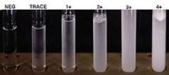
Sulfosalicyclic acid
Trichloracetic acid
Benzethonium chloride
Chemicals for turbidimetric method for urinary protein measurement
Dye-Binding/Colorimetric
Dye binds to albumin and causes a shift in the max absorption; resulting color change is measured
Methyl orange, Bromcresol green (BCG), or Bromcresol purple (BCP) may be used
Methyl Orange
Bromcremsol Green
Bromcresol Purple
Dye-binding chemicals used to measure urinary albumin
BCG most common
Uric Acid
Produced from the cellular breakdown products of the purine nucleosides
Filtered through the renal glomerulus and is almost completely reabsorbed in the proximal tubules, but some end up in urine (6-12%)
adenosine and guanine
purine nucleosides that uric acid is derived from
Urate crystals
At acidic urine pH levels above 5.7, what type of crystals are formed?
chemical form that is soluble in urine

Uric acid crystals
At acidic urine pH levels below 5.7, what type of crystals are formed?
painful accumulation in joints or present in kidney stones
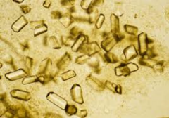
leukemia and gout
Increased levels of uric acid are seen in (2):
Hyperuricemia
Increased uric acid in the blood; > 7.0 mg/dL
may be asymptomatic, but are at increased risk for associated renal problems
Gout
a disease in which deposits of urates are deposited in body fluids (especially in synovial fluid - joints)
caused by: over-production of purines or defects in the metabolic pathways of purine metabolism
big toe
Where is the most common place we see gout in the body?
gout arthritis
Classic symptom of gout
**hint: uric acid is deposited in joints causing pain & inflammation
causes of Hyperuricemia
over-production of purines
renal retention of uric acid
acidemia (low pH = uric crystals)
defects in the enzymes of the purine metabolic pathway (Lesch-Nyhan syndrome)
Lesch-Nyhan syndrome
a deficiency of a transferase enzyme in the purine metabolic pathway, leading to tophi deposits
hyperuricemia
can be:
minor - depending on location
major - accumulation in brain tissue which can lead to intellectual disability, signs of cerebral palsy, and self-injurious behavior
Hypouricemia
decreased uric acid
causes of Hypocuricemia
reduced purine synthesis
defective reabsorption of uric acid in the kidney (Fanconi's syndrome)
Fanconi's syndrome
a disorder of the proximal tubule of the nephron, resulting in poor transport
hypouricemia

Uricase Method (Enzymatic)
Phosphotungstate Method (Colorimetric)
What are the 2 methods used to measure Uric acid?
Uricase Method
Enzymatic method for Uric Acid measurement
Rxn measured in 2 ways:
1. Allantoin is produced and measured as a decrease in absorbance at 293nm due to a loss of urate
2. Hydrogen peroxide may be converted to a chromogen using 4-aminophenazone & peroxidase in a couple reaction

Uricase
enzyme that converts uric acid to allantoin
interference with Uricase method
False pos: excessive amounts of protein and lipids; hemoglobin and bilirubin
False neg: Xanthine (competitive inhibitor -- keeps uric acid from binding & reacting) & anti-coagulants containing fluoride or citrate
Phosphotungstate Method
Colorimetric method for Uric Acid measurement

protein-free filtrate for serum or plasma
Phosphotungstate method for uric acid requires:
**hint: proteins are interfering substances
interference of Phosphotungstate Method
False pos: Glucose, Ascorbic acid, Glutathione, Hemoglobin and drugs (Acetaminophen)
False neg: Caffeine (competitive inhibitor -- keeps uric acid from binding & reacting) & anti-coagulants containing fluoride or citrate
serum, heparinized plasma and urine
Specimen requirements for Uricase Method & Phosphotungstate Method
Anticoagulants containing fluoride or citrate should not be used