ANATOMY OF THE HEART
1/89
There's no tags or description
Looks like no tags are added yet.
Name | Mastery | Learn | Test | Matching | Spaced |
|---|
No study sessions yet.
90 Terms
Where is the heart located?
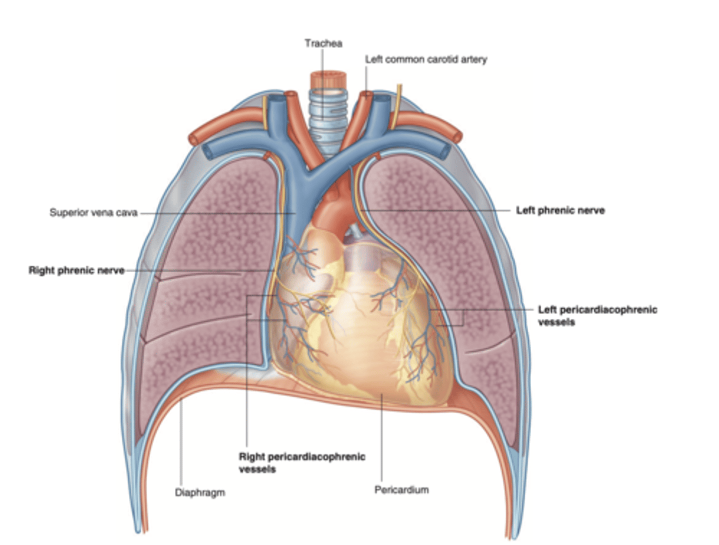
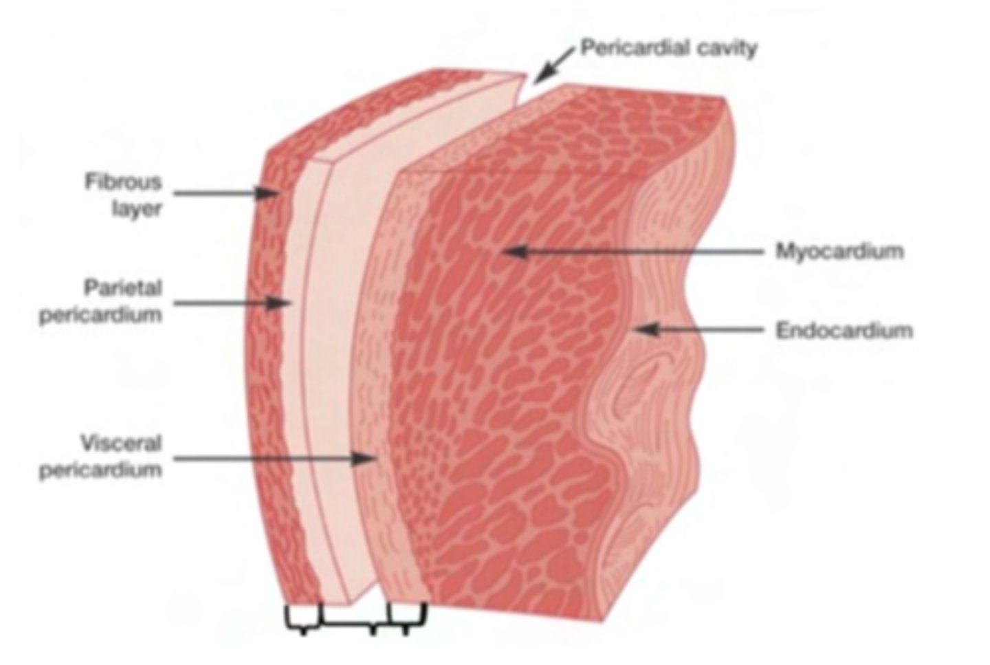
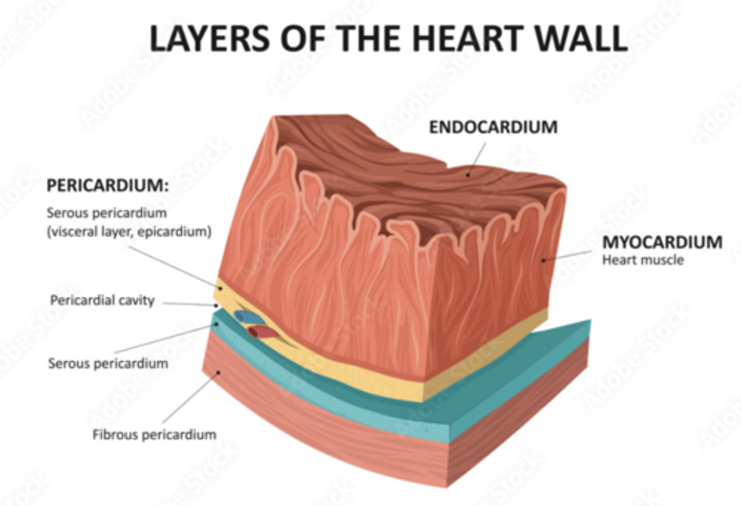
The heart is located in the middle mediastinum, surrounded by the pericardium.
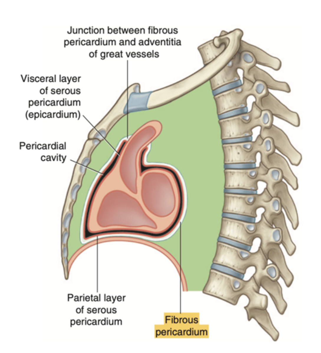
What are the layers of the pericardium?
The pericardium consists of two main layers:
- the fibrous pericardium
- the serous pericardium
PERICARDIUM : FIBROUS SAC SURROUNDING THE HEART AND THE ROOT OF THE GREAT VESSELS

How is divided the serous pericardium?
. Visceral layer or Epicardium (innermost). Adheres to the heart
. Parietal layer (outermost)
Between these layers is the pericardial fluid (pericardium cavity )
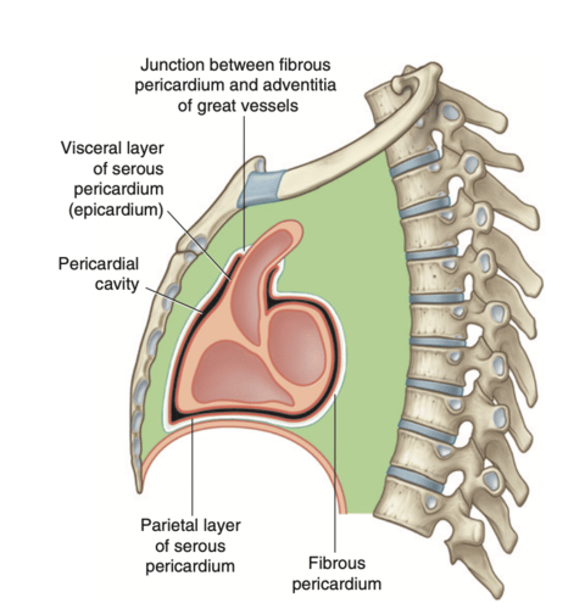
What is the fibrous pericardium?
the tough, outer layer
protects the heart
maintains its position in the thorax.
defines the boundaries of the medium mediastinum
What are the reflections (transitions) of the serous pericardium?
. spaces known as pericardial sinuses
. occur in the transition from visceral to parietal
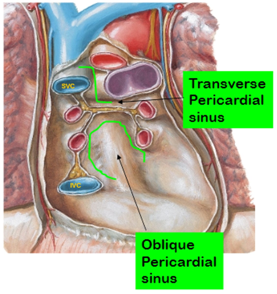
What are the pericardial sinuses and their relevance?
. transverse sinus
. oblique sinus
. Allow for the passage of structures such as blood vessels
. Clinically significant as potential sites of fluid accumulation.
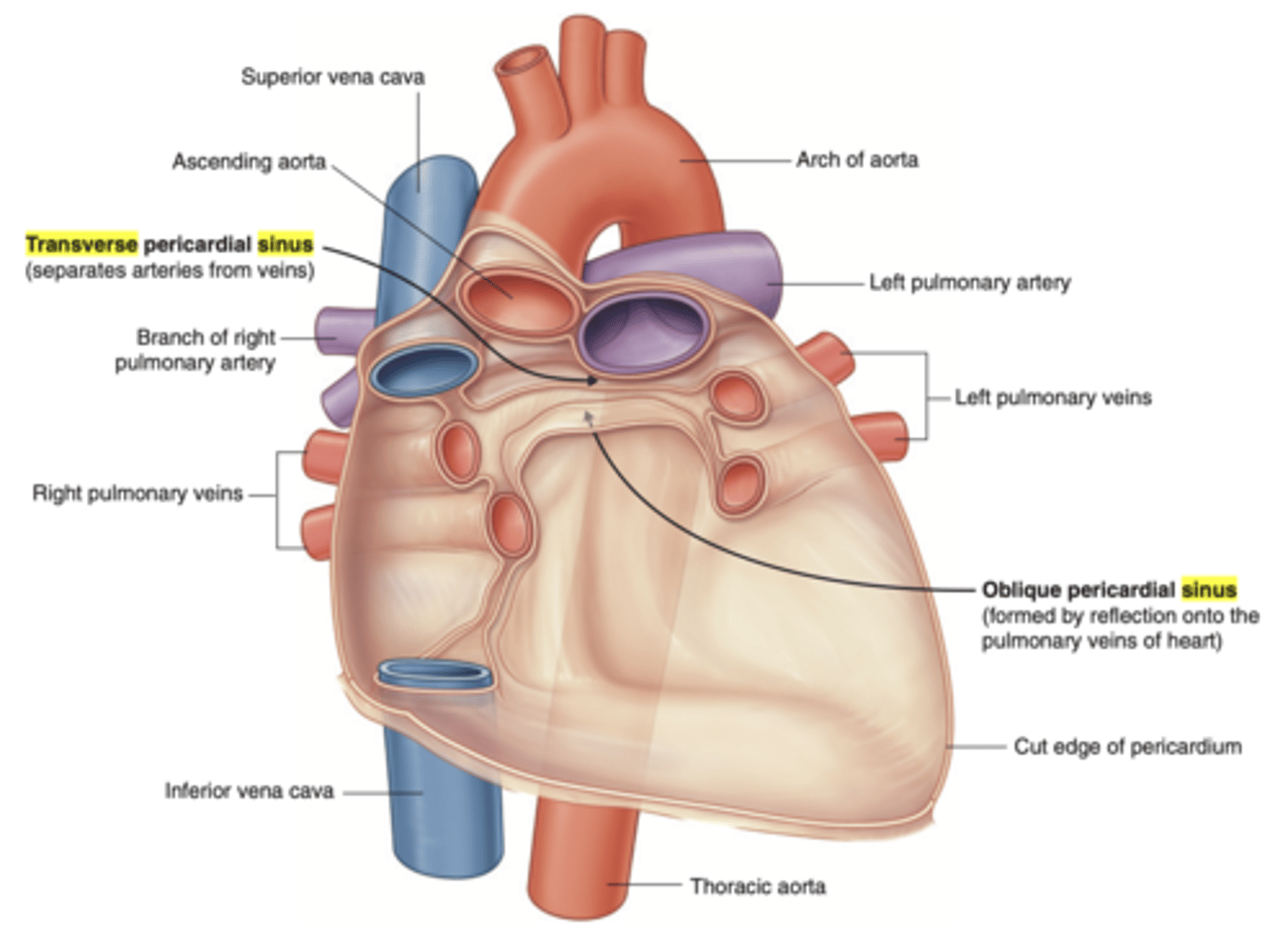
What is the transverse sinus of the pericardium ?
. space posterior to the aorta and pulmonary trunk
. anterior to vena cava
EMBRIOLOGICALLY COMES FROM THE DISINTEGRATION OF THE DORSAL MESOCARDIUM
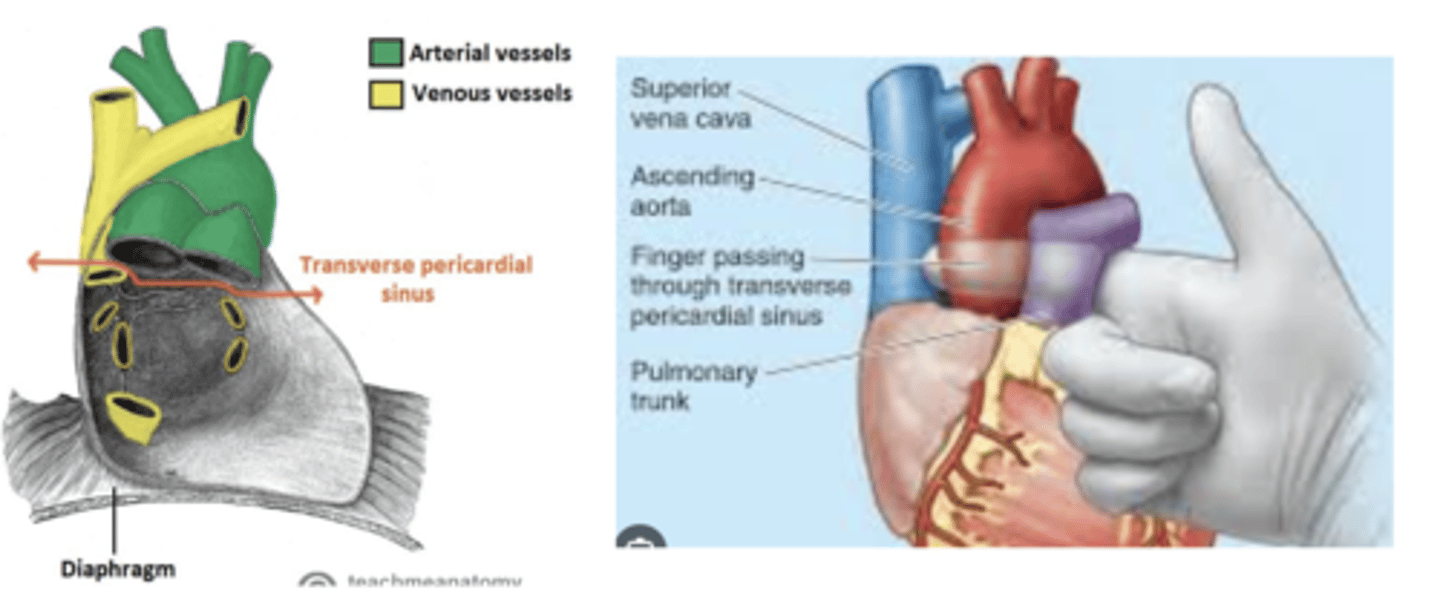
Clinical significance of the transverse sinus?
Surgeons use it during cardiac procedures.
By passing a finger or clamp through the sinus they can isolate these arteries and control blood flow to the heart during surgeries like coronary artery bypass grafting (CABG).

What is the oblique sinus of the pericardium?
Area where the parietal pericardium reflects (transition) into the visceral pericardium.
Posterior to the heart, cradled by the left atrium and the pulmonary veins.
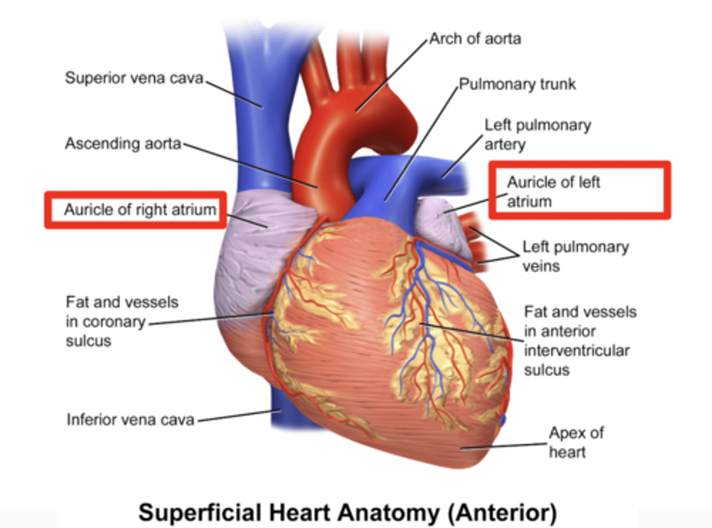
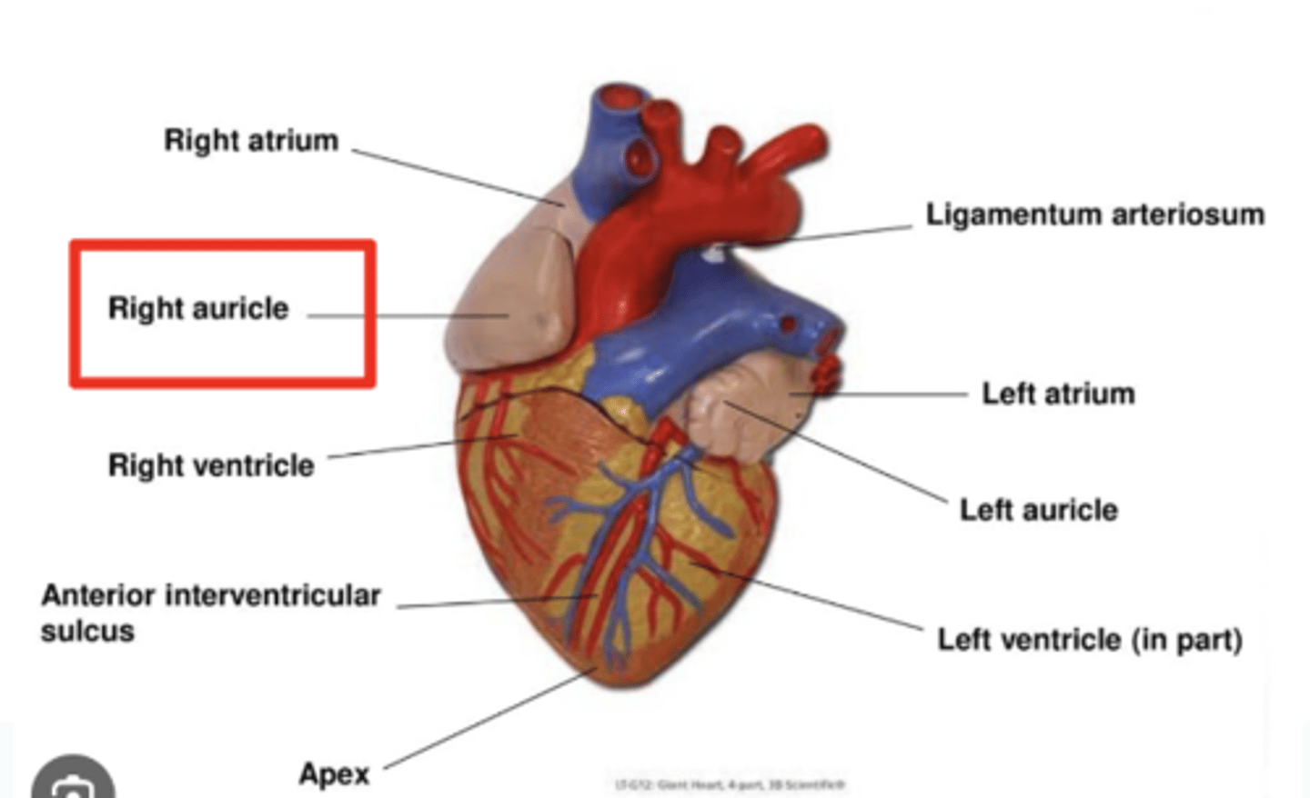
How is the heart's shape described and what are its surfaces?
The heart has an inverted pyramidal shape lying in one side.
Surfaces:
. Inferior diaphragmatic surface
. Sternocostal surface or anterior surface (corresponds mainly to the right ventricle)
. Posterior surface (corresponds to the ventricles, mainly to the left one)
. Right pulmonary surface
. Left pulmonary surface
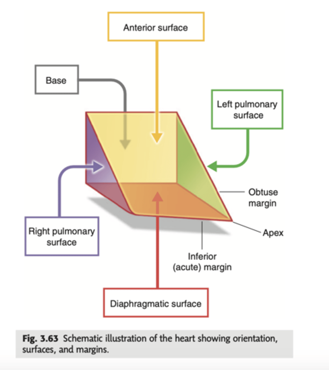
Describe the apex in the heart's anterior view
. Point to left side, downwards.
. At the level of the 5th intercostal space (we will auscultate the apex of the left ventricle)
APEX: HIGHEST POINT, VERTEX.
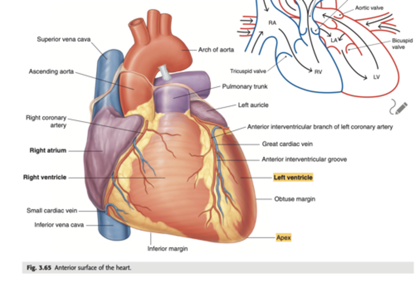
Describe the Sternocostal Surface
Most of the anterior surface of the heart corresponds to the right ventricle.
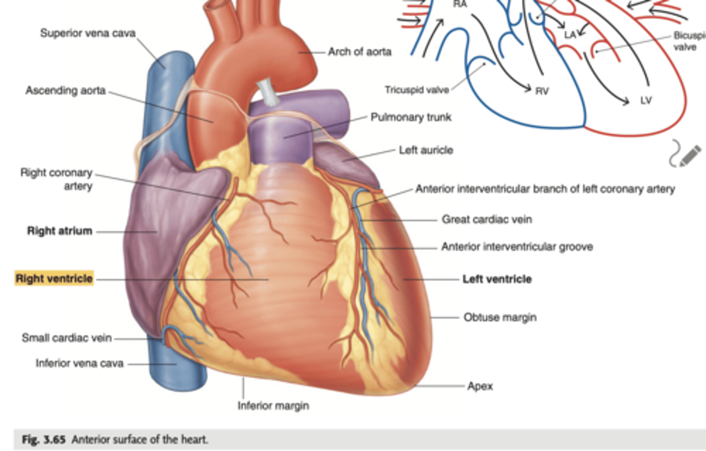
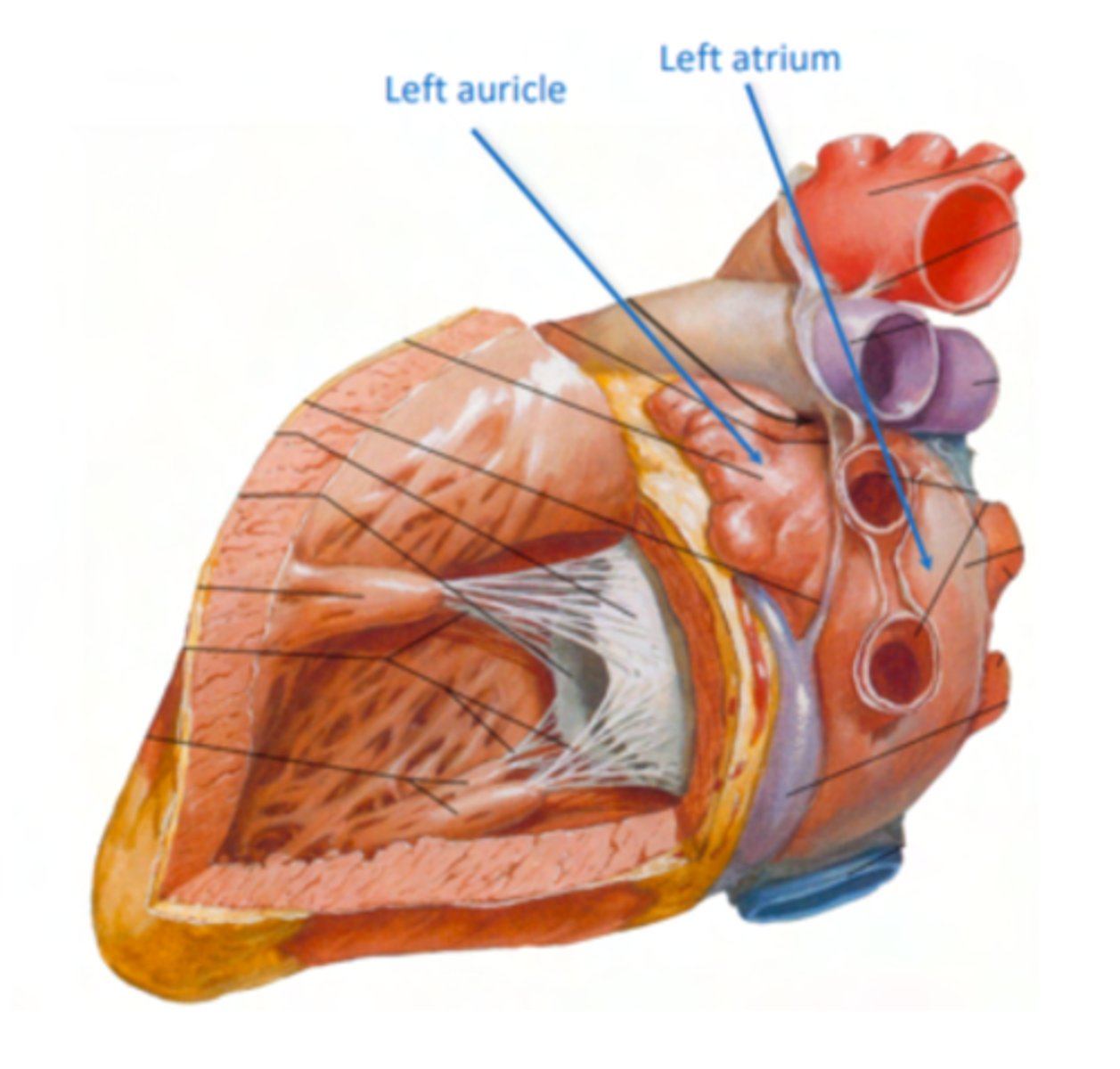
What are the atrial appendages?
Rough trabeculated region of the atria (Extensions of the atria)
External portions of the atria, (Real cavities of the right and left atrium being posterior to these auricles)
ALSO CALLED AURICLES
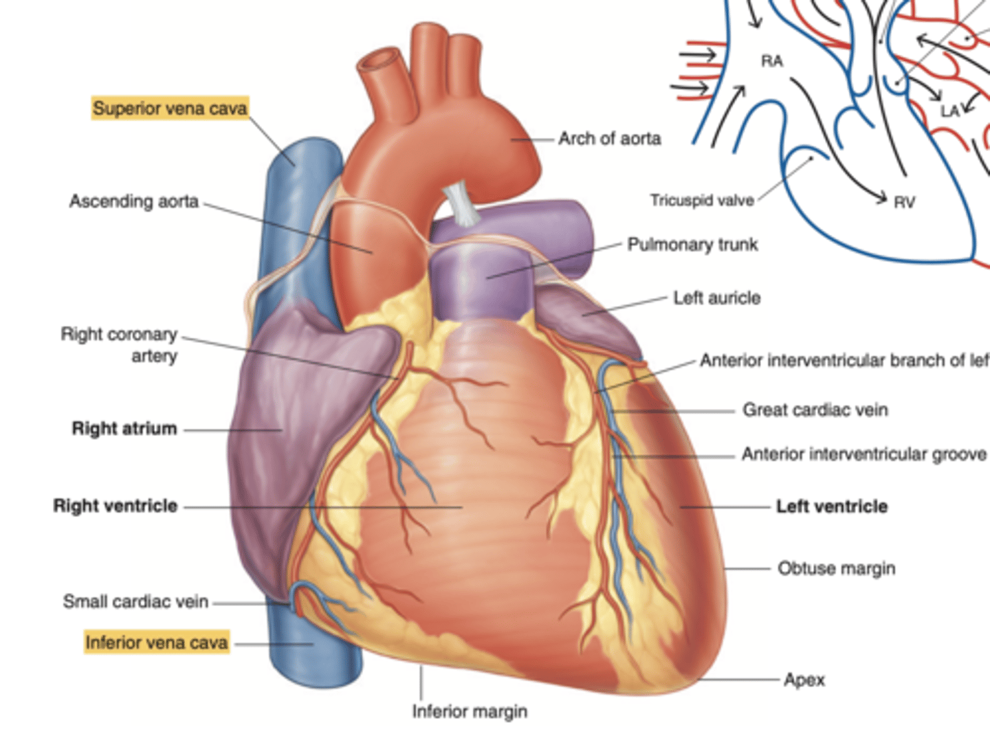
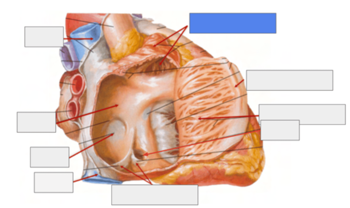
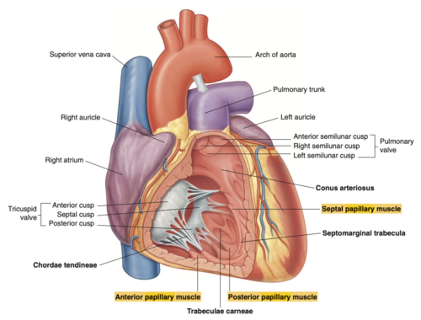
Describe the location of the main arteries in the anterior view of the heart
The pulmonary trunk exits the right ventricle
. aorta exits the left ventricle.
During early heart development, the pulmonary artery is on the left and the aorta on the right because they develop from a common tube (truncus arteriosus).

What is the pulmonary trunk?
. major blood vessel that plays a crucial role in the circulatory system by carrying deoxygenated blood from the heart to the lungs.
. Divides into left and right pulmonary arteries
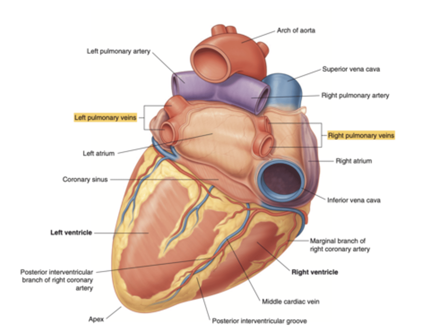
What structures enter the right atrium posteriorly?
. Superior Vena Cava (SVC)
. Inferior Vena Cava (IVC)
. Coronary sinus (visible only in the posterior view)
Show the position of the coronary sinus in the posterior view of the heart and its function
collects deoxygenated blood from most of the heart's venous system
delivers it to the right atrium for reoxygenation.
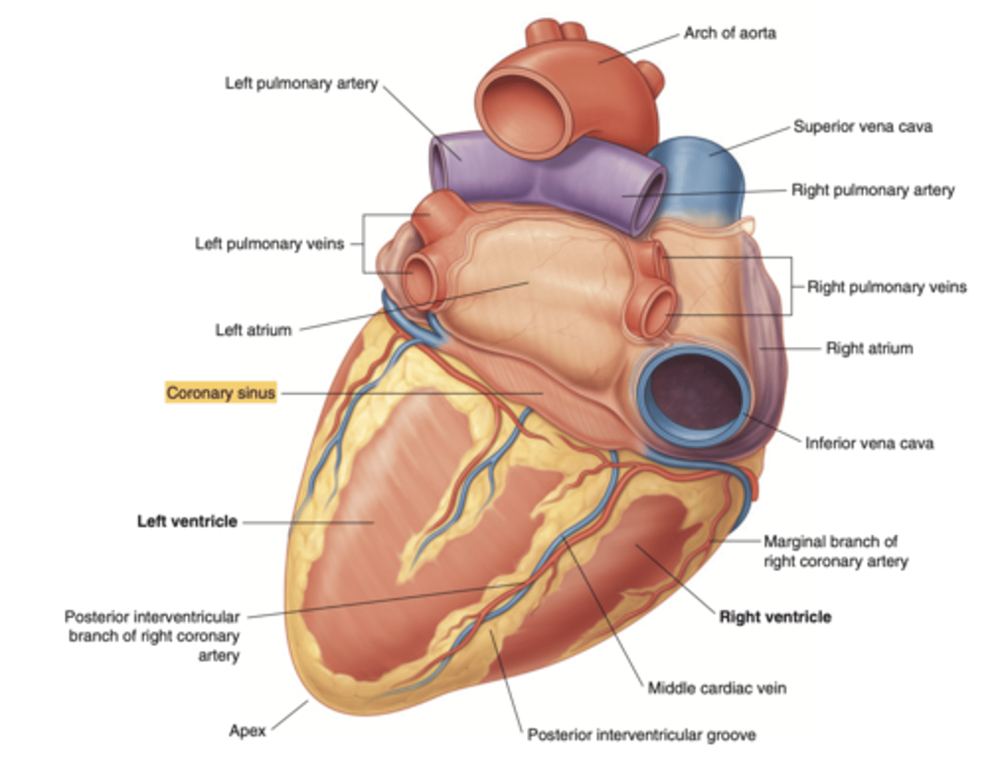
Show the position of the ventricles and auricles in the posterior view
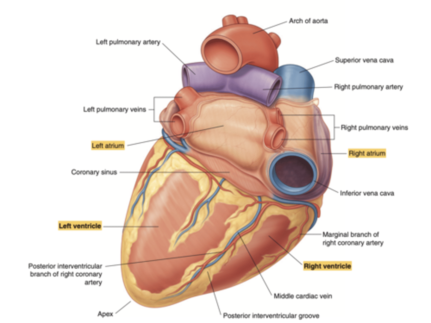
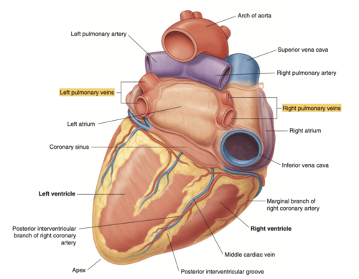
Which veins enter the left atrium?
left pulmonary veins
right pulmonary veins
ALLOWS OXIGENATED BLOOD FROM THE LUNGS TO ENTER THE HEART
How is generated the atria?
. Auricles, form the primitive atria → rough portion of the atrium
. Real cavities as such originate from the incorporated vessels
TWO EMBRYOLOGICAL SOURCES (ATRIA AND PRIMITIVE VEINS)
What are the vessels incorporated to the atria?
cava veins (sinus venosus embryo origin, for the right one)
pulmonary veins (4, for the left one)
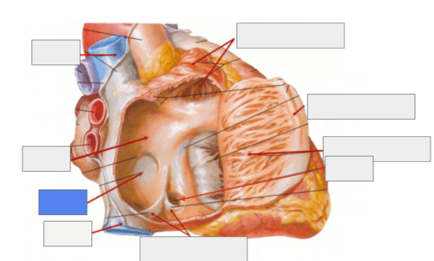
What are the six surfaces that form the internal structure of the right atrium?
. superior
. inferior
. posterior
. medial
. anterior
. lateral walls.
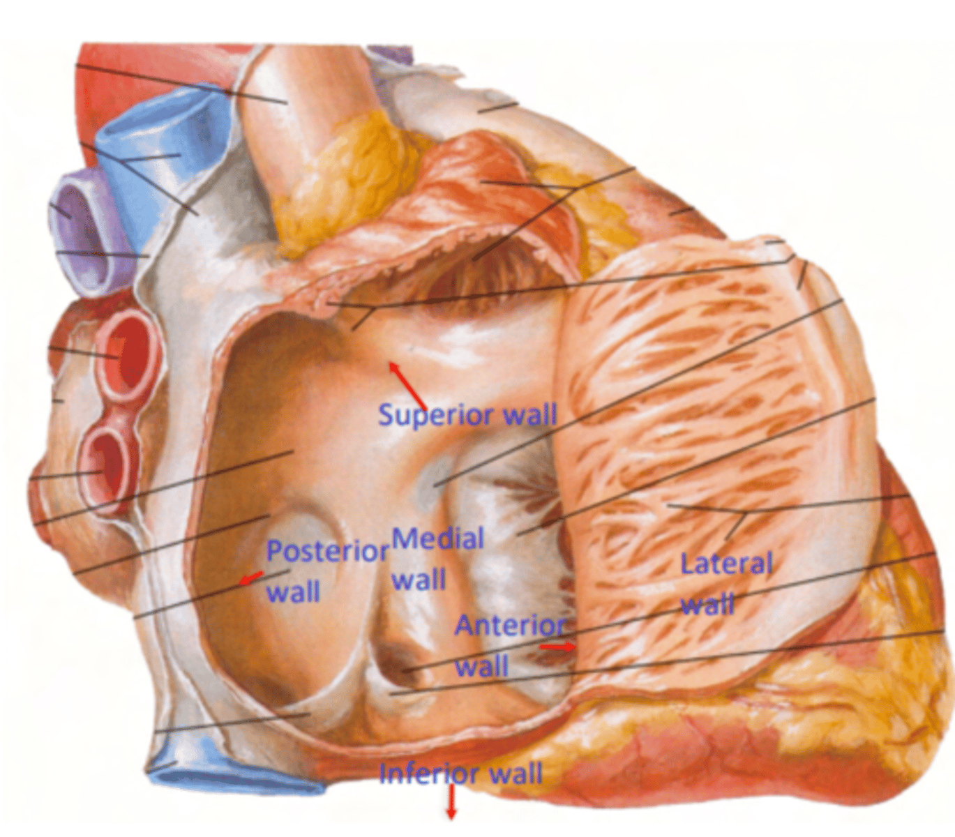
What is the vein entering without valve in the right atrium?
Superior Cava vein
(No valve for entry)
What are the veins entering with valves in the right atrium?
Inferior cava vein (left)
Coronary sinus (right)
Both of them with a valve for entry
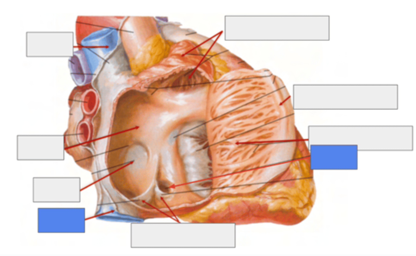
What is pointing the blue square?
valves of inferior vena cava and coronary sinus
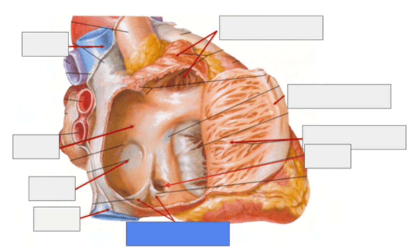
What is the trabeculated wall in the atrium?
Right Auricle
What are the name of the musculi of the right auricle?
Musculi pectinati
TRABECULATED WALL VS THE SMOOTH INTERIOR PART OF THE ATRIA
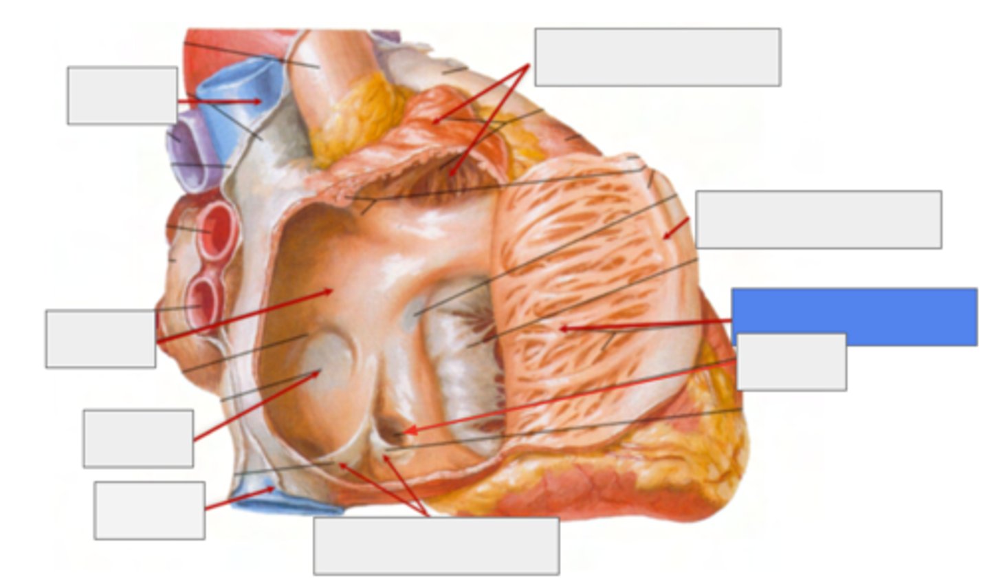
What separates the trabeculated from the smooth walls in the atrium?
Crista terminallis
ANATOMICAL LANDMARK indicates the transition between the original embryological parts of the atrium.
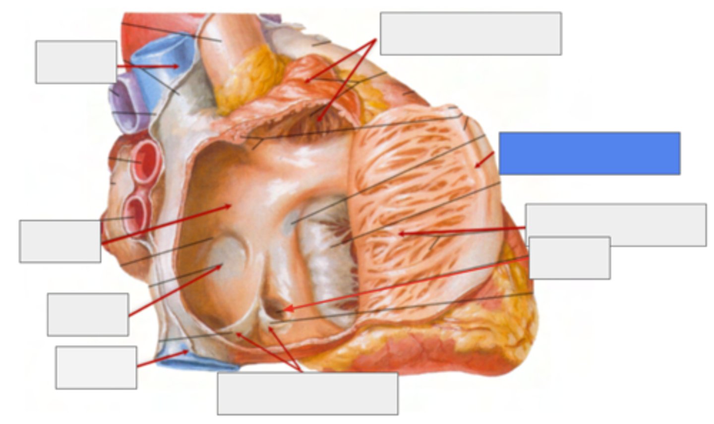
What is the remnant of the foramen ovale?
Fossa Ovalis
belongs to the Septum Primum (thinner atrium region)
during fetal live it serves as a passage of blood from let to right
What are the main roles of the right auricle in the atrium's structure?
1. Reservoir for Blood
During atrial relaxation accommodate fluctuations in venous return, especially during exercise or increased cardiac demand.
2. Assist in Atrial Contraction
Propel additional blood into the ventricles
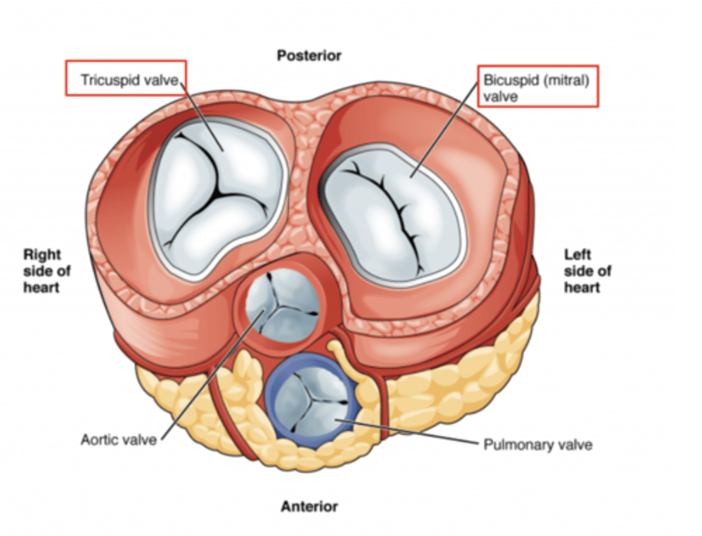
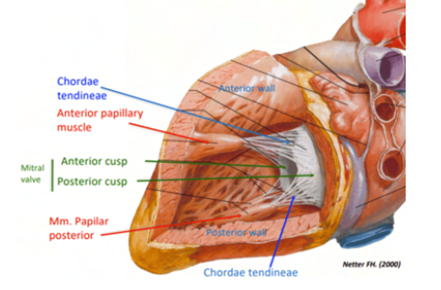
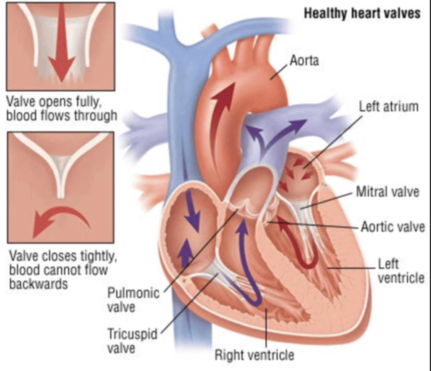
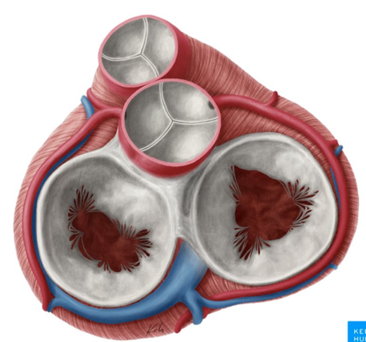
What are the valves separating atrium and ventricle?
. Tricuspid valve (right atrioventricular valve, 3 cusps)
. Mitral or bicuspid valve (left atrioventricular valve, 2 cusps)
AXIAL VIEW OF THE HEART
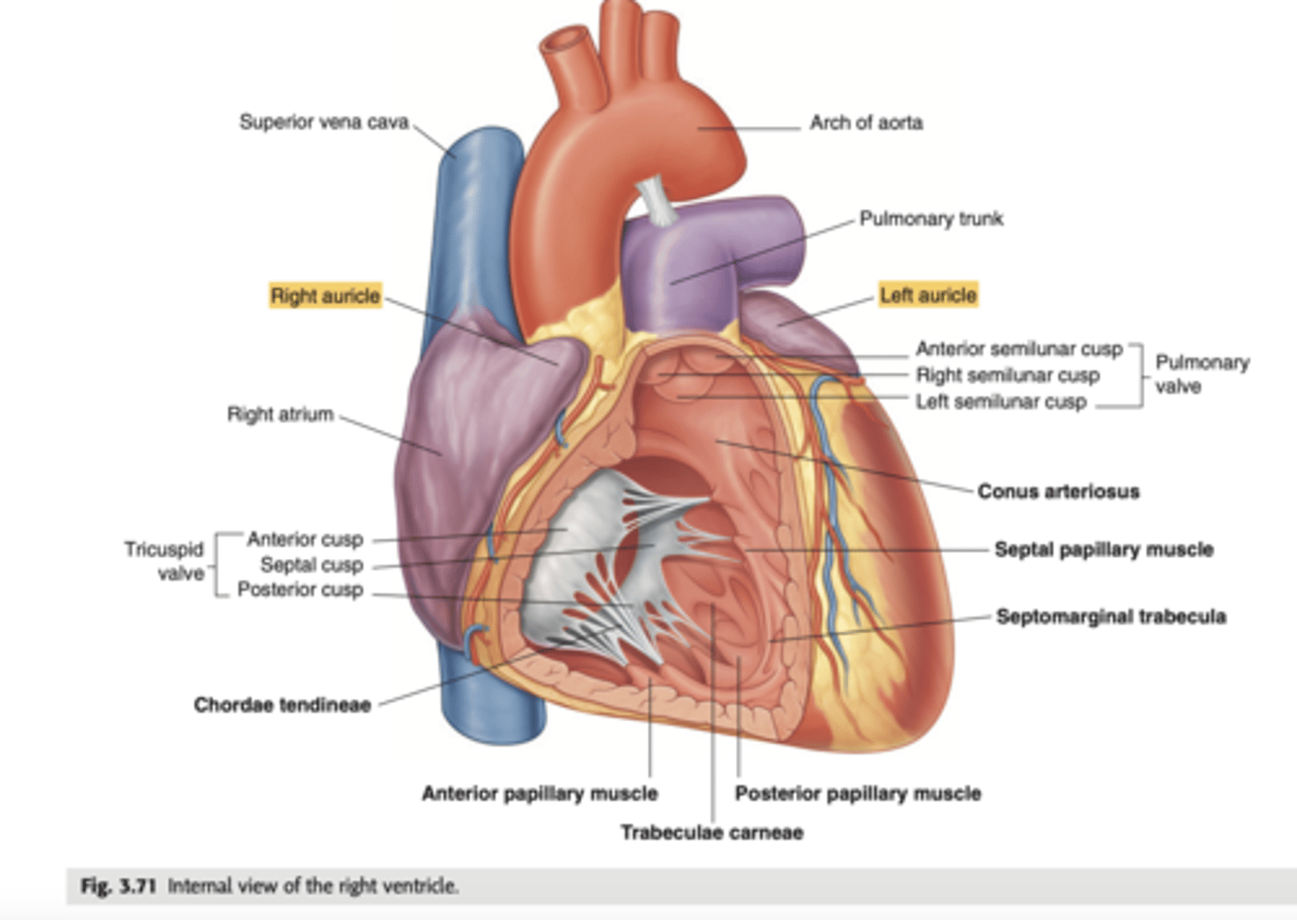
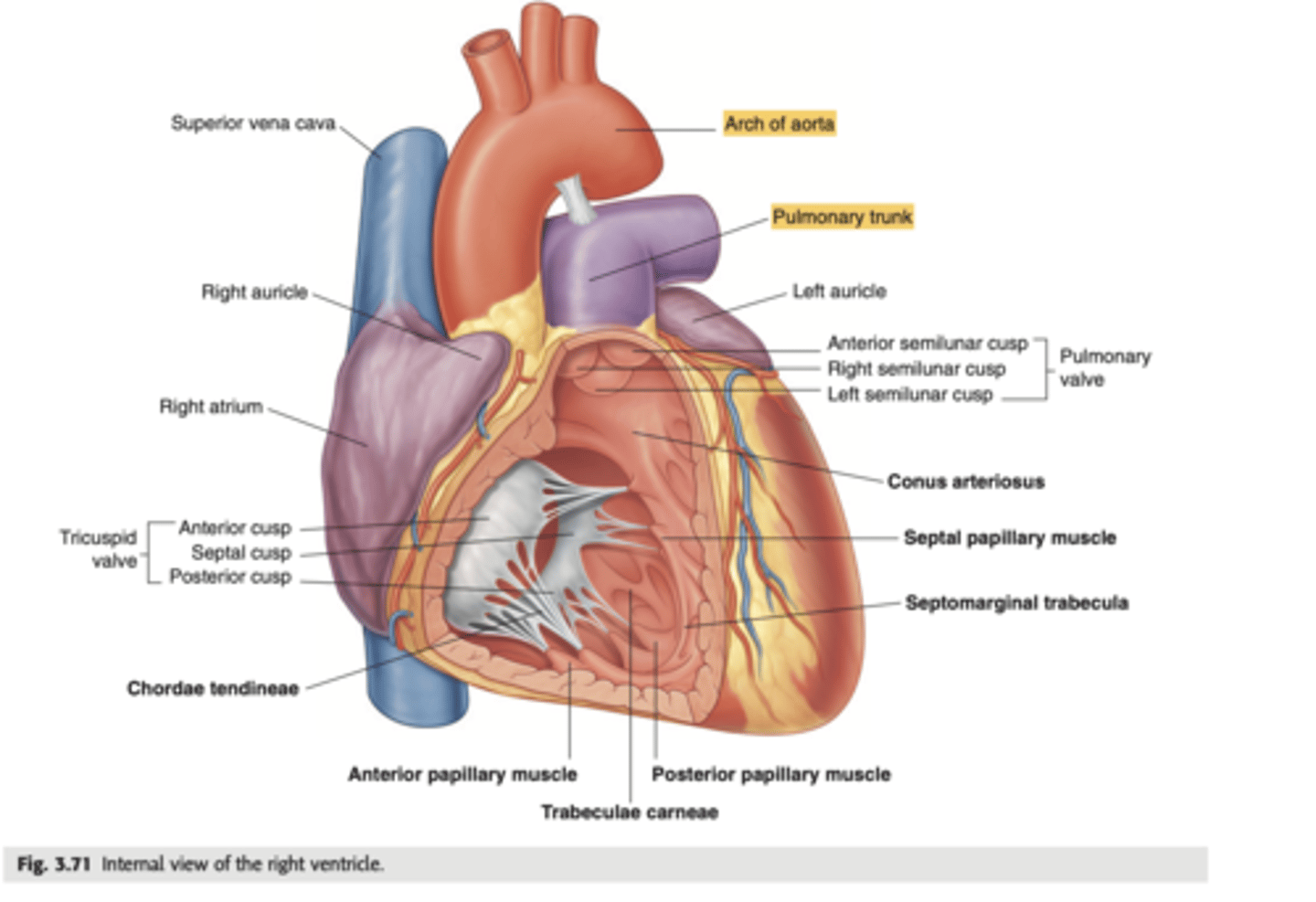
What are the walls of the right ventricle?
. Anterior wall
. Posterior wall
. Septal wall
Right ventricle has a conical shape with 2 o 3 walls
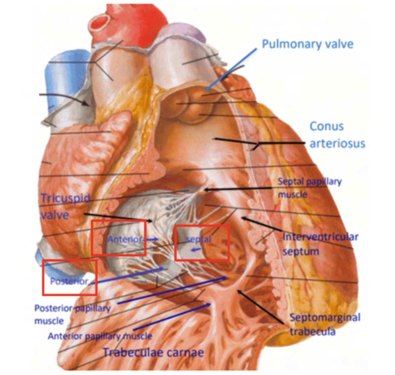
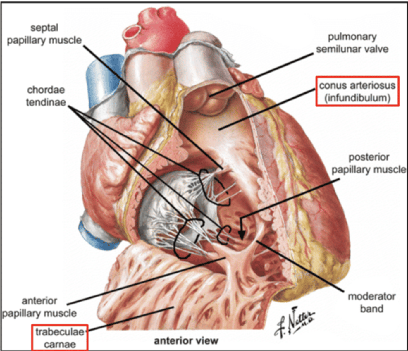
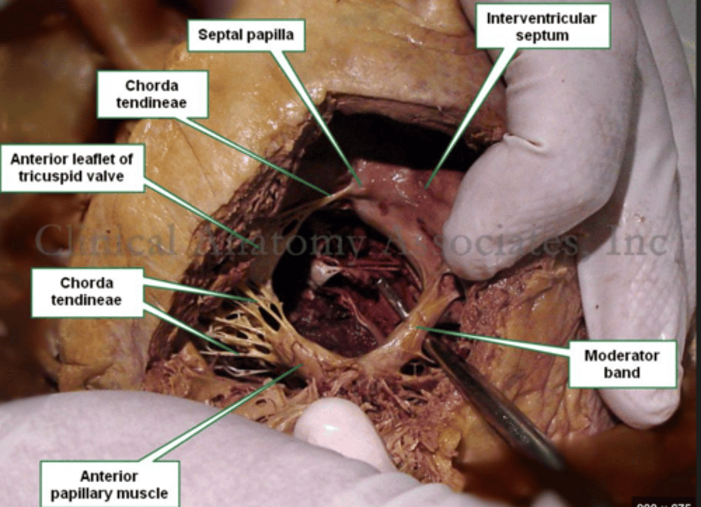
Describe the two portions of the medial/septal face/wall of the right ventricle.
infundibulum or outflow region or conus arteriosus (smooth and membranous)
trabeculae carnae (muscular and irregular)
What are the 3 cusps of the tricuspid valve?
Anterior
Posterior
Septal
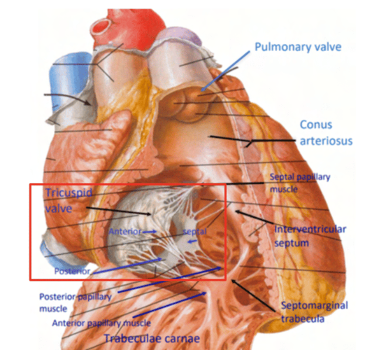
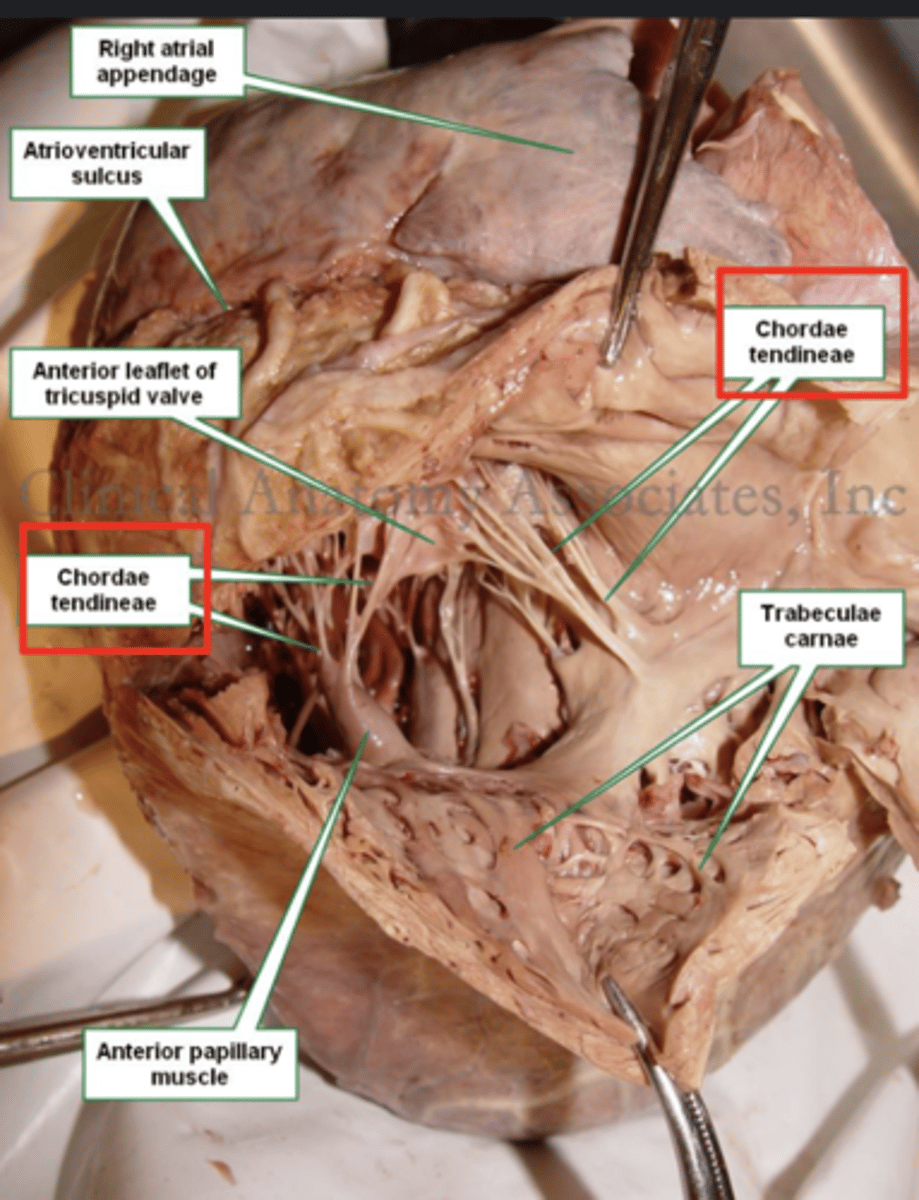
What connect the cusps of the valve a to papillary muscles in the
right ventricle?
Chordae tendinae
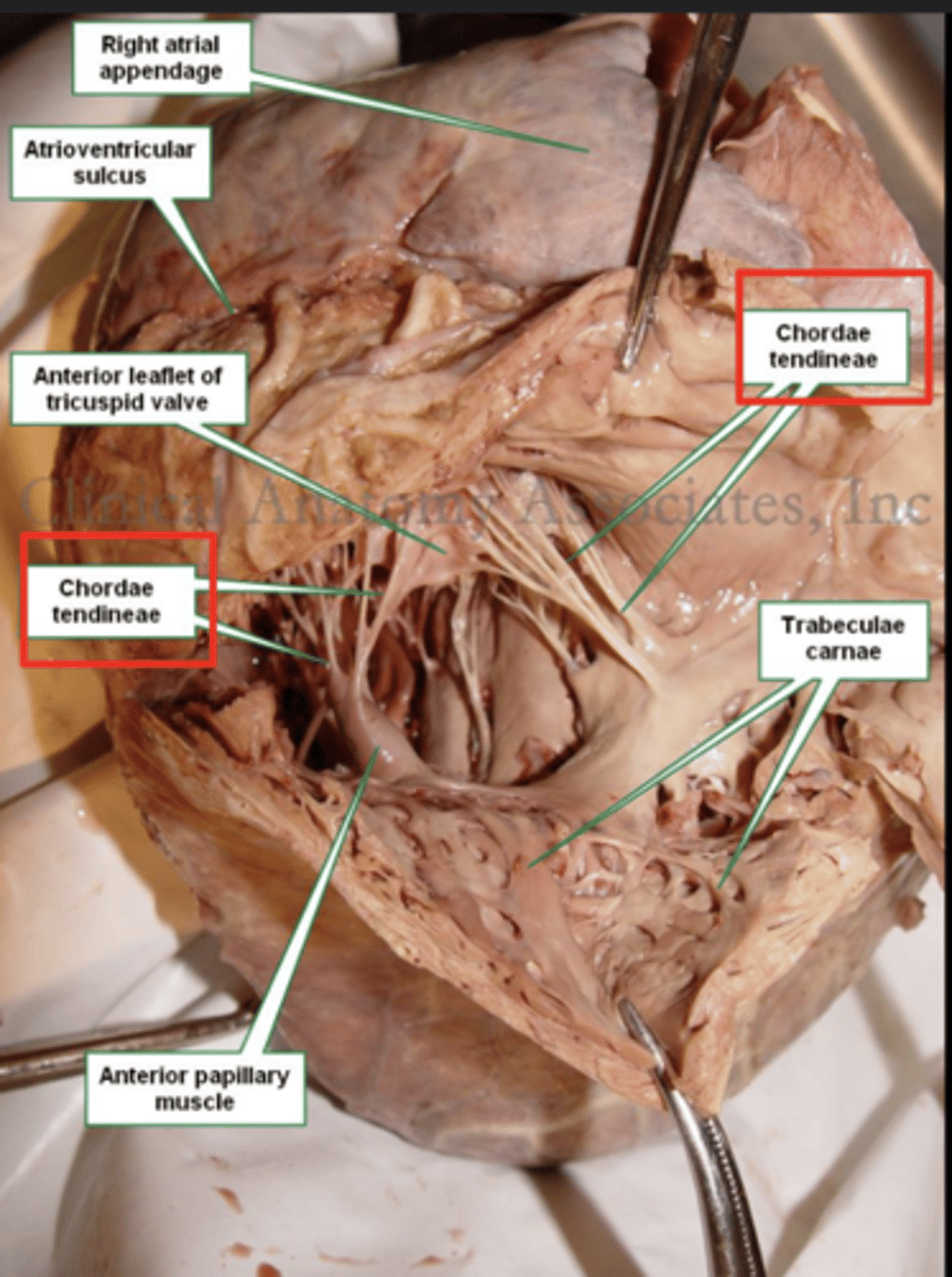
With the contraction of the walls of the ventricle what's the action of the papillary muscles?
contract as well
With the contraction of papillary muscles what will be happen with the valve?
Close

What is the septomarginal trabeculae (or moderator band)?
Part of the conduction system of the heart.
electrical waves pass through this band to reach the papillary muscles.
LANDMARK
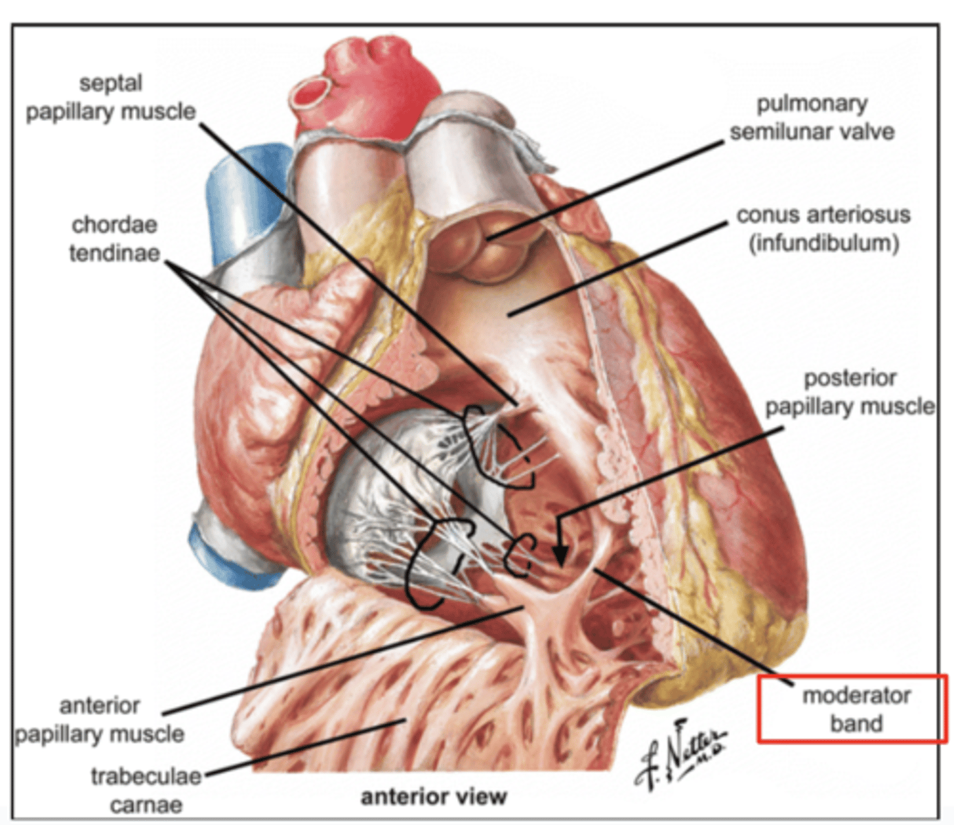
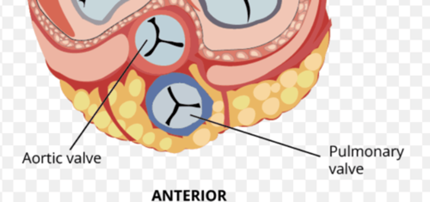
What are the positions of the pulmonary valve cusps?
Anterior
Right
Left
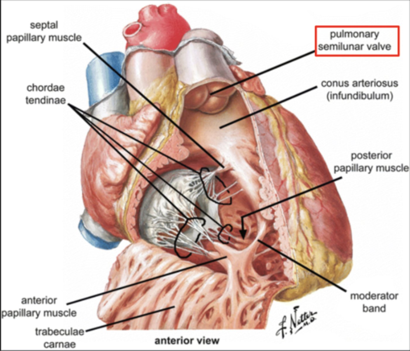
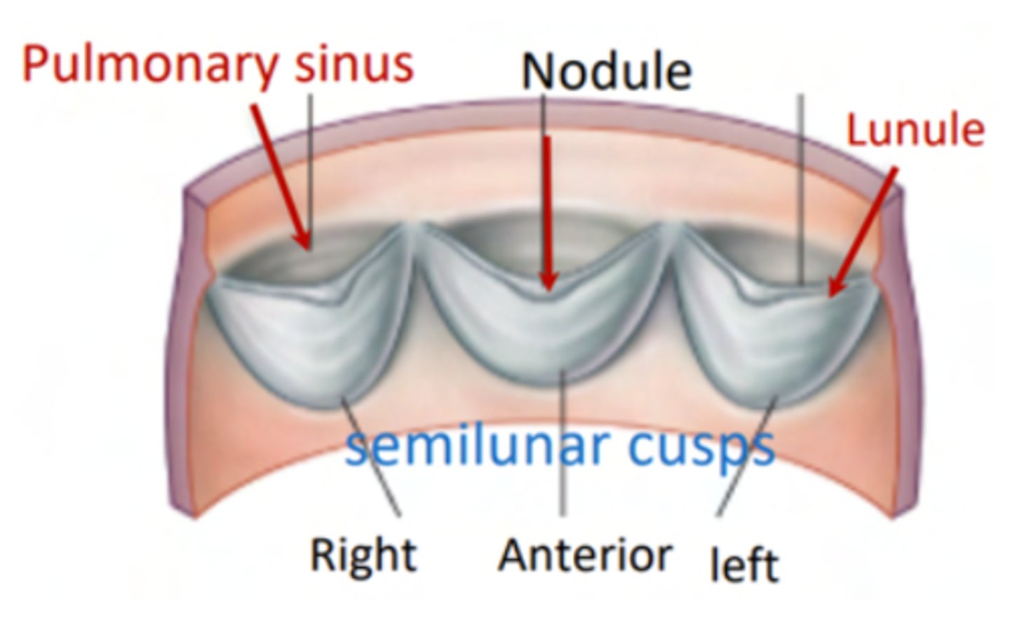
What are the 3 parts of the pulmonary valve?
. Nodules. (little point at the center)
. Lunules (from the nodule to the wall)
. Sinus (left in between)
What are the two portions of the left atrium?
Smooth portion, originated from the incorporation of the
pulmonary veins. The proper atrium itself
Rough portion, the auricle or appendage,
SAME AS RIGHT ATRIUM

What is the valve in the AV left canal?
. Mitral valve
2 cusps (anterior and posterior) and 2 associated papillary muscles
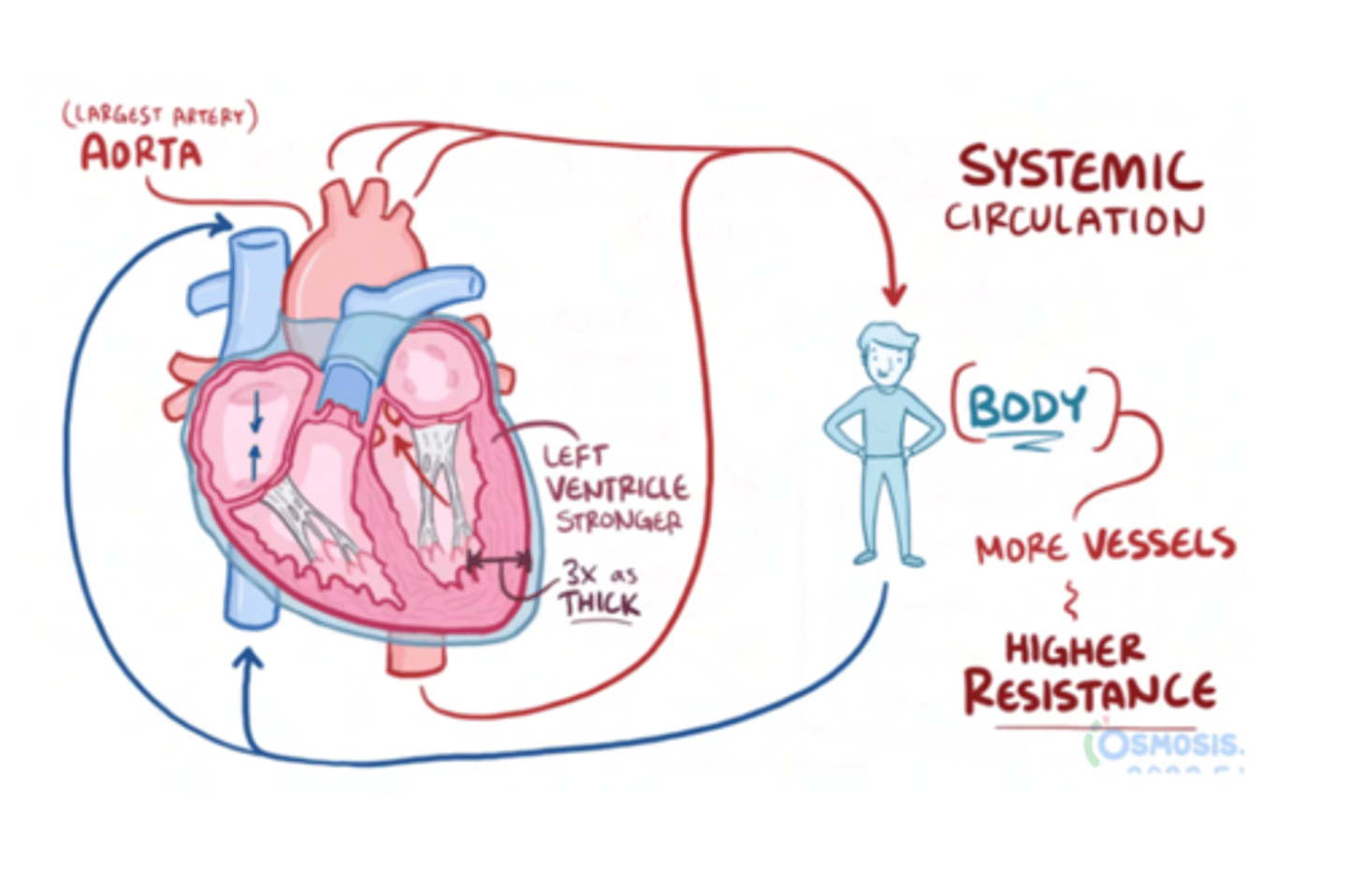
Why is the left ventricle wall thicker than the right?
It is 3 times thicker than the one on the right, in order to pump blood into the aorta at high pressures
SAME THICKNESS AT BIRTH, IT IS DEVELOPED DUE TO THE HIGH PRESSURE OF THIS CAVITY
What prevents backflow of blood from the left ventricle to the left atrium?
mitral valve or biscuspid valve
consisting of two cusps connected to two papillary muscles,
What is the valve to prevents reflux in the aorta?
Aortic valve

What are the 3 parts of the aortic valve?
3 semilunar cusps
posterior
right
left
POSITION IS DIFFERENT THAT IN THE PULMONARY (posterior vs anterior)
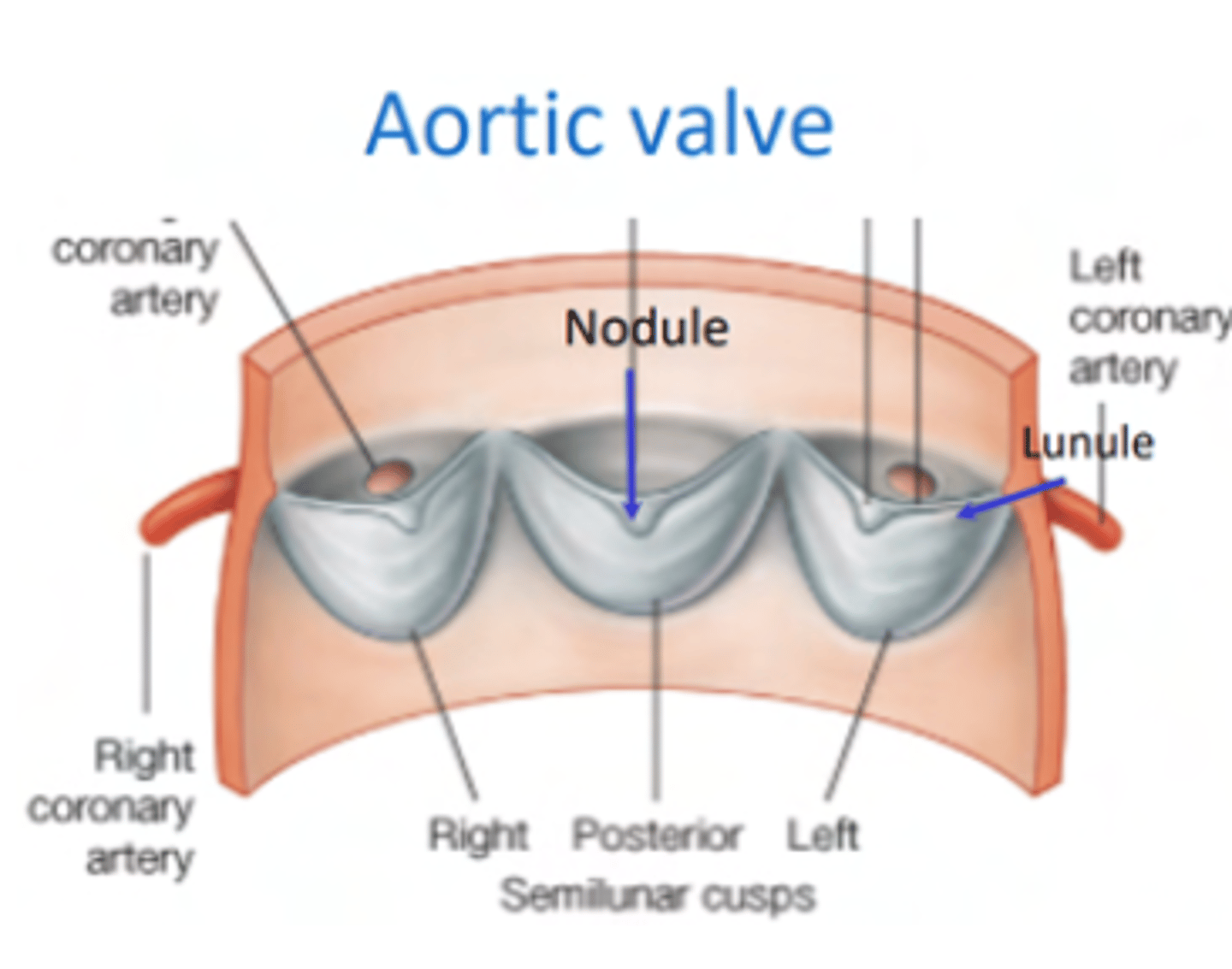
What is the structure of the cusps of the aortic valve?
. Nodules
. Lunules
. Small holes for the coronary arteries
MAIN DIFFERENCE WITH PULMONARY VALVES ARE THE SMALL HOLES FOR THE CORONARY ARTERIES.
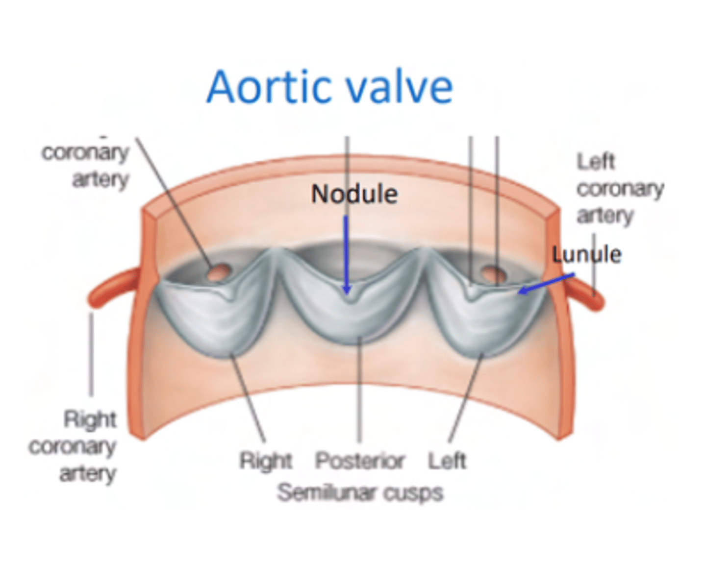
What is the 1st branch of the aorta?
The small holes in the aortic valve cusps to irrigate the coronary arteries.
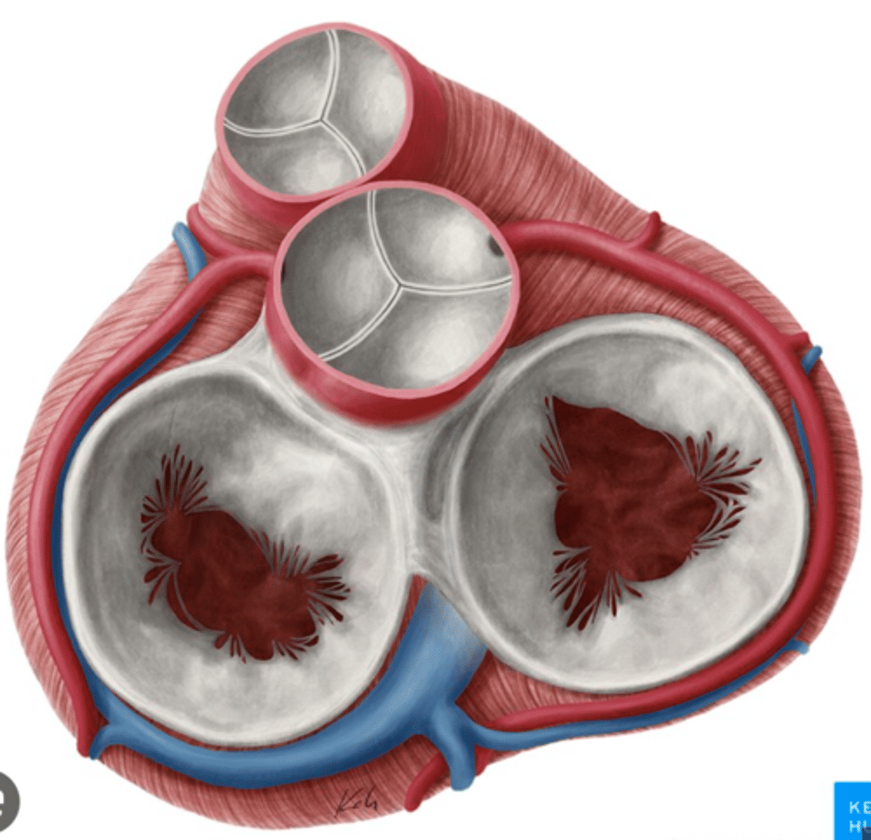
What are the four valves in the heart?
Tricuspid
Mitral or bicuspid
Pulmonary
Aortic
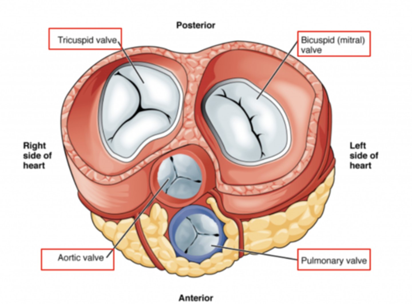
What valves are assuring the AV unidirectional flow?
Tricuspid (right)
Mitral or bicuspid (left)
How do the AV valves respond during the cardiac cycle?
AV valves open during diastole
AV valves close in systole
OPEN : ALLOW FLOW FROM ATRIA TO VENTRICLES
CLOSE : PREVENT BACKFLOW

What role do the papillary muscles and chordae tendineae play in the function of the tricuspid valve?
During ventricular systole (contraction), the tricuspid valve closes
The papillary muscles contract at the same time as the ventricle.
This contraction tightens the chordae tendineae, pulling the valve leaflets taut and holding them in place.
This mechanism prevents the valve leaflets from prolapsing (bulging back) into the atrium under the high pressure of ventricular contraction.
What are other functions of the rings in the cardiac skeleton?
maintain the shape of the openings between the heart's chambers
insertion site for the cusps of the valves and muscular heart tissue.
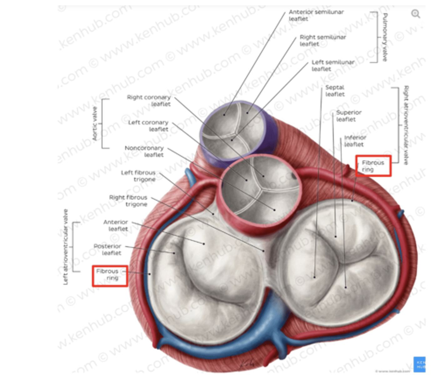
What are the components of the cardiac skeleton?
. pulmonary ring,
. aortic ring
. AV rings
RINGS ARE IN THE SAME PLANE AROUND THE VALVES
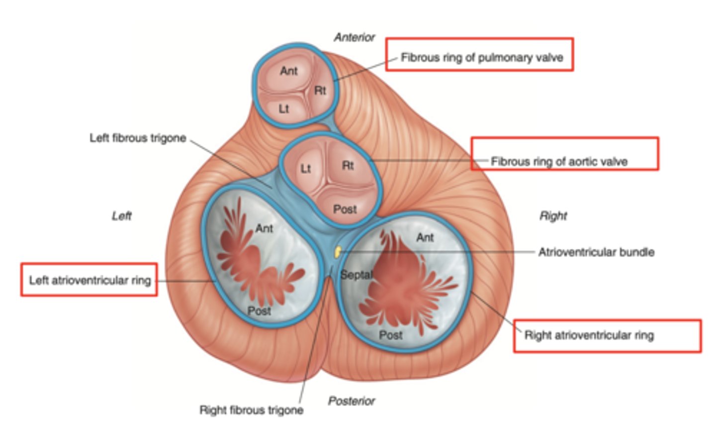
What makes the cardiac skeleton?
Connective tissue
What is the most important function of the rings of the cardiac skeleton?
isolate the atrium from the ventricle,
so conduction stops at the level of these rings +
ALLOWS SEPARATE RHYTHMIC CONTRACTION (ATRIA AND VENTRICLE CAN CONTRACT INDEPENDENTLY)

How are the valves open in diastole?
AV valves are open,
Semilunar valves are closed
How are the valves open in systole?
AV valves are closed
Semilunar valves are open

Why is electrical isolation between the atria and ventricles important?
Ensures that the atria and ventricles contract at different times,
maximizing the heart's pumping efficiency during the cardiac
cycle.
How does the cardiac skeleton influence the heart's conduction system?
By isolating the electrical activity between the atria and ventricles,
coordinating the timing of heart muscle contractions,
ensuring a proper sequence of atrial and ventricular activation.

What structural feature of the heart ensures efficient blood ejection?
The semilunar valves, supported by the cardiac skeleton,
preventing backflow.
How does the cardiac skeleton facilitate heart valve repair or replacement?
By offering a defined structure for valve attachment,
aids surgeons in precisely locating and securing prosthetic
valves or repairing existing valves during cardiac surgery.
What are the layers of the heart?
Epicardium: outermost covering.
Myocardium (right << left)
Endocardium or endothelium: inner thin layer of simple squamous epithelium
What is the primary function of the myocardium?
To contract, pumping blood throughout the body and
maintaining the circulation of blood within the heart
chambers.

What distinguishes the myocardium from other muscle tissues?
Striated appearance,
involuntary control,
intercalated discs that facilitate synchronized contraction.
What are type A fibers in myocardium?
Start at one ring of the cardiac skeleton and finish at the other.
Form the interventricular septum
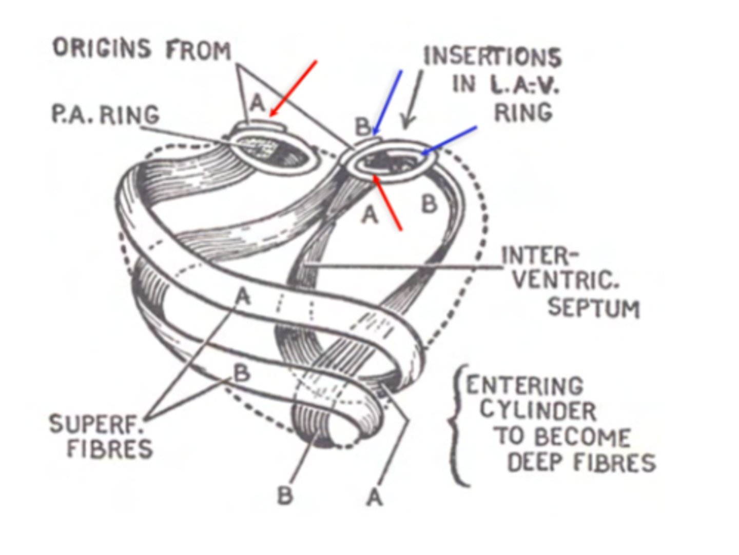
What are type B fibers in myocardium?
Begin at one ring and finish at the same ring where they started (either right or left).
Reinforce the ventricles

What are the layers of myocardium that divide the chambers of the heart?
left : superficial, middle and deep
right : superficial and deep
REMEMBER LEFT IS THICKER DUE TO THE HIGH PRESSURE OF THE AORTA

What is the clinical significance of the myocardium's structure?
Alterations in its thickness, such as hypertrophy or dilatation, can indicate or lead to cardiac diseases.

What allows the heart to contract by itself?
The heart possesses a specialized conduction system that gives it the autonomous capacity to contract, allowing it to pump by itself in a correct buffer.

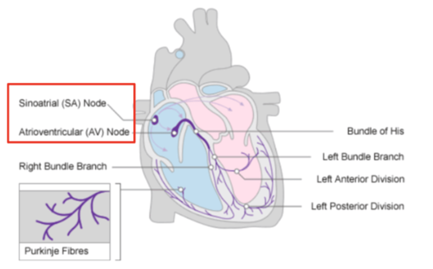
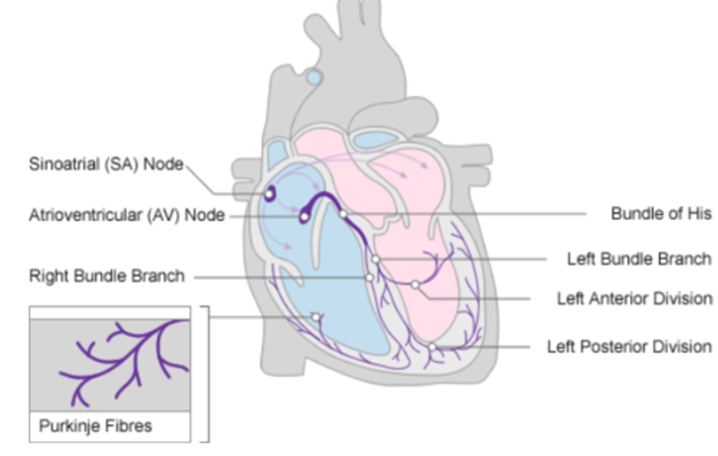
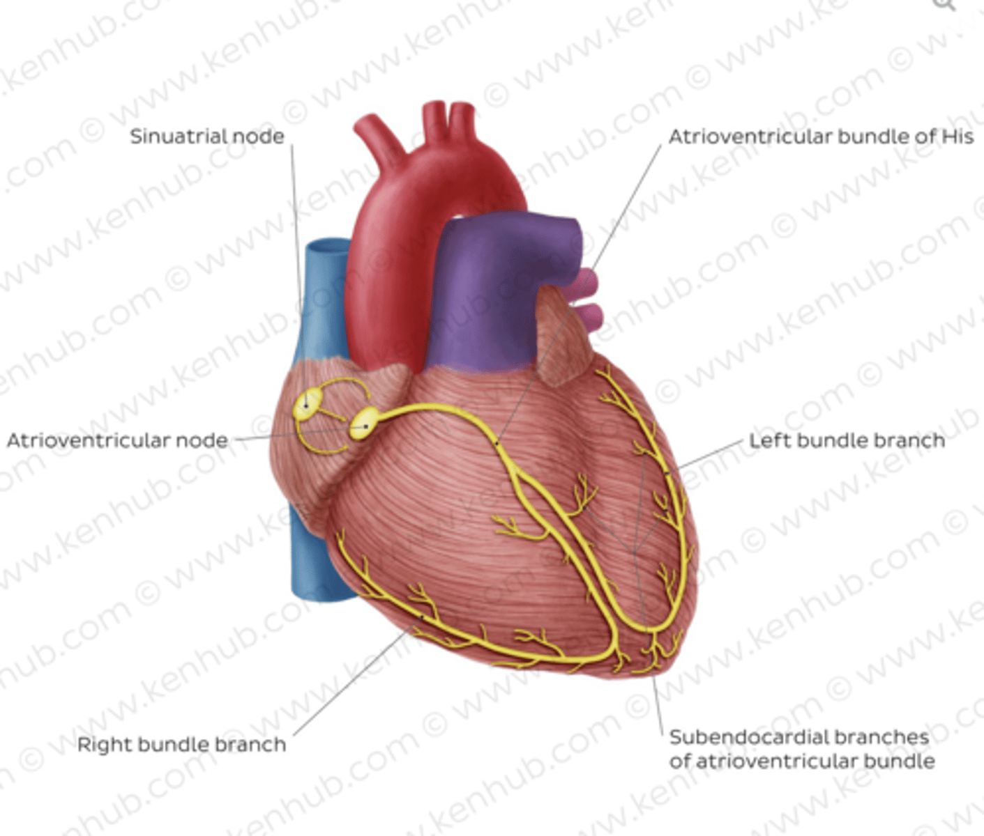
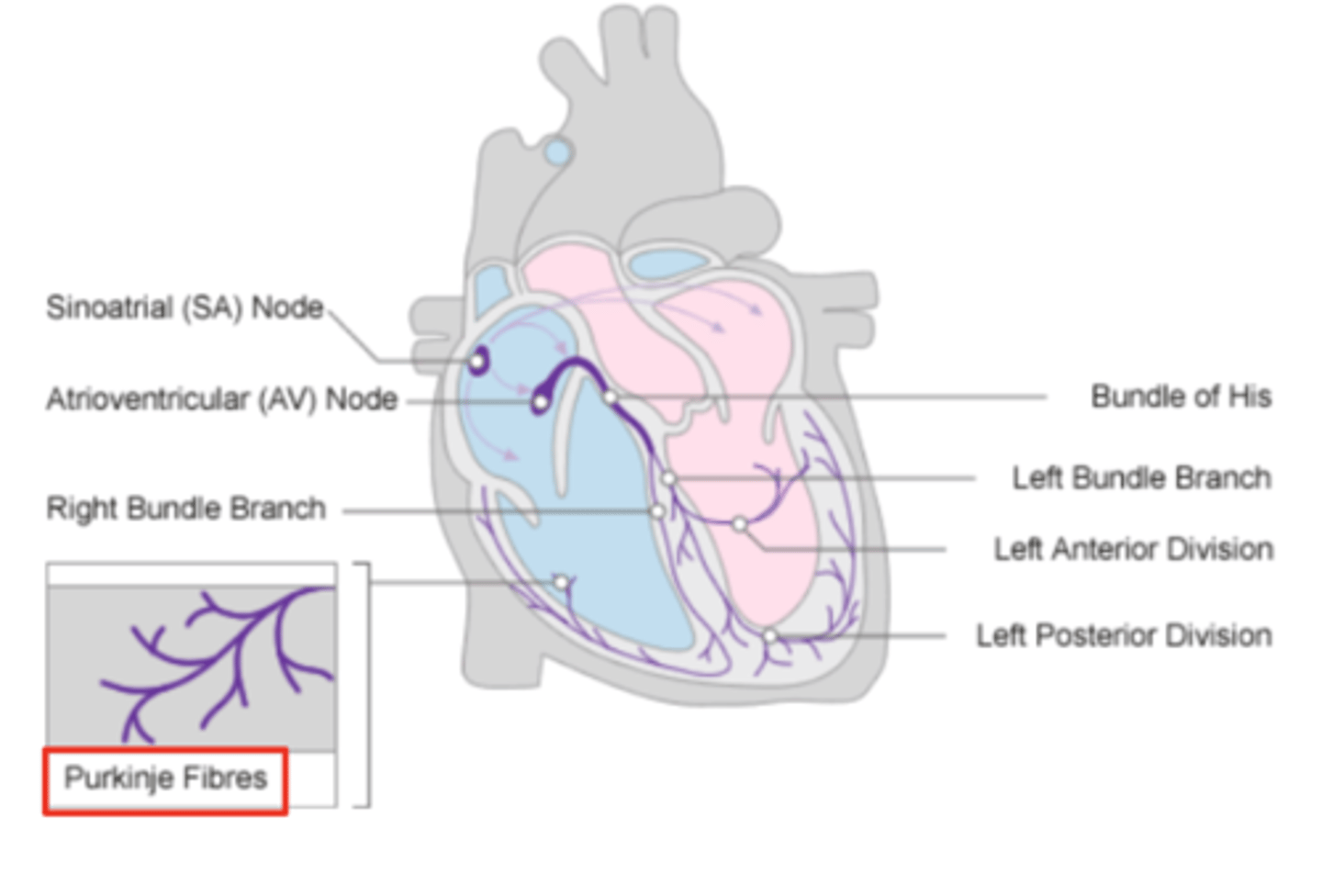
What are the two conduction nodes of the heart conduction system?
. SA node (sinoatrial node)
. AV node (atrioventicular node)

What is the function of the sinoatrial (SA) node?
Pacemaker of the heart

Where is initiated the contraction of the SA node?
SVC (Superior Vena Cava)
passing through the atrium to the AV node
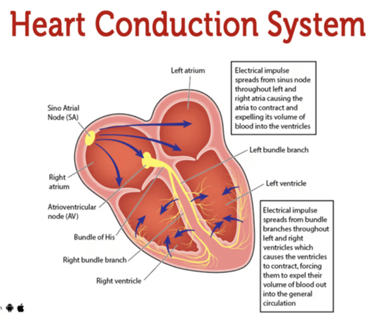
Where is the SA located?
upper wall of the right atrium,
at the junction where the superior vena cava enters
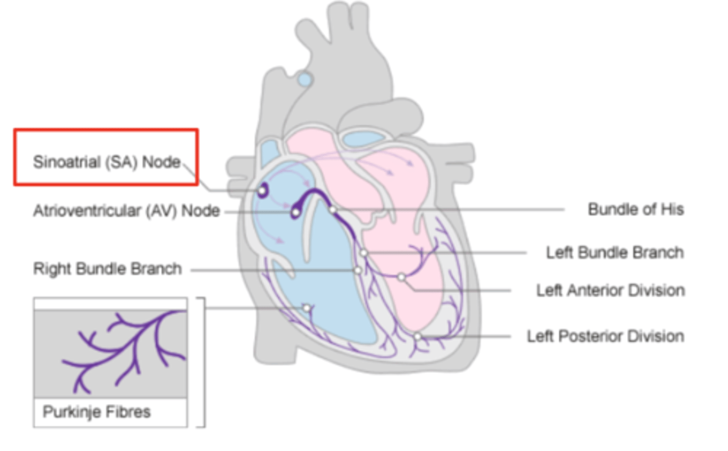
Where is the atrioventricular (AV) node located, and what follows it?
At the level of the atrioventricular septum,
AV node is followed by?
Bundle of Hiss that divides into two branches
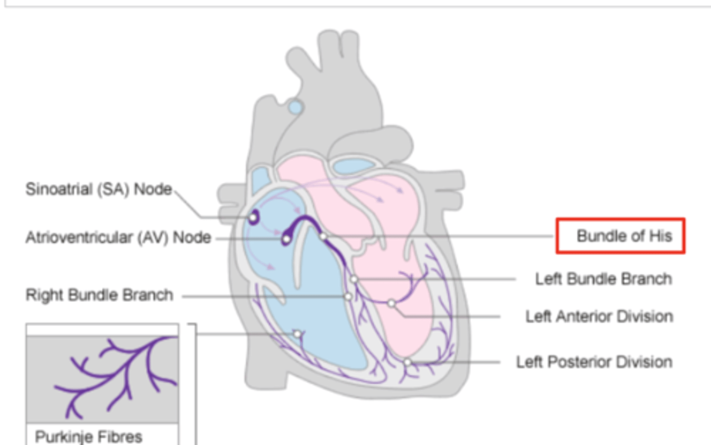
Where does Bundle of Hiss ends?
Purkninje fibers
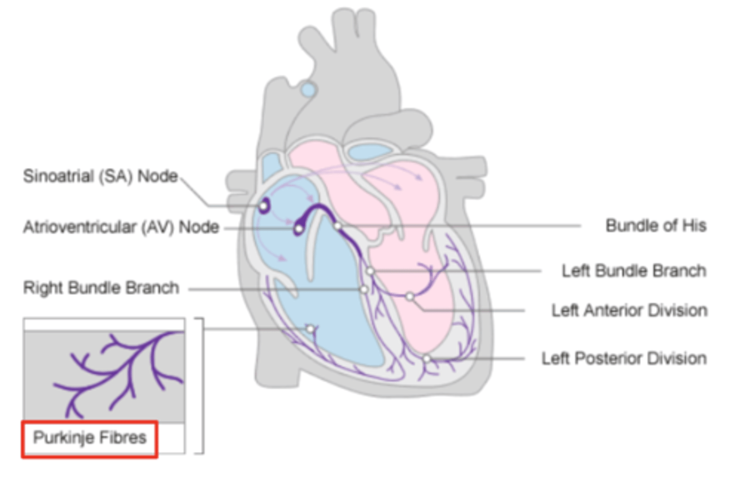
Describe the propagation of waves in the conduction system ?
. SA node sends the initial wave
Passes through the walls of the atria
. Ends at the AV node in the AV septum
. Travels along the Bundle of His
. Reaches Purkinje fibers in the walls of the ventricles
What role does the moderator band play in the conduction system?
Allows the wave to reach the papillary muscles, inducing their contraction simultaneously with that of the ventricles.
REMEMBER: PAPILLARY MUSCLES ACT THROUGH THE CHORDAE TENDINAE TO CONTROL THE TRICUSPID AND MITRAL VALVES.
What is the significance of the Bundle of His in heart conduction?
Branching to the right and left to ensure coordinated contraction of the ventricles.
What are the Purkinje fibers, and what is their function?
specialized conductive fibers in the walls of the ventricles that
facilitate rapid transmission of electrical impulses,
ensuring efficient and synchronized ventricular contraction.

How does the AV node contribute to heart rhythm?
Delays the electrical impulse from the SA node,
ensuring that the atria contract fully to transfer blood to the
ventricles before the ventricles contract.
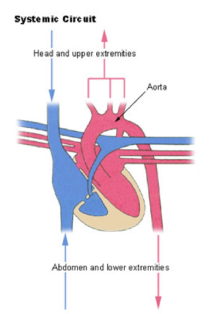
SYSTEMIC CIRCULATION
flow of blood from body tissue to the heart and then from the heart back to body tissues
It carries oxygen and nutrients to the cells and picks up carbon dioxide and waste products
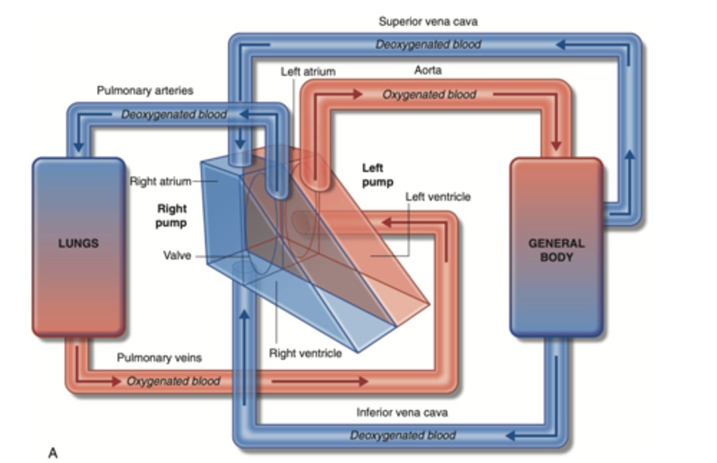
Describe in detail systemic circulation
From the left ventricle, oxygenated blood,
through the arteries,
to the capillaries in the tissues of the body.
From the tissue capillaries, the deoxygenated blood
returns through a system of veins to the right atrium of the heart. (superior and inferior cava veins)
PULMONARY CIRCULATION
Circulation of blood between the heart and the lungs
transports oxygen-poor blood from the right ventricle to the lungs,
where blood picks up a new oxygenated blood
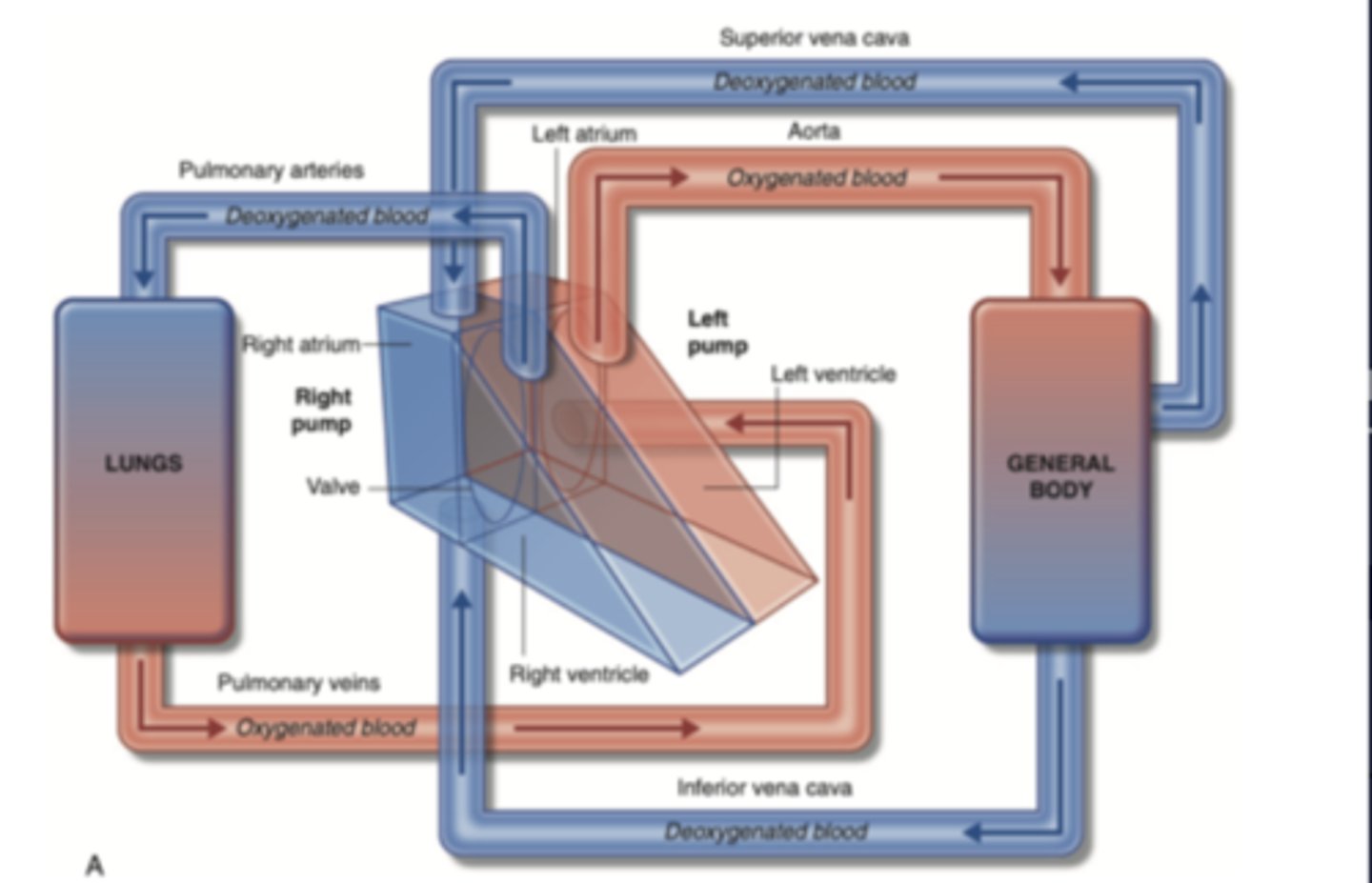
Describe in detail pulmonary circulation
From the right ventricle poor oxygenated blood is impulsed
to the lungs, through the pulmonary artery
blood is oxygenated
returns to the left atria through the pulmonary veins
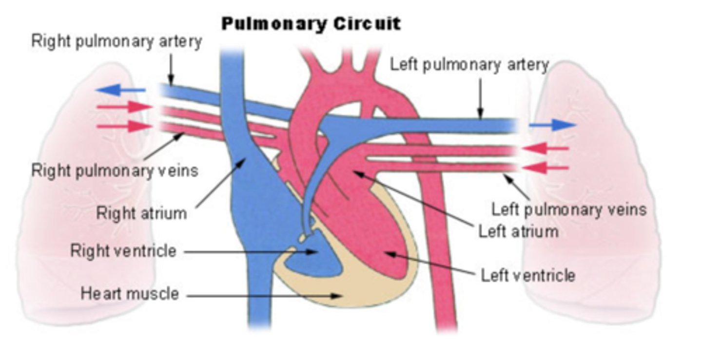
from where is the right atrium receiving the blood?
From the whole body
Right ventricle pumps blood to?
pulmonary artery
from where receives blood the left atrium?
pulmonary veins
Where pumps blood the left ventricle?
into the aorta