equine and swine
 Call Kai
 Learn
Learn Practice Test
 Spaced Repetition
 Match
Match Flashcards
 Knowt Play
1/20

There's no tags or description
Looks like no tags are added yet.
Name | Mastery | Learn | Test | Matching | Spaced | Call with Kai | Chat |
|---|
No analytics yet
Send a link to your students to track their progress
21 Terms
horse overview
small stomach with glandular and non glandular areas
enlarged caecum produce lots of VFAs
enlarged complex ascending colon
horse stomach
2 regions
what separates
type of epithelium
what is the blood supply and where does this drain into?
non glandular- keratinised stratified squamous epithelium
margo plicatus
glandular- ruggae and gastric pits, simple columnar epithelium
gastric artery- blood supply.
all gastric veins drain into hepatic portal vein
small intestine
what are the 2 types of papillae and what empty
has lesser papllea- lesser pancreatic duct empties
greater papillae- greater pancreatic duct and bile duct empty
caecum
what side
main fermentation chamber
right hand side of the body (rumen is left)
‘comma’ shape
blind sac apex points towards the head
folds
caecum development and what is the caecocolic orific
strippled part is homologous with other species
non strippled part is annexed first part of colon
caecocolic orifice is a constriction of the ascendng colon
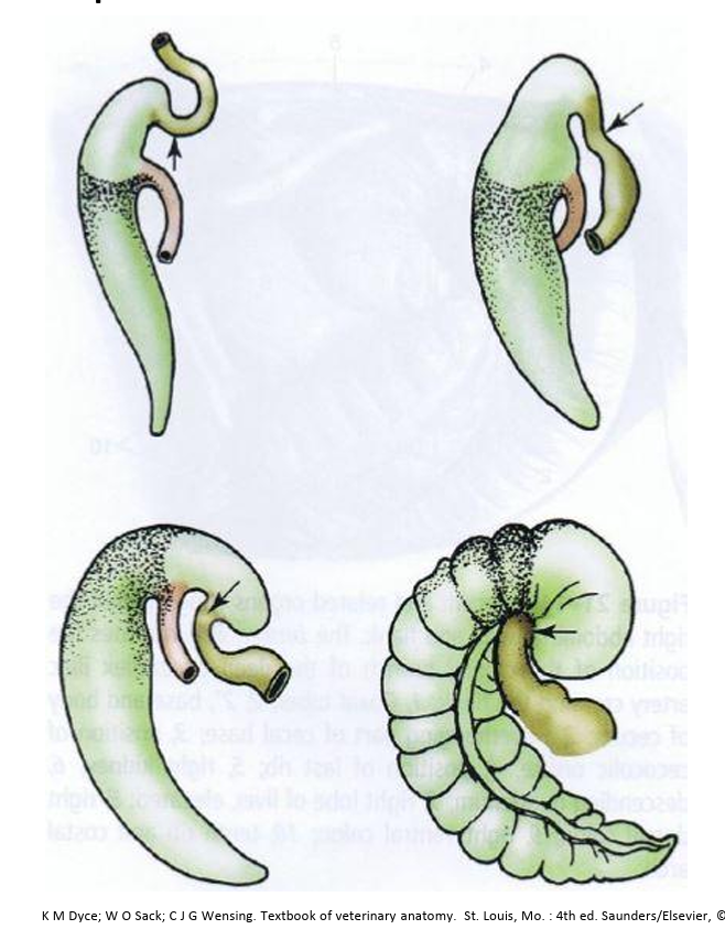
ileocaecocolic junction comparison
Horses: separated ileocaecal orifice and caecocolic orifice
Carnivores: major is ileocolic orifice and ileocaecal fold
liver
bile is released slowly throughout the day
no gall bladder
only 4 lobes instead of 64 Lobes: Right, Left (Medial & Lateral), Quadrate, Caudate Lobe
evident renal impression
spleen
structure
blood supply
which part of the kidney is is connected to through which ligament
elongated
broad at base and narrow
recieves blood from splenic artery which is a branch of the celiac artery
part of spleen is connected to left kidney through the nephrosplenic ligament/renosplenic ligament
pancreas
where does it open
where is it embedded
compare to carnivores
opens in lesser and greater papilla
embedded in mesentery- sits on top/within
more lobulated than carnivores
what can be felt on renal palpation
descending colon- ball of faeces
caudal border of spleen
nephrosplenic ligament
caudal pole left kidney
root of mesntery
caeccum- base and ventral taenia
pelvic flexture- a sharp, narrow hairpin turn in the horse's large colon where the left ventral colon folds back on itself to connect to the left dorsal colon, creating a bottleneck where food (digesta) slows down and can become impacted, leading to painful colic
small intestine loops normally too soft but can feel ifdistended
right kidney cannot be felt
colic and causes
abdominal pain
stretch of gut wall
ischaemia
inflammation
build up of gas due to blockage of bloakcga itself
torsion leading to tension in mesentery and cut off blood supply
cn x damage can cause limited motility leading to gas accumulation
pig digestive tract overview
simple stomach
somewhat enlarged caecum
complex ascending colon
omnivores
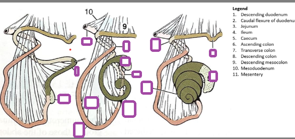
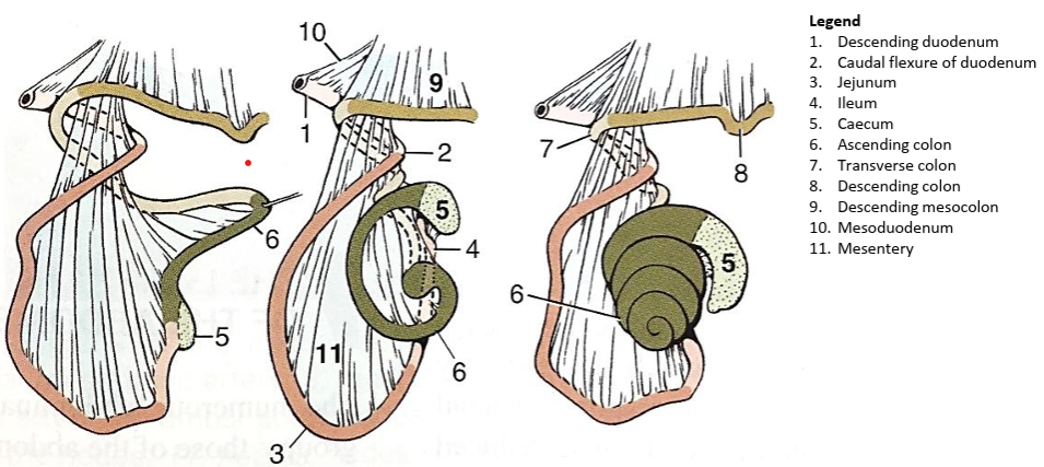
development of the ascending colon in the pig
sphincters in equine stomachs- 2 types
cardiac sphincter prevents reflux and vomiting
pyloric sphincter controls outflow of the small intestine
what is the ansa spiralis
coiled shape of the ascending colon
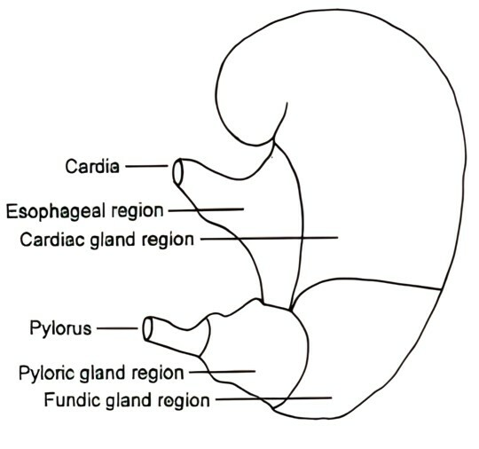
swine stomach anatomy
4 distinct areas
oesophageal, cardiac, fundic and pyloric regions
in oesophageal, ulcer formation occurs
in cardiac portion mucus is secreted and mixed with digested food
fundic region- gastric glands secrete hcl and also pepsinogen is secreted
pyloric region- secrete mucus to line digestive membrane and regulates chyme secretion
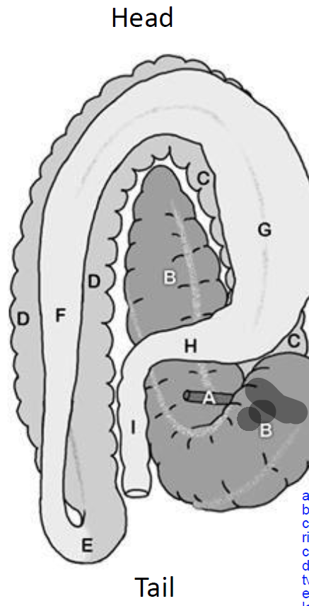
A- the ileum
B- caecum
C- right ventral colon
This moves cranially and twists over to the left side to form a flexure called the sternal flexure.
D- left ventral colon
this then twists upwards from ventra to dorsal
E- pelvic flexure
F- left dorsal colon
this moves from the pelvic region cranially again to the right to form the diaphragmatic flexure
G- right dorsal colon which sits on top of the right ventral colon
H- transverse colon
I- descending colon
caecocolic orifice
cecum into ascending colon (right ventral colon)
caecal tympany
dilation of the caecum due to accumulation of gases
caecal impaction
fluid filled or solid filled in the caecum
solid filled: impaction of dehydrated ingesta.
fluid filled: functional failure of the cecum to empty
caecocolic intussucception
base of the caecum invaginates into the proximal part of the right ventral colon (ascending colon)
another simular condition to this is the caeco caecal, which intussusception results from uncoordinated peristalsis allowing the apex of the caecum to invaginate into the base. this is a relatively rare cause of colic, accounting for only 3-5 percent caecal disease in the horse