OVARIES
 Call Kai
 Learn
Learn Practice Test
 Spaced Repetition
 Match
Match Flashcards
 Knowt Play
1/47

There's no tags or description
Looks like no tags are added yet.
Name | Mastery | Learn | Test | Matching | Spaced | Call with Kai | Chat |
|---|
No analytics yet
Send a link to your students to track their progress
48 Terms
_____ phase many follicles develop and increase in size (one becomes dominant)
proliferative (day 6-14)
what are the most common solid serious ovarian tumours
cystadenoma and cystadenocarcinoma
doppler of ovary
cut off for PI is 1.0
cut off for RI is 0.4
endometriosis
functioning endometrial tissue is present outside the uterus
negative sliding sign
organs done move, stuck together (frozen pelvis)
obliterated PCDS
kissing ovaries
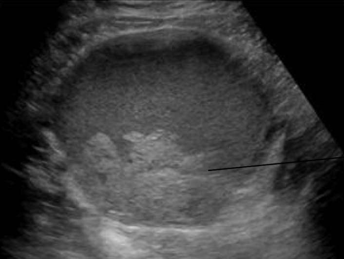
endometrioma
endometrioma sono
well-defined unilocular or multilocular
predominantly cystic mass
diffuse homogenous, low-level internal echoes
polycystic ovarian syndrome
includes Stein-Leventhal syndrome
bilaterally enlarged polycystic ovaries
clinic of polycystic ovarian syndrome
amenorrhea
obesity
infertility
hirsutism
sono of polycystic ovarian syndrome
multiple tiny cysts around periphery of ovary
ovary may be normal size or enlarged

ovarian torsion symptoms
severe pain
nausea/vomit
fever
palpable mass
(similar to appendicitis)
ovarian torsion sono
enlargement of ovary
decreased or absent blood flow
twisted coiled vessels within vascular pedicle (whirlpool sign)

what is the m/c cause of ovarian enlargement in young women
functional ovarian cysts
functional ovarian cysts include
follicular
corpus luteum
hemorrhagic
theca-lutein cysts
follicular cysts
usually unilateral
thin walled, translucent, watery fluid
simple cyst

corpus luteum cysts
result from hemorrhage within persistently mature corpus luteum
filled with blood and cystic fluid
may accompany IUP
> 4cm

corpus luteum sono
small, < 4cm
hypoechoic
cyst with low level internal echoes
thicker wall
ring of fire
involutes before menstruation

hemorrhagic cysts
internal hemorrhage in cyst
acute onset of pelvic pain
acute: hyperechoic, smooth posterior walls, enhancement
internal pattern becomes more complex

theca-lutein cysts
large, bilateral multiloculated cysts
high level of hCG
nausea and vomit
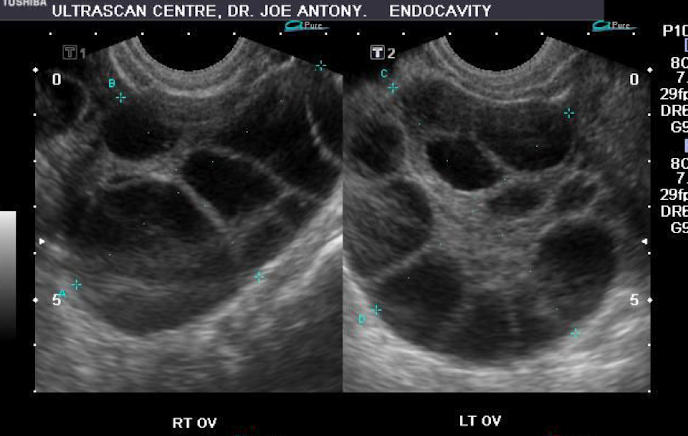
ovarian hyperstimulation syndrome
complication of ovulation induction
mild
pelvic discomfort, ovaries enlarged, < 5cm in diameter
severe
severe pelvic pain, abdominal distention, ovaries > 10cm diameter

ovarian remnant syndrome
pt undergone bilateral oophorectomy
cystic mass may develop
residual tissue left behind
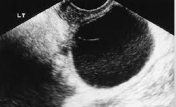
peritoneal inclusion cysts
lined with mesothelial cells
when adhesions trap peritoneal fluid around ovaries

paraovarian cysts
located in broad ligament
simple cyst adjacent to ovary

ovarian carcinoma
masses > 10cm more likely to be malignant
increasing pt age correlates with increased incidence of malignancy
strongest risk factor is family hx of ovarian/breast cancer
ovarian carcinoma clinical
vague abdominal pain
swelling
indigestion
frequent urination
constipation
weight change (ascites)
most common type of ovarian malignancy
epithelial tumours
SEROUS (m/c)
mucinous
mucinous cystadenoma sono
can be very large
usually unilateral
simple or septate thin walled multilocular cysts
contains internal echoes with compartments differing in echogenicity


mucinous cystadenocarcinoma sono
can be very large
bilateral
thick, irregular walls and septations
papillary projections and echogenic material
ascites

what is m/c benign tumour of ovary
dermoid cyst
serous cystadenoma sono
second m/c benign tumour of ovary
unilateral
unilocular or multiocular with thin septations
smaller than mucinous
borders irregular with loss of capsular definition
small amount of solid tissue
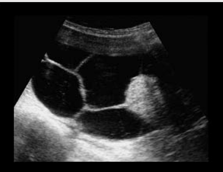
serous cystadenocarcinoma sono
bilateral
loss of capsular definition, tumour fixation calcifications
septations / papillary projections
internal and external papillomas

m/c germ cell tumours
benign cystic teratomas (dermoid)
germ cell tumours are associated with elevated
AFP and hCG
germ cell tumours include
teratomas
dysgerminoma
embryonal cell carcinoma
choriocarcinoma
transdermal sinus tumour
teratoma/dermoid tumours
fatty, sebaceous material, hair, cartilage, bone, teeth

sono for dermoid/teratoma
completely cystic
echogenic nodule along mural wall, dermoid plug “tip of the iceburg”
fat-fluid level
high amplitude echoes with shadowing
complex mass with internal septations

what are the two m/c ovarian neoplasms seen in pregnancy
dysgerminoma, serous cystadenoma
endodermal sinus tumour
yolk sac tumour
< 20ys
unilateraal
increase AFP
poor prognosis
sex cord stromal tumour include
granulosa cell tumour
thecoma
fibroma
sertoli leydig cell tumours
thecomas
abundance of thecal cells
usually unilateral
postmenopausal women
shows signs of estrogen production
fibroma
rarely associated with estrogen production
postmenopausal women
associated with ascites and pleural effusion
MEIGS SYNDROME
sono of fibroma
thecoma appears similar
unilateral
variable sono appearance
hypoechoic with post enhancement
larger are prone to torsion

meigs syndrome
presence of
ascites
pleural effusion
benign, ovarian mass
most common hormone-active estrogenic tumour of ovary
granulosa
clinical of granulosa
precocious puberty
vaginal bleeding
full breasts
sono of granulosa
variable
similar to endometrioma/cystadenoma

Krukenberg tumours
“drop” metastases to ovaries from GI tract
sono of metastatic disease
bilateral
ascites
completely solid or solid with “moth-eaten” cystic pattern

benign sono
well defined anechoic lesions
thin septations
more peripheral flow
higher resistive flow
malignant sono
irregular walls
thick septations
mural nodules
solid echogenic elements
centralized flow
lower PI
lower RI
higher diastolic flow