Movements of Region 1110
 Call Kai
 Learn
Learn Practice Test
 Spaced Repetition
 Match
Match Flashcards
 Knowt Play
1/412

Earn XP
Description and Tags
Final Lab Exam
Name | Mastery | Learn | Test | Matching | Spaced | Call with Kai | Chat |
|---|
No analytics yet
Send a link to your students to track their progress
413 Terms
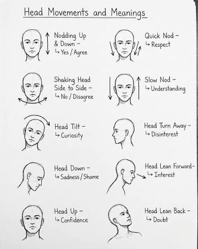
Movements of Head?
Flexion
Extension
Hyperextension
Lateral Flexion
Rotation
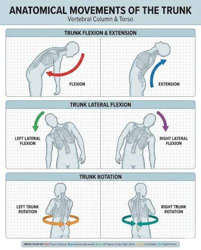
Movement of Trunk?
Flexion
Extension
Hyperextension
Lateral Flexion (L/R)
Rotation (L/R)
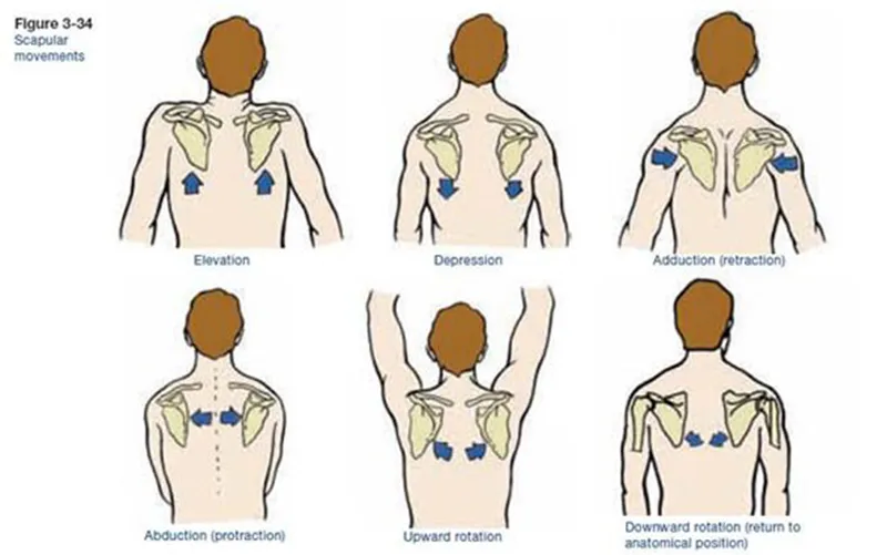
Movements of Scapula
Elevation
Depression
Protraction
Retraction
Upward Rotation
Downward Rotation
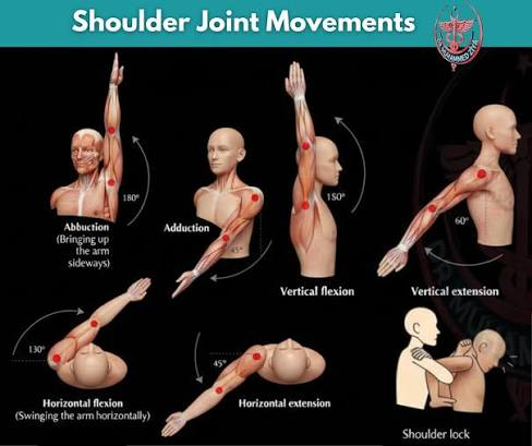
Movements of Arm (Humerus)?
Flexion
Extenson
Abduction
Adduction
Internal Rotation
External Rotation
Circumduction
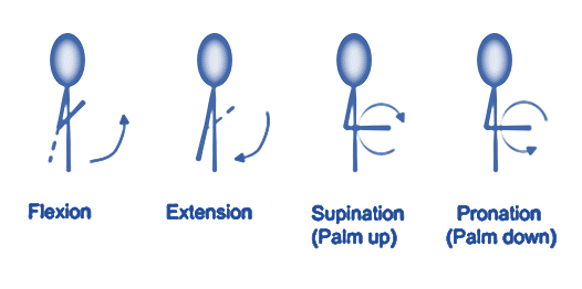
Movements of Forearm (Elbow)
Flexion
Extension
Supination
Pronation
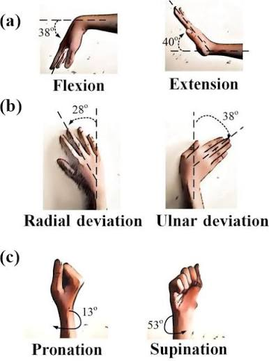
Movements of Wrist
Flexion (Palmar Flexion)
Extension (Dorsiflexion)
Radial Deviation (Abduction)
Ulnar Deviation (Adduction)
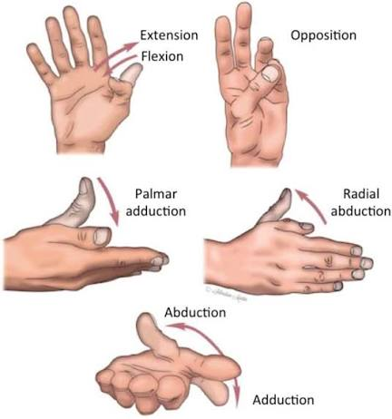
Movements of Thumb (Pollex)
Flexion
Extension
Abduction
Adduction
Opposition
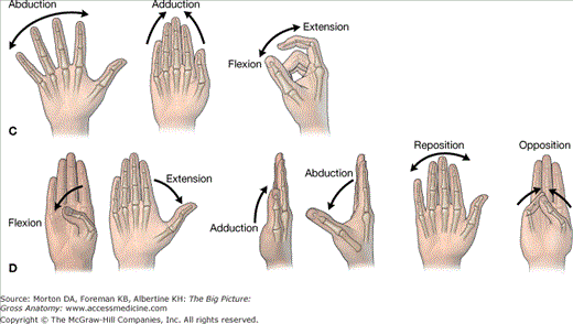
Movements of Fingers (Entire)
Flexion
Extension
Abduction
Adduction
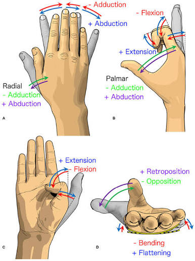
Movements of Fingers (Each)
Flexion
Extension
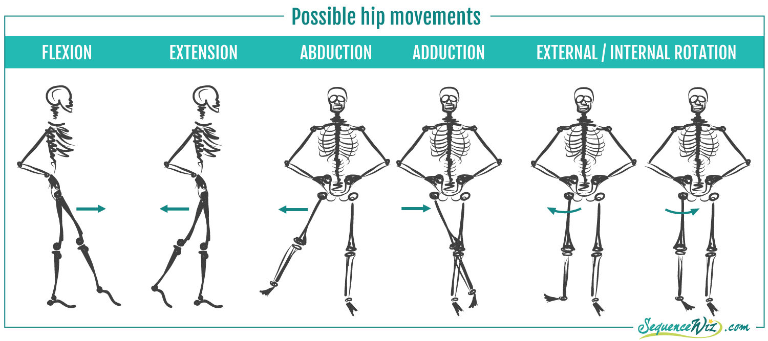
Movements of Hip (Femur)
Flexion
Extension
Abduction
Adduction
Internal Rotation
External Rotation
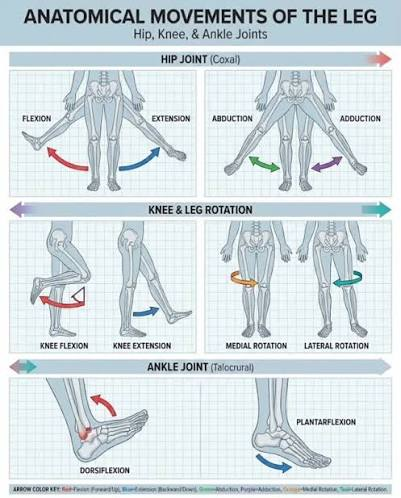
Movements of Knee (Lower Leg)
Flexion
Extension
Internal Rotation (When flexed)
External Rotation (When flexed)
Movements of Ankle?
Dorsiflexion
Plantar Flexion
Inversion
Eversion
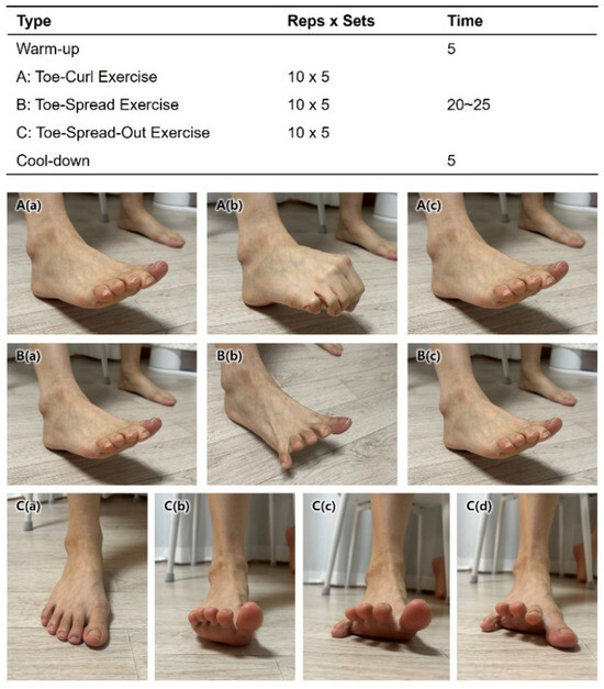
Movements of Toes (Entire)
Flexion
Extension
Abduction
Adduction
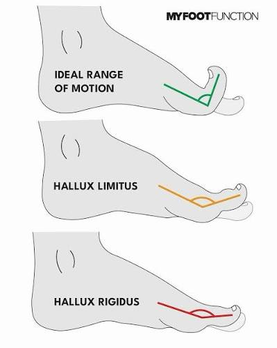
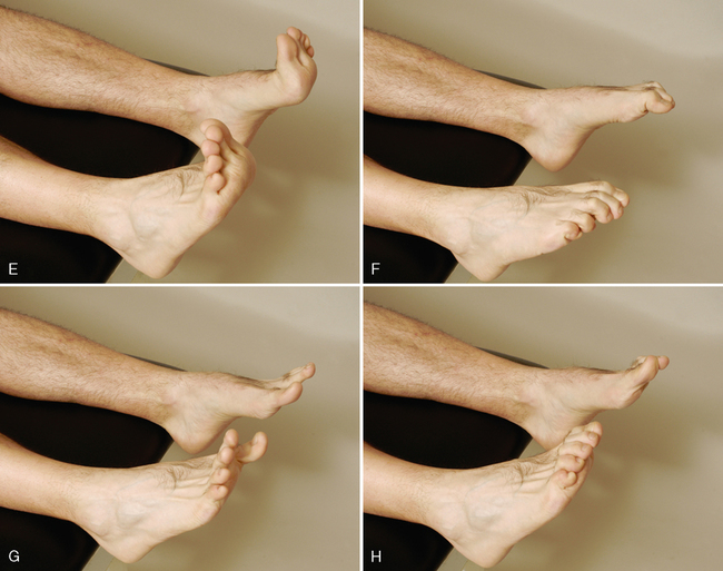
Movements of (Toes) each
Flexion
Extension
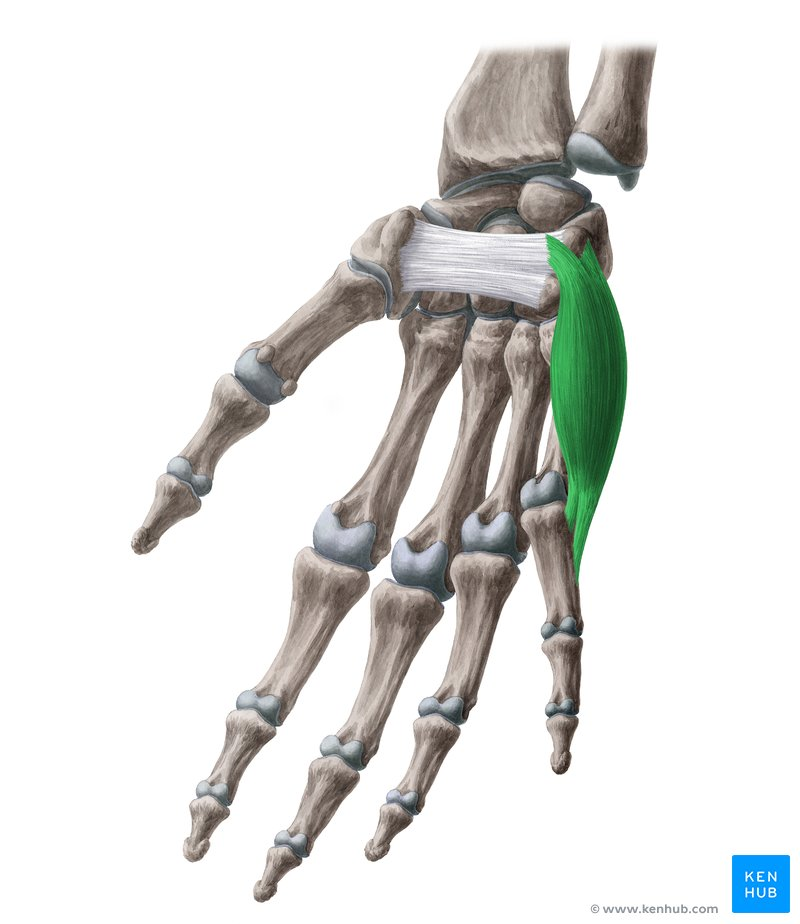
What Muscle?
Abductor Digiti Minimi (Hand)
O: Pisiform bone, pisohamate ligament, and the tendon of flexor carpi ulnaris.
I: Medial (ulnar) side of the base of the proximal phalanx of the 5th digit (little finger)
A: Abducts the 5th digit at the metatarsophalangeal joint and assists in flexing its proximal phalanx
In: : Deep branch of the ulnar nerve (spinal roots C8, T1).
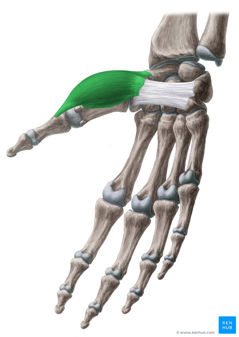
What Muscle?
Abductor Pollicis Brevis
O: Flexor retinaculum, scaphoid tubercle, and trapezium tubercle
I: Radial (lateral) side of the base of the proximal phalanx of the 1st digit (thumb).
A: Abducts the thumb at the carpometacarpal and metatarsophalangeal joints
In: Recurrent branch of the median nerve (spinal roots C8, T1). [1, 2, 3, 4, 5]
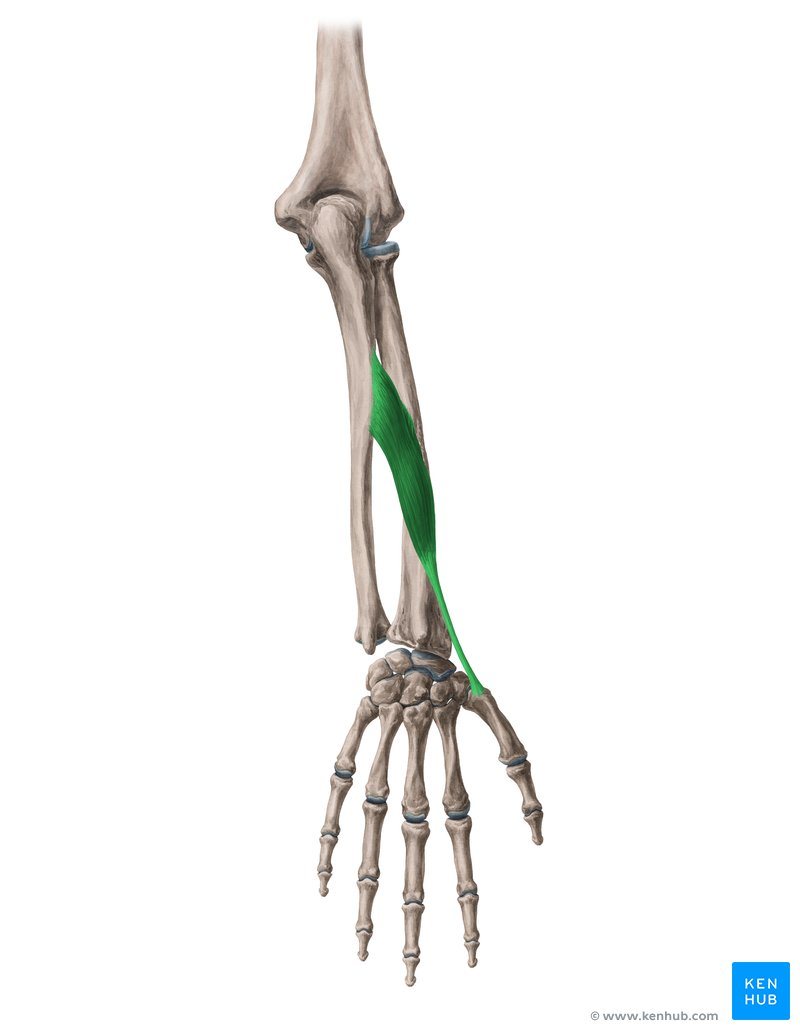
What Muscle?
Abductor Pollicis Longus
O: Posterior surfaces of the ulna, radius, and the interosseous membrane.
I: Radial (lateral) side of the base of the 1st metacarpal bone.
A: Abducts and extends the thumb at the carpometacarpal joint, and abducts (radially deviates) the wrist.
In: Posterior interosseous nerve (a continuation of the deep branch of the radial nerve, spinal roots C7, C8). [1, 2, 3, 4, 5]
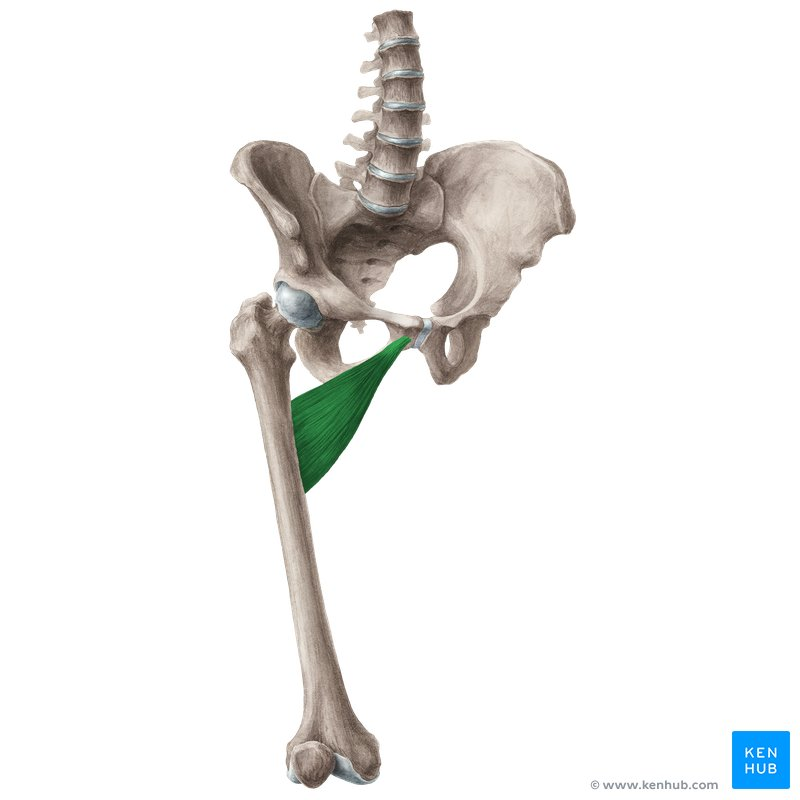
What Muscle?
Adductor Brevis
O: Body and inferior ramus of the pubis.
I: Pectineal line and upper part of the linea aspera on the posterior surface of the femur.
A: Adducts and flexes the thigh at the hip joint; assists in lateral rotation of the thigh
In: Obturator nerve (spinal roots L2, L3, L4). [1, 2, 3, 4, 5]
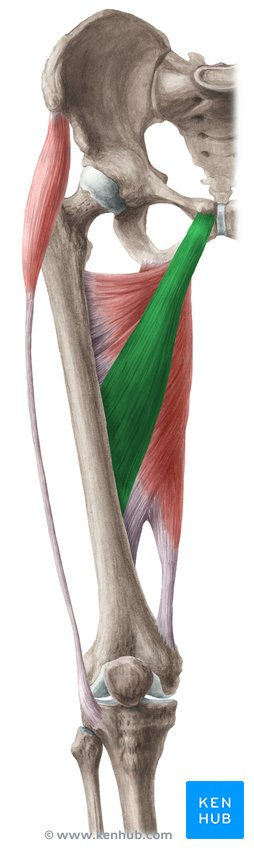
What Muscle?
Adductor Longus
O: Body of the pubis, just inferior to the pubic crest and lateral to the pubic symphysis.
I: Middle third of the linea aspera on the posterior surface of the femur.
A: Adducts and flexes the thigh at the hip joint; assists in lateral rotation of the thigh.
In: Anterior division of the obturator nerve (spinal roots L2, L3, L4). [1, 2, 3, 4, 5]
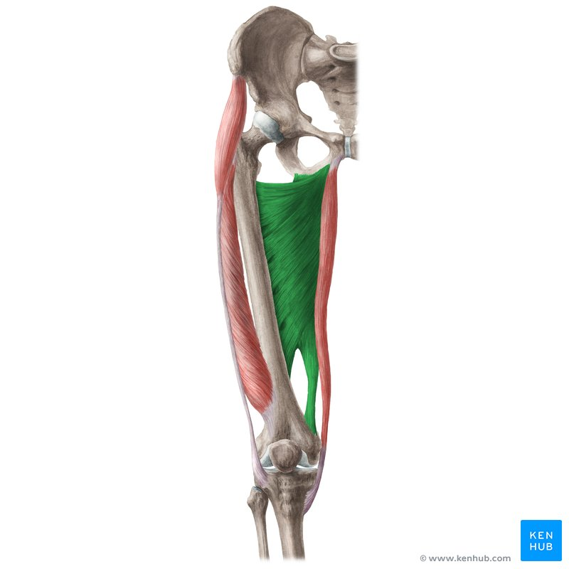
What Muscle?
Adductor Magnus
O: Adductor (pubofemoral) part: Inferior ramus of the pubis and ramus of the ischium.
Hamstring (ischiocondylar) part: Ischial tuberosity. [1, 2, 3, 4, 5]
I: Adductor part: Gluteal tuberosity, linea aspera, and medial supracondylar line of the femur.
Hamstring part: Adductor tubercle of the femur.
A: Adductor part: Adducts and flexes the thigh at the hip joint.
Hamstring part: Extends the thigh at the hip joint. [1, 2, 3, 4, 5]
In: Adductor part: Obturator nerve (spinal roots L2, L3, L4).
Hamstring part: Tibial division of the sciatic nerve (spinal root L4). [1, 2, 3, 4, 5]
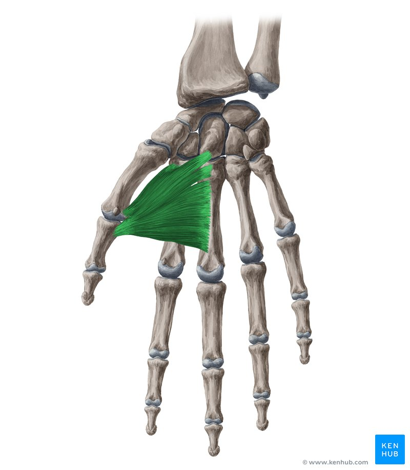
What Muscle?
Adductor Pollicis
O: Oblique head: Bases of the 2nd and 3rd metacarpals, capitate, and adjacent carpal bones.
Transverse head: Anterior surface of the shaft of the 3rd metacarpal bone.
I: Medial (ulnar) side of the base of the proximal phalanx of the 1st digit (thumb). [1, 2, 3]
A: Adducts the thumb at the carpometacarpal and metatarsophalangeal joints, bringing it toward the palm. [1, 2, 3]
In: Deep branch of the ulnar nerve (spinal roots C8, T1). [1, 2]
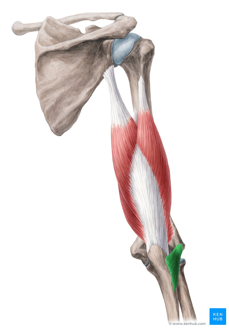
What Muscle?
Aconeus
O: Posterior aspect of the lateral epicondyle of the humerus.
I: Lateral surface of the olecranon and superior part of the posterior surface of the ulna.
A: Assists the triceps brachii in extending the elbow joint; stabilizes the elbow and abducts the ulna during forearm pronation.
In: Radial nerve (spinal roots C7, C8, T1). [1, 2, 3, 4, 5]
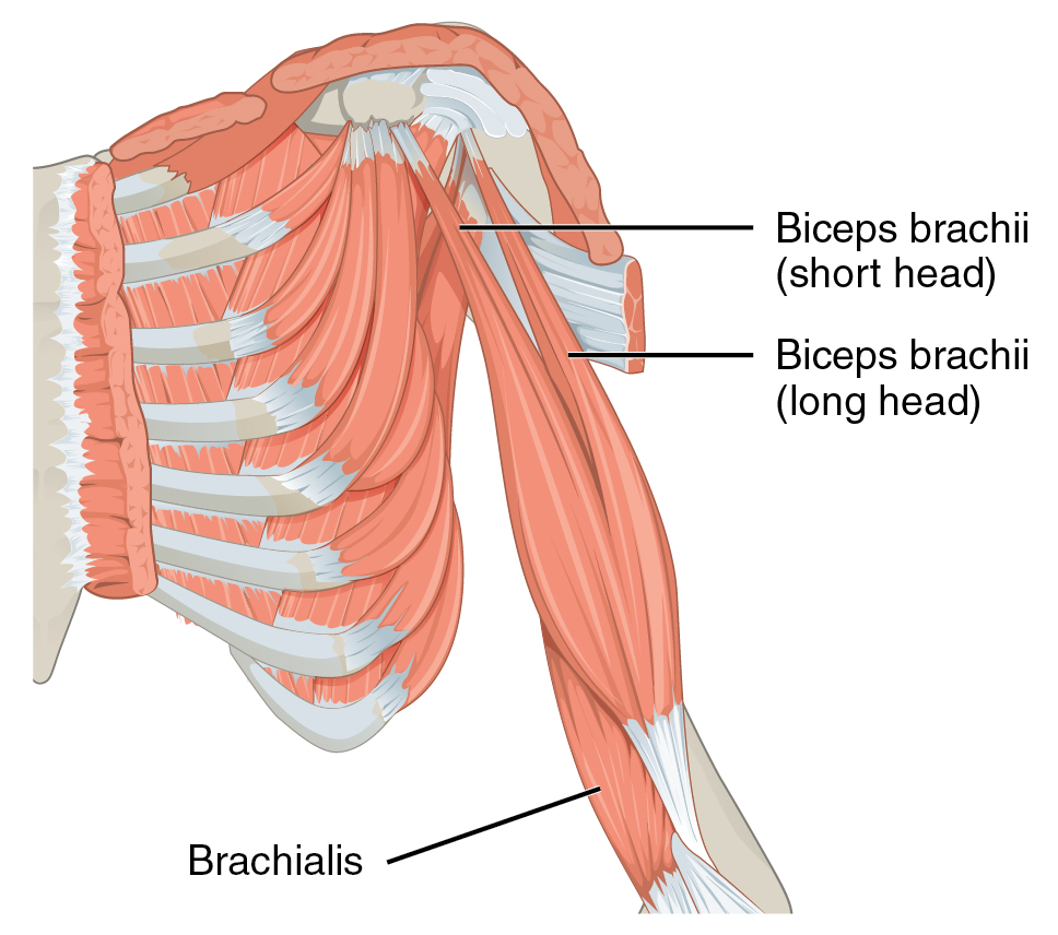
What Muscle?
Biceps Brachii (Long/Short heads)
O: Long head: Supraglenoid tubercle of the scapula.
Short head: Apex of the coracoid process of the scapula. [1, 2, 3]
I: Radial tuberosity and the bicipital aponeurosis (into the deep fascia of the medial forearm).
A: Flexes the elbow joint, powerfully supinates the forearm (when the elbow is flexed), and weakly flexes the glenohumeral (shoulder) joint. [1, 2, 3, 4, 5]
In: Musculocutaneous nerve (spinal roots C5, C6). [1, 2, 3]
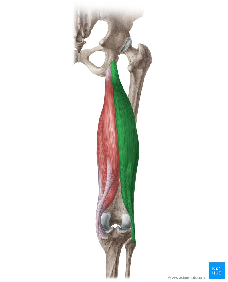
What Muscle?
Biceps Femoris (Long/Short heads)
O: Long head: Ischial tuberosity (via a common tendon with the semitendinosus muscle).
Short head: Lateral lip of the linea aspera and lateral supracondylar line of the femur. [1, 2, 3, 4, 5]
I: Lateral side of the head of the fibula and the lateral condyle of the tibia. [1, 2, 3]
A: Flexes and laterally rotates the leg at the knee joint; the long head also extends the thigh at the hip joint. [1, 2, 3]
In: Long head: Tibial division of the sciatic nerve (spinal roots L5, S1, S2).
Short head: Common fibular (peroneal) division of the sciatic nerve (spinal roots L5, S1, S2). [1, 2, 3, 4, 5]
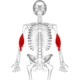
What Muscle?
Brachilais
O: Distal half of the anterior surface of the humerus.
I: Coronoid process and tuberosity of the ulna.
A: Flexes the elbow joint in all forearm positions (pronated, supinated, or neutral).
In: Musculocutaneous nerve (spinal roots C5, C6); may also receive a minor contribution from the radial nerve (C7) for its lateral portion. [1, 2, 3, 4, 5]
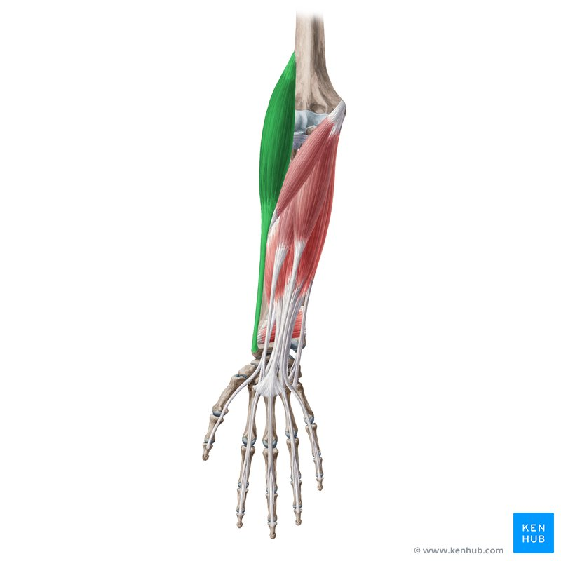
What Muscle?
Brachioradilais
O: Proximal two-thirds of the lateral supracondylar ridge of the humerus.
I: Lateral surface of the distal radius, just proximal to the styloid process.
A: Flexes the elbow joint, especially when the forearm is in a mid-pronated (neutral) position.
In: Radial nerve (spinal roots C5, C6). [1, 2, 3, 4, 5]
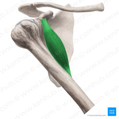
What Muscle?
Coracobrachialis
O: Apex of the coracoid process of the scapula (sharing a tendon with the short head of the biceps brachii)
I: Middle third of the medial surface and border of the shaft of the humerus.
A: Flexes and adducts the arm at the glenohumeral (shoulder) joint; assists in stabilizing the humeral head.
In: Musculocutaneous nerve (spinal roots C5, C6, C7), which pierces through the belly of the muscle. [1, 2, 3, 4, 5]
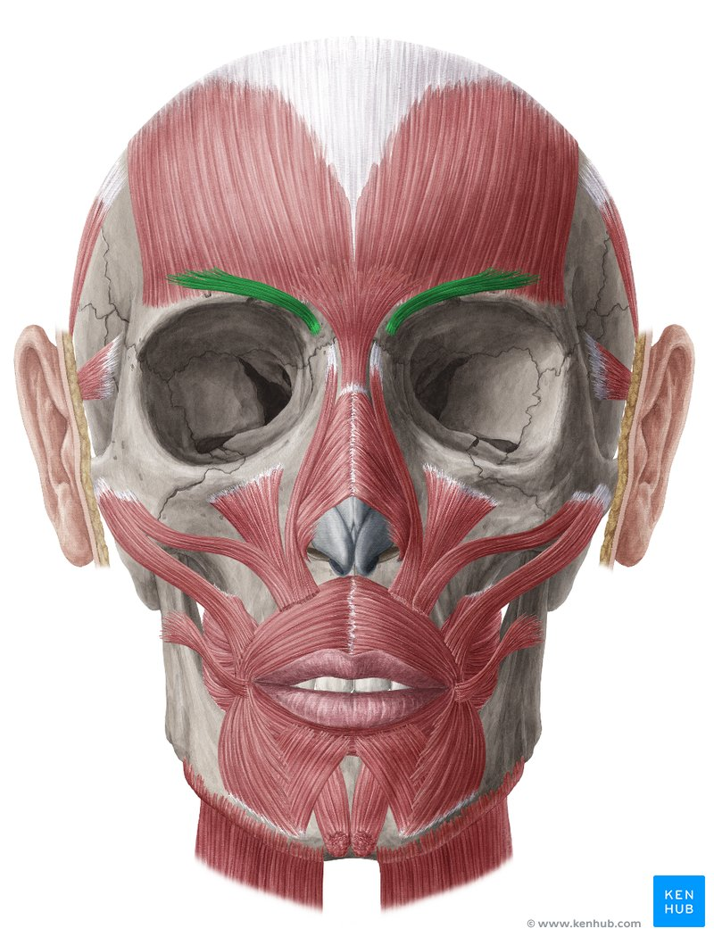
What Muscle?
Corruagator Supercicli
O: Medial end of the superciliary arch of the frontal bone.
I: Skin of the medial half of the eyebrow.
A: Draws the eyebrows medially and inferiorly, creating vertical wrinkles above the nose (frowning/worry expression) and shielding the eyes from bright light
In: Temporal and zygomatic branches of the facial nerve (CN VII).
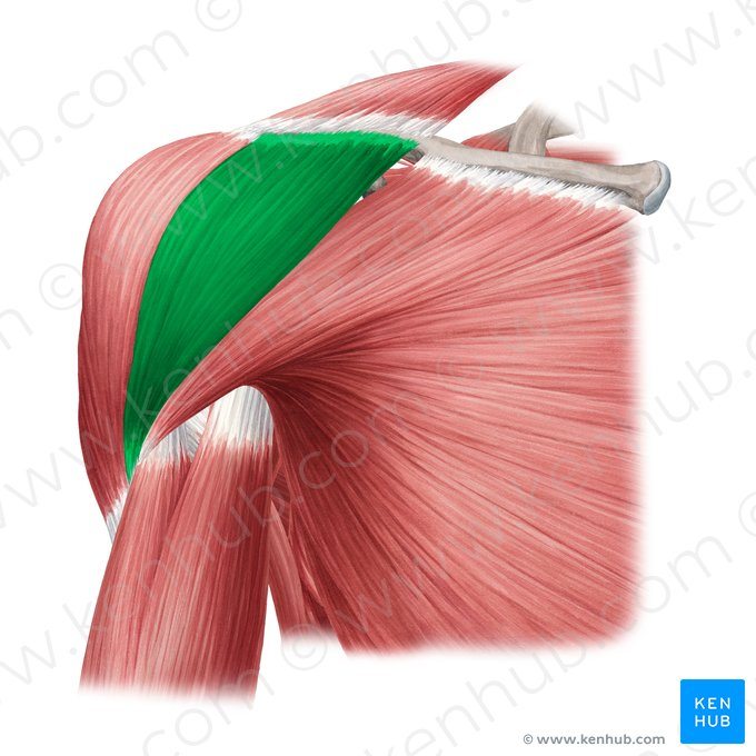
What Muscle?
Deltoid (Anterior)
O: Anterior border and superior surface of the lateral third of the clavicle
I: Deltoid tuberosity on the lateral aspect of the shaft of the humerus.
A: lexes, medially (internally) rotates, and assists the middle deltoid in abducting the arm at the glenohumeral (shoulder) joint.
In: Axillary nerve (spinal roots C5, C6). [1, 2, 3, 4, 5]
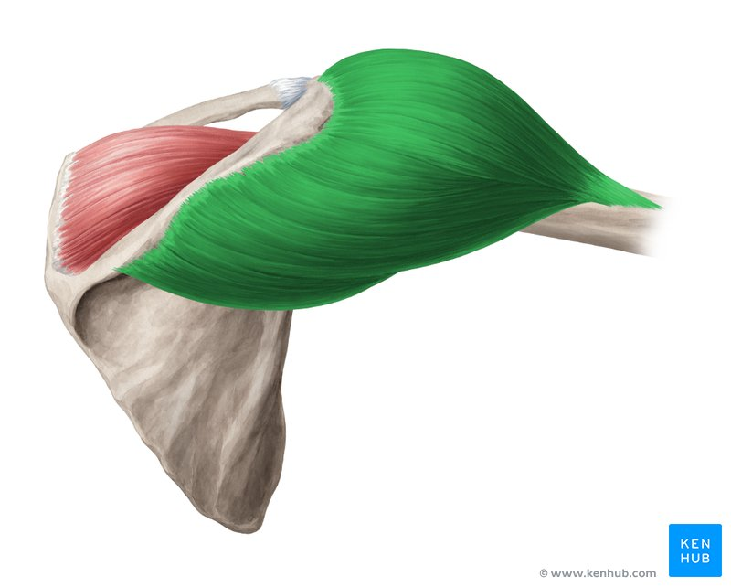
What Muscle?
Deltoid (Middle/Lateral)
O: Lateral border and superior surface of the acromion of the scapula.
I: Deltoid tuberosity on the lateral aspect of the shaft of the humerus.
A: Abducts the arm at the glenohumeral (shoulder) joint (primarily after the first 15 degrees of movement initiated by the supraspinatus).
In: Axillary nerve (spinal roots C5, C6). [1, 2, 3, 4, 5]
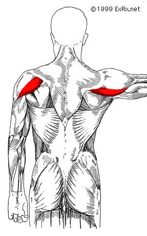
What Muscle?
Deltoid (posterior)
O: Inferior lip of the posterior border of the spine of the scapula.
I: Deltoid tuberosity on the lateral aspect of the shaft of the humerus.
A: Extends, laterally (externally) rotates, and horizontally abducts the arm at the glenohumeral (shoulder) joint.
In: Axillary nerve (spinal roots C5, C6).
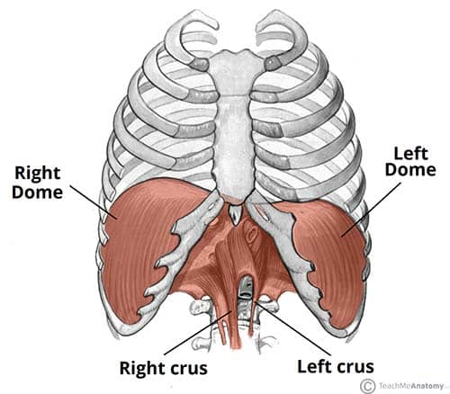
What Muscle?
Diaphragm
O: Sternal part: Posterior surface of the xiphoid process.
Costal part: Internal surfaces of the lower six ribs and their costal cartilages (ribs 7–12).
Lumbar part: Medial and lateral arcuate ligaments, and the lumbar vertebrae via the left crus (L1–L2) and right crus (L1–L3). [1, 2, 3, 4, 5]
I: Central tendon of the diaphragm (a trifoliate aponeurosis with no bony attachment). [1, 2]
A: : Draws down its central tendon during contraction, flattening the dome; this increases the vertical volume of the thoracic cavity, decreases intrathoracic pressure to inspire air, and increases intra-abdominal pressure. [1, 2, 3, 4, 5]
In: Phrenic nerve (spinal roots C3, C4, C5—"C3, 4, 5 keep the diaphragm alive"). [1, 2]
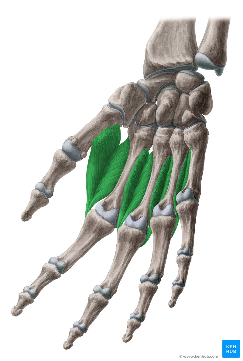
What Muscle?
Dorsal Interossei
O: Adjacent sides of two metacarpal bones (1st through 5th
I: Bases of the proximal phalanges and the extensor expansions of digits 2–4 (index, middle, and ring fingers).
A: Abducts digits 2, 3, and 4 from the axial line (middle finger); assists in flexing the metatarsophalangeal (MCP) joints and extending the interphalangeal (IP) joints
In: Deep branch of the ulnar nerve (spinal roots C8, T1). [1, 2, 3, 4, 5]
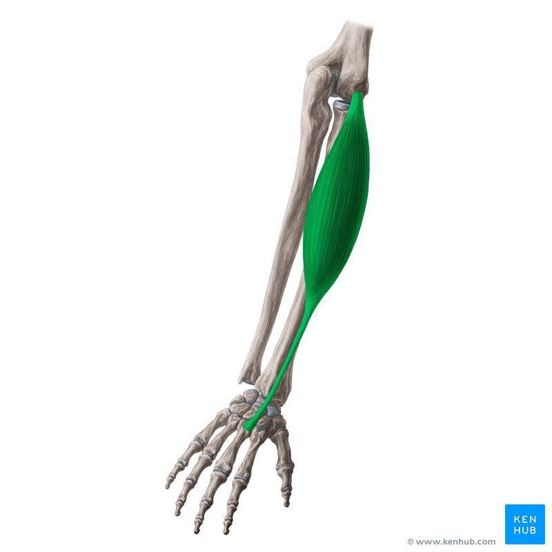
What Muscle?
Extensor Carpi Radialis Brevis
O: Medial surface of the lateral epicondyle of the humerus (via the common extensor tendon).
I: Posterior (dorsal) surface of the base of the 3rd metacarpal bone.
A: Extends and abducts (radially deviates) the hand at the wrist joint.
In: Deep branch of the radial nerve (spinal roots C7, C8). [1, 2, 3, 4, 5]
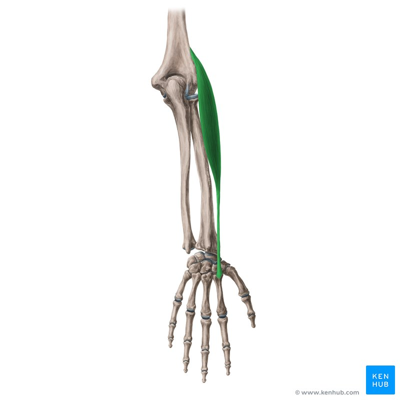
What Muscle?
Extensor Carpi Radialis Longus
O: Distal third of the lateral supracondylar ridge of the humerus.
I: Posterior (dorsal) surface of the base of the 2nd metacarpal bone.
A: Extends and abducts (radially deviates) the hand at the wrist joint.
In: Radial nerve (spinal roots C6, C7). [1, 2, 3, 4, 5]
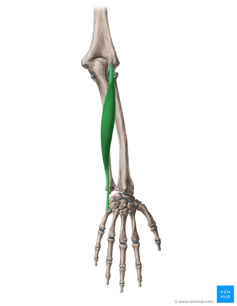
What Muscle?
Extensor Carpi Ulnaris
O: Humeral head: Lateral epicondyle of the humerus (via the common extensor tendon).
Ulnar head: Posterior border of the ulna (via a shared aponeurosis). [1, 2, 3, 4, 5]
I: Posterior (dorsal) surface of the base of the 5th metacarpal bone. [1, 2]
A: Extends and adducts (ulnar deviates) the hand at the wrist joint. [1, 2, 3]
In: Posterior interosseous nerve (a continuation of the deep branch of the radial nerve, spinal roots C7, C8). [1, 2, 3]
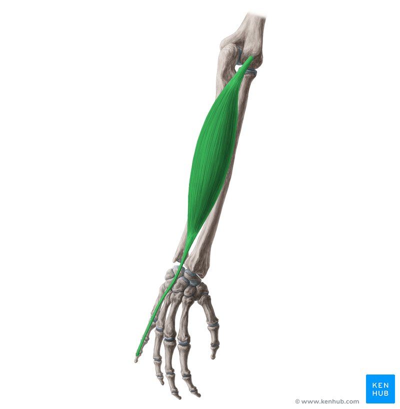
What Muscle?
Extensor Digiti Minimi
O: Lateral epicondyle of the humerus (via the common extensor tendon).
I: Extensor expansion of the 5th digit (little finger) at the dorsum of the proximal phalanx.
A: Extends the 5th digit (little finger) at all joints (metatarsophalangeal and interphalangeal) and assists in wrist extension.
In: Posterior interosseous nerve (a continuation of the deep branch of the radial nerve, spinal roots C7, C8). [1, 2, 3, 4, 5]
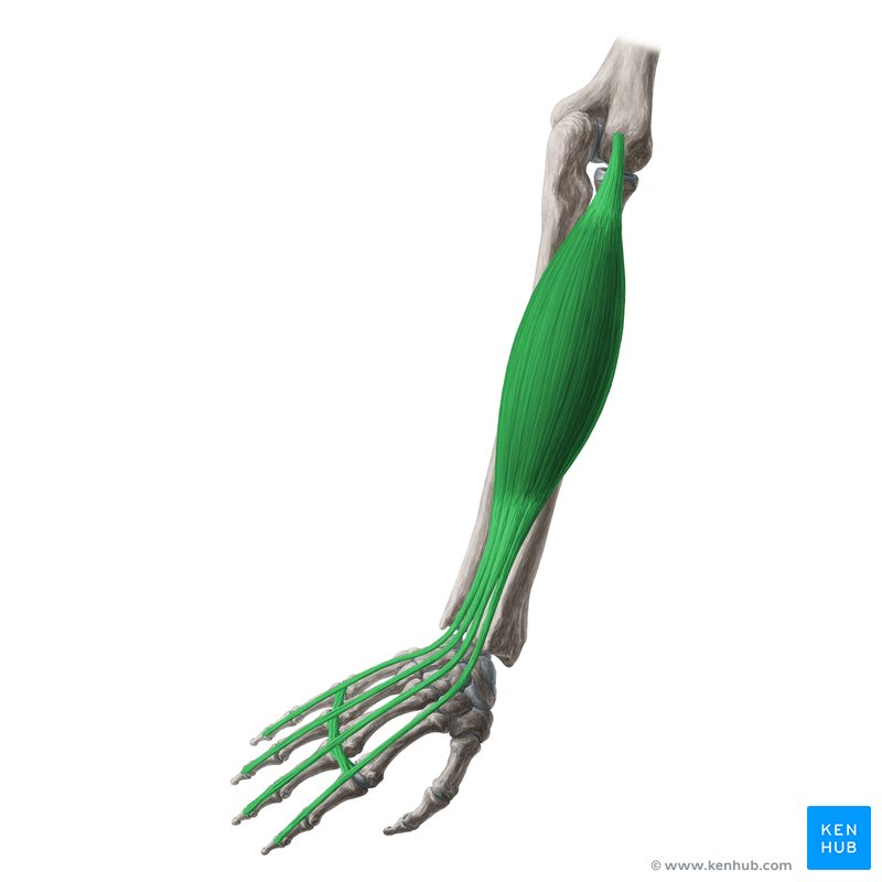
What Muscle?
Extensor Digitorum
O: Lateral epicondyle of the humerus (via the common extensor tendon).
I: Extensor expansions of the medial four digits (index, middle, ring, and little fingers).
A: : Extends the medial four digits at the metatarsophalangeal and interphalangeal joints; assists in wrist extension.
In: Posterior interosseous nerve (a continuation of the deep branch of the radial nerve, spinal roots C7, C8). [1, 2, 3, 4, 5]
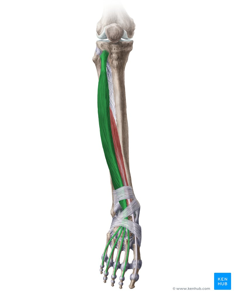
What Muscle?
Extensor Digitorum Longus
O: Lateral condyle of the tibia, proximal three-fourths of the anterior surface of the fibula, and the interosseous membrane. [1, 2, 3, 4]
I: Extensor expansions of the lateral four digits, inserting onto the bases of the middle and distal phalanges of the 2nd through 5th toes. [1, 2]
A: Extends the lateral four digits (toes 2–5) at the metatarsophalangeal and interphalangeal joints; dorsiflexes and everts the foot at the ankle joint. [1, 2, 3, 4]
In: Deep fibular (peroneal) nerve (spinal roots L4, L5, S1). [1, 2, 3, 4]
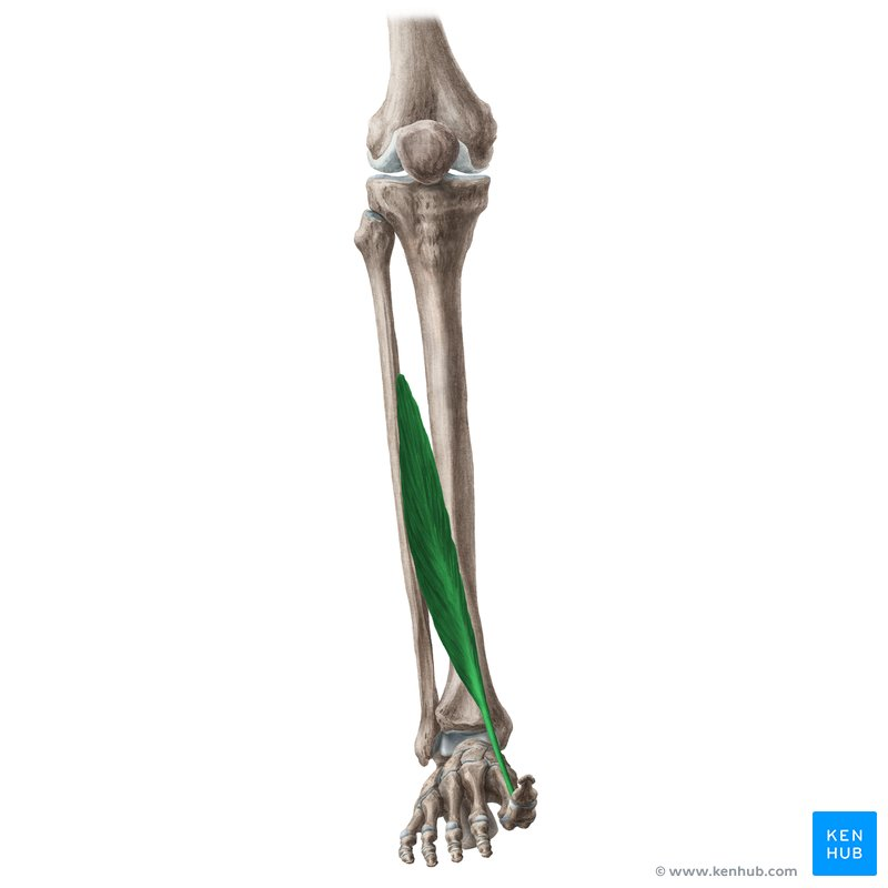
What Muscle?
Extensor Hallucis Longus
O: Middle half of the anterior surface of the fibula and the adjacent interosseous membrane.
I: Posterior (dorsal) surface of the base of the distal phalanx of the great toe (hallux).
A: Extends the great toe at the metatarsophalangeal and interphalangeal joints; assists in dorsiflexion of the foot at the ankle joint.
In: Deep fibular (peroneal) nerve (spinal roots L4, L5, S1). [1, 2, 3, 4, 5]
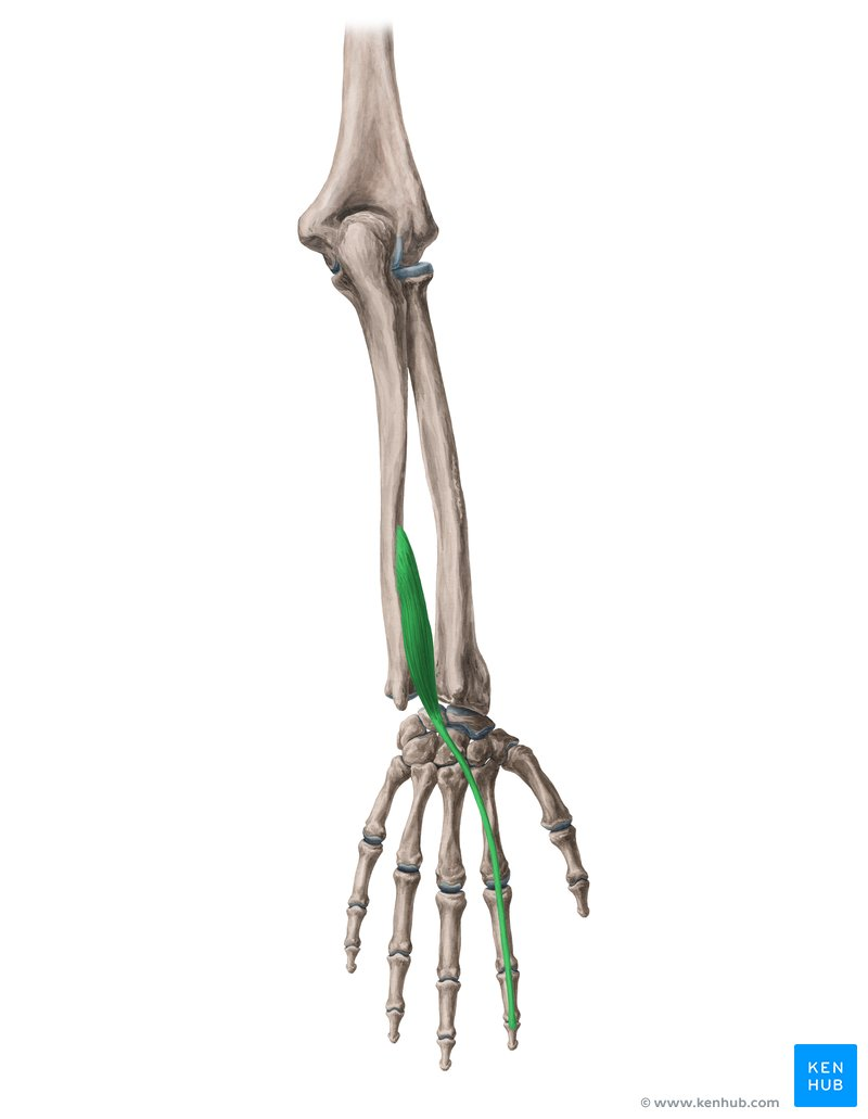
What Muscle?
Extensor Indicis
O: Posterior surface of the distal third of the ulna and the adjacent interosseous membrane.
I: Extensor expansion of the 2nd digit (index finger), joining the tendon of the extensor digitorum.
A: Extends the 2nd digit (index finger) at all joints (metatarsophalangeal and interphalangeal) and assists in wrist extension.
In: osterior interosseous nerve (a continuation of the deep branch of the radial nerve, spinal roots C7, C8). [1, 2, 3, 4, 5]
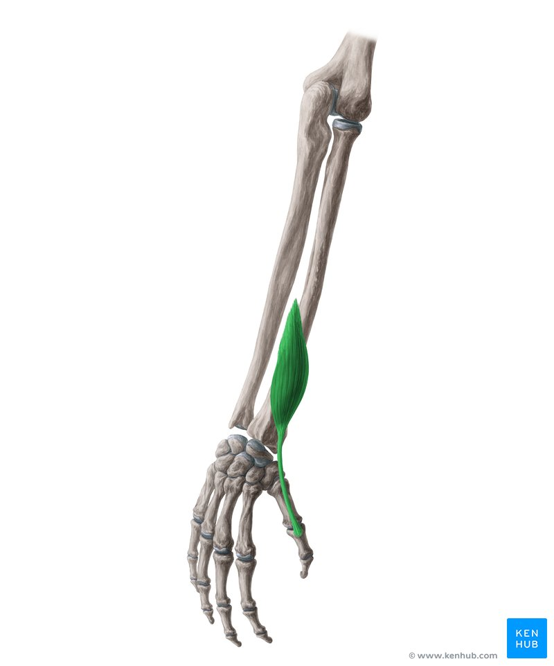
What Muscle?
Extensor Pollicis Brevis
O: Posterior surface of the radius and the adjacent interosseous membrane.
I: Posterior (dorsal) surface of the base of the proximal phalanx of the 1st digit (thumb).
A: Extends the proximal phalanx of the thumb at the metatarsophalangeal joint; assists in extending the carpometacarpal joint of the thumb and extending the wrist.
In: Posterior interosseous nerve (a continuation of the deep branch of the radial nerve, spinal roots C7, C8).
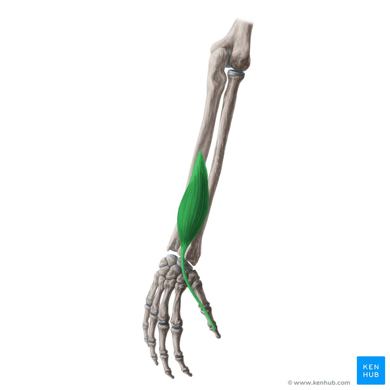
What Muscle?
Extensor Pollicis Longus
O: Posterior surface of the middle third of the ulna and the adjacent interosseous membrane.
I: Posterior (dorsal) surface of the base of the distal phalanx of the 1st digit (thumb)
A: Extends the distal phalanx of the thumb at the interphalangeal joint; assists in extending the metatarsophalangeal and carpometacarpal joints of the thumb and extending the wrist
In: Posterior interosseous nerve (a continuation of the deep branch of the radial nerve, spinal roots C7, C8).
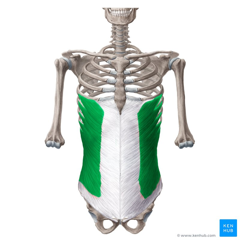
What Muscle?
External Oblique
O: External surfaces of the lower eight rib s (ribs 5–12).
I: : Linea alba, pubic tubercle, and the anterior half of the iliac crest.
A: Bilaterally compresses the abdomen and flexes the vertebral column; unilaterally rotates the trunk to the opposite (contralateral) side and laterally flexes the trunk to the same side.
In: Anterior rami of the lower six thoracic spinal nerves (thoraco-abdominal nerves T7–T11) and the subcostal nerve (T12).
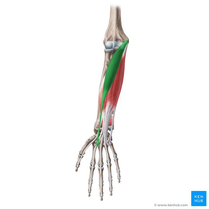
What Muscle?
Flexor Carpi Radialis
O: Medial epicondyle of the humerus (via the common flexor tendon).
I: Bases of the 2nd and 3rd metacarpals.
A: Flexes and abducts (radially deviates) the wrist joint.
In: Median nerve (spinal roots C6, C7). [1, 2, 3, 4, 5]
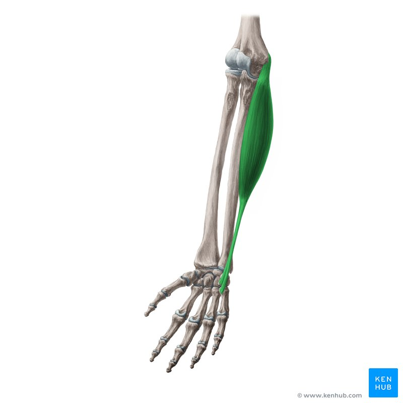
What Muscle?
Flexor Carpi Ulnaris
O: Humeral head: Medial epicondyle of the humerus.
Ulnar head: Olecranon and posterior border of the ulna. [1, 2, 3, 4, 5]
I: Pisiform bone, hook of hamate, and base of the 5th metacarpal. [1, 2, 3, 4]
A: Flexes and adducts (ulnar deviates) the wrist joint.
In: Ulnar nerve (spinal roots C7, C8)
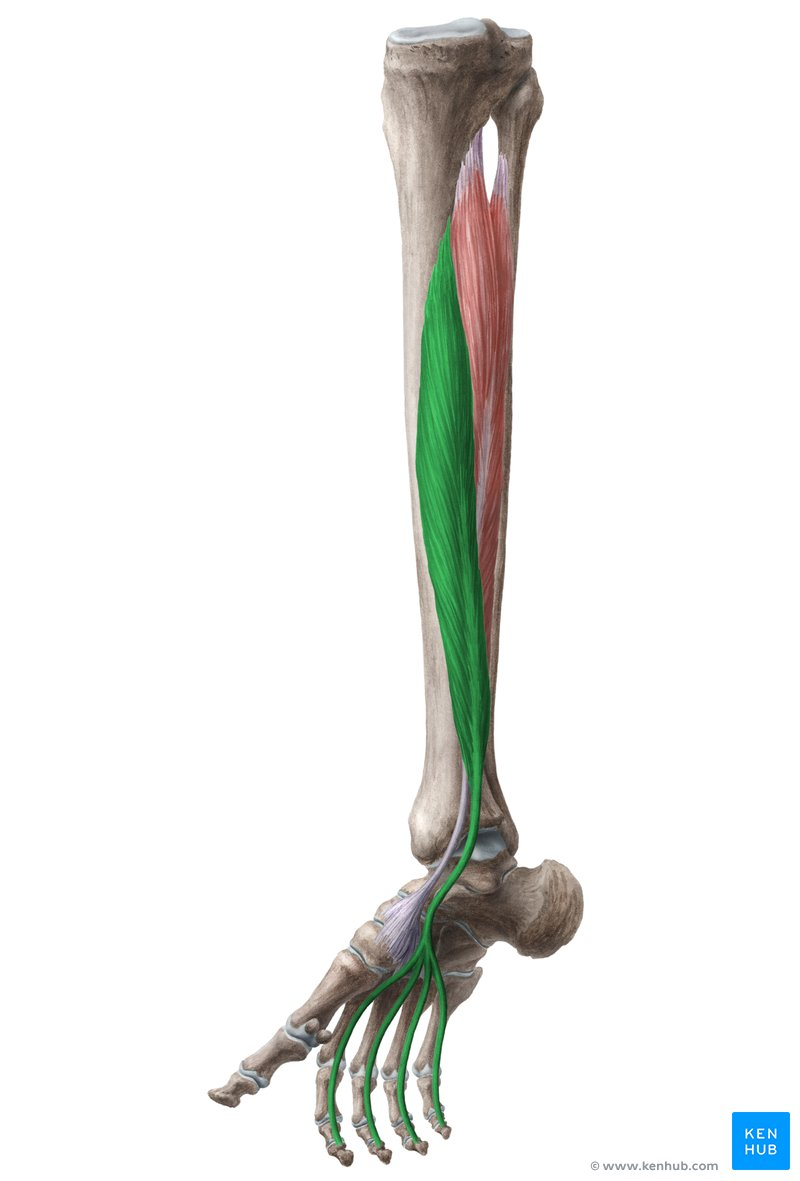
What Muscle?
Flexor Digitorum Longus
O: Medial part of the posterior surface of the tibia (below the soleal line)
I: : Bases of the distal phalanges of the second through fifth (lateral four) toes.
A: Flexes toes 2–5 (at metatarsophalangeal, proximal interphalangeal, and distal interphalangeal joints), assists in plantarflexion of the ankle, and supports the medial and lateral longitudinal arches of the foot.
In: Tibial nerve (spinal roots L5, S1, S2
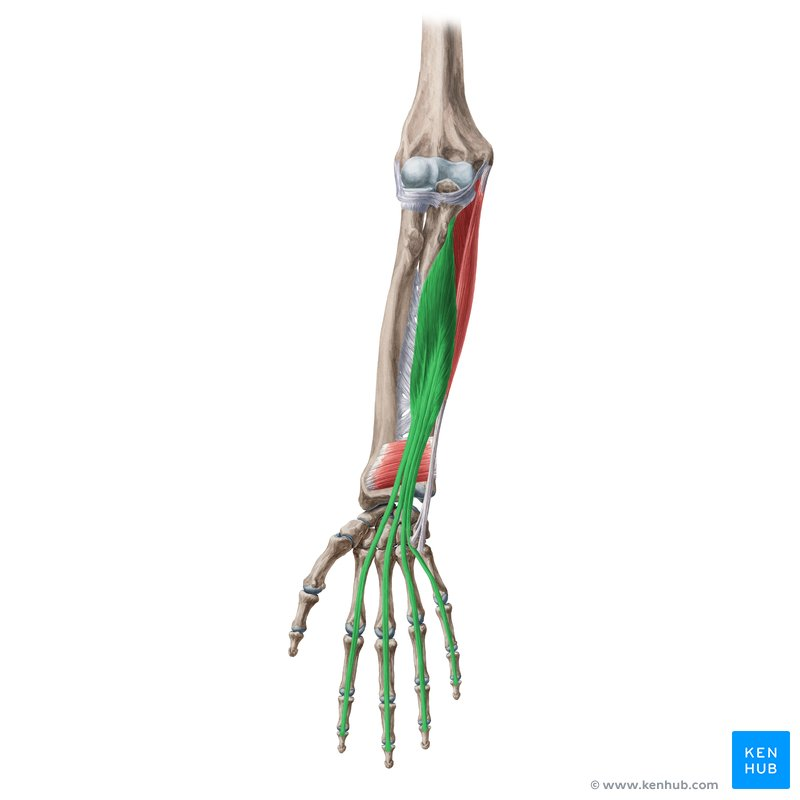
What Muscle?
Flexor Digitorum Profundus
O: Originates from the proximal three-quarters of the anterior and medial surfaces of the ulna and the adjacent interosseous membrane. [1, 2]
I:
A: Flexes the distal interphalangeal joints, proximal interphalangeal joints, and metacarpophalangeal joints of the fingers (digits 2-5), while also weakly assisting with wrist flexion. [1]
In: Features a dual nerve supply. The lateral portion (index and middle fingers) is innervated by the anterior interosseous branch of the median nerve, while the medial portion (ring and little fingers) is supplied by the ulnar nerve
What Muscle?
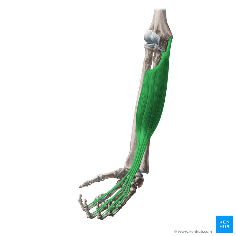
Flexor Digitorum Superficialis
O: Humeroulnar head: Medial epicondyle of the humerus (common flexor tendon) and the coronoid process of the ulna.
Radial head: Superior half of the anterior border of the radius. [1, 2, 3, 4, 5]
I: Inserts onto the bodies of the middle phalanges of the four medial fingers (digits 2–5).
A: Flexes the proximal interphalangeal (PIP) joints of digits 2–5. It also assists with flexing the metacarpophalangeal (MCP) joints and the wrist joint. [1, 2, 3, 4]
In: Median nerve (C7, C8, T1)
What Muscle?
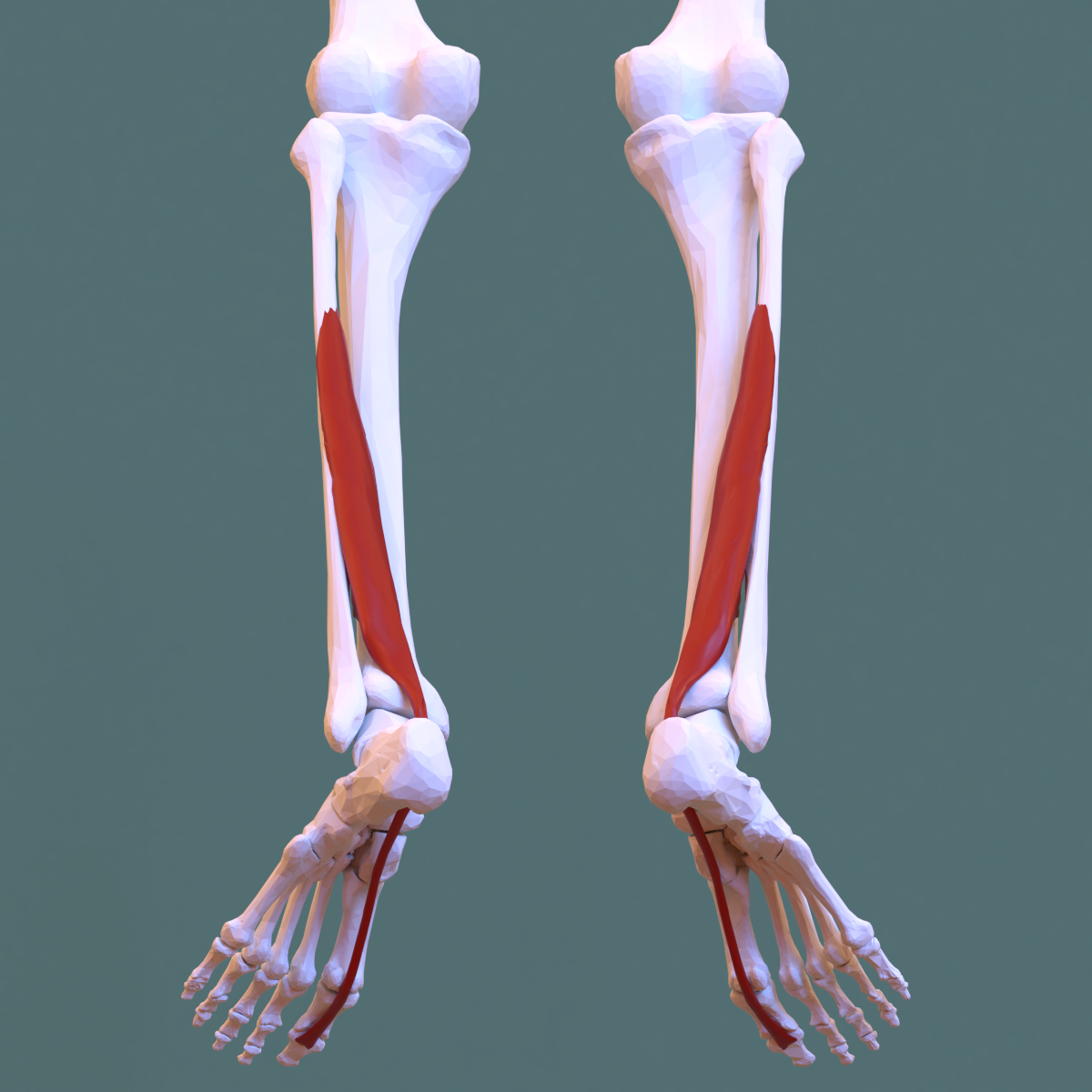
Flexor Hallucis Longus
O: Inferior two-thirds of the posterior surface of the fibula and the lower part of the interosseous membrane
I: Base of the distal phalanx of the great toe (hallux / digit 1).
A: Flexes the distal phalanx of the great toe. It also assists with plantarflexion of the foot at the ankle joint and inversion of the foot.
In: Tibial nerve (S1, S2). [1, 2, 3, 4, 5]
What Muscle?
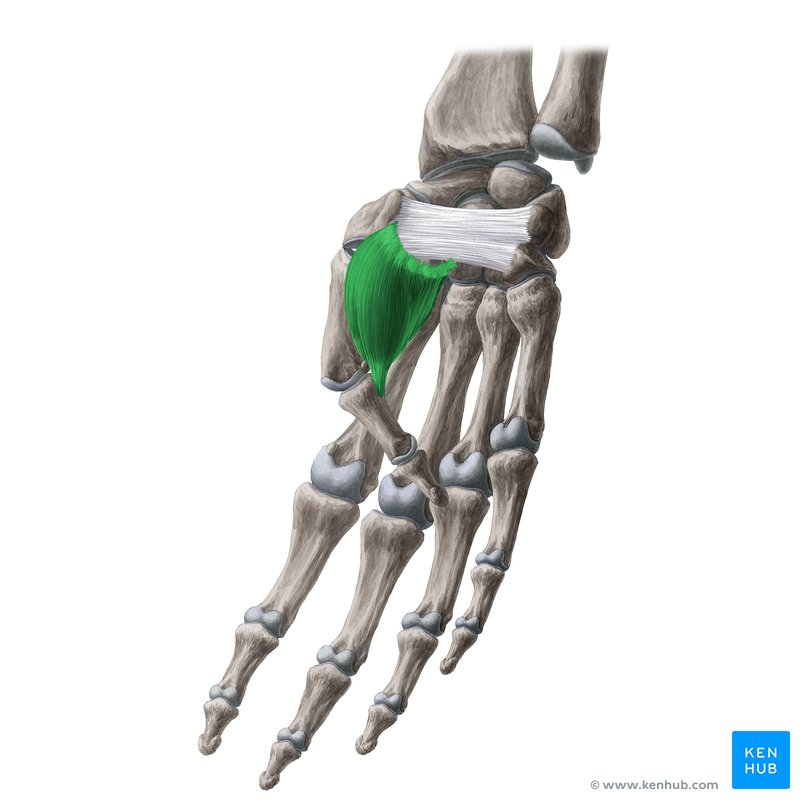
Flexor Pollicis Brevis
O: Superficial head: Flexor retinaculum and the tubercle of the trapezium bone.
Deep head: Trapezoid and capitate bones, as well as the palmar ligaments of the distal row of carpal bones.
I: Base of the proximal phalanx of the thumb (digit 1), via the radial sesamoid bone.
A: Flexes the thumb at the metacarpophalangeal (MCP) and carpometacarpal (CMC) joints. It also assists with opposition of the thumb. [1, 2, 3, 4]
In: eatures a dual nerve supply corresponding to its heads:
Superficial head: Recurrent branch of the median nerve (C8, T1).
Deep head: Deep branch of the ulnar nerve (C8, T1). [1, 2, 3, 4, 5]
What Muscle?
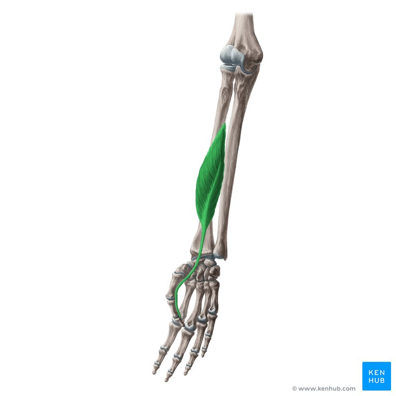
Flexor Pollicis Longus
O: Anterior surface of the radius (distal to the radial tuberosity) and the adjacent interosseous membrane.
I: Base of the distal phalanx of the thumb (digit 1).
A: Flexes the joints of the thumb, specifically the interphalangeal (IP) joint, metacarpophalangeal (MCP) joint, and carpometacarpal (CMC) joint. It also assists with weak flexion of the wrist.
In: Anterior interosseous nerve, which is a branch of the median nerve (C7, C8). [1, 2, 3, 4, 5]
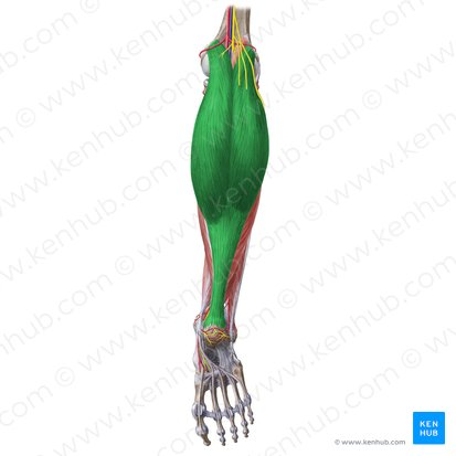
What Muscle?
Gatrocenmius (Medial Head)
O: Popliteal surface of the femur, specifically superior to the medial condyle.
I: Posterior surface of the calcaneus (heel bone) via the Achilles tendon (calcaneal tendon), sharing a common insertion with the lateral head and soleus.
A: Plantarflexes the foot at the ankle joint and flexes the leg at the knee joint.
In: Tibial nerve (S1, S2
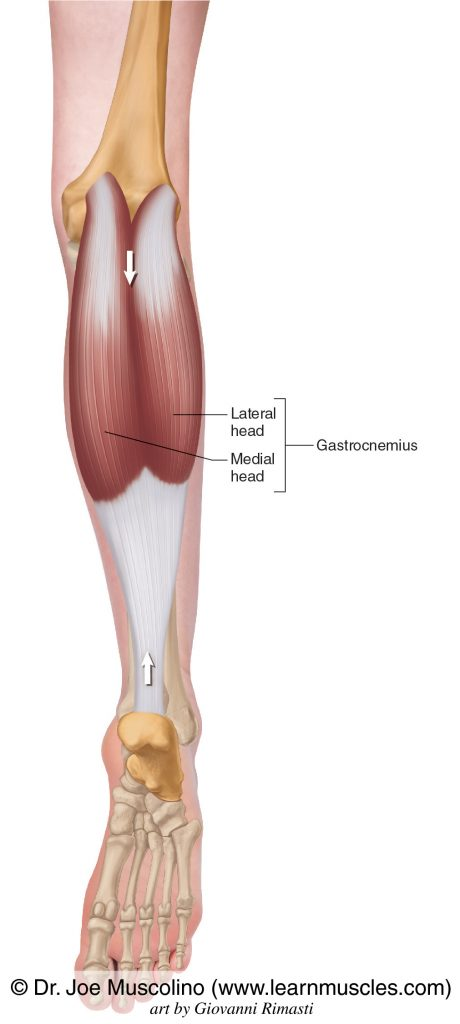
What Muscle?
Gatrocenmius (Lateral head)
O: Lateral aspect of the lateral condyle of the femur.
I: Posterior surface of the calcaneus (heel bone) via the Achilles tendon (calcaneal tendon).
A: Plantarflexes the foot at the ankle joint and flexes the leg at the knee joint.
In: Tibial nerve (S1, S2). [1, 2, 3, 4, 5]
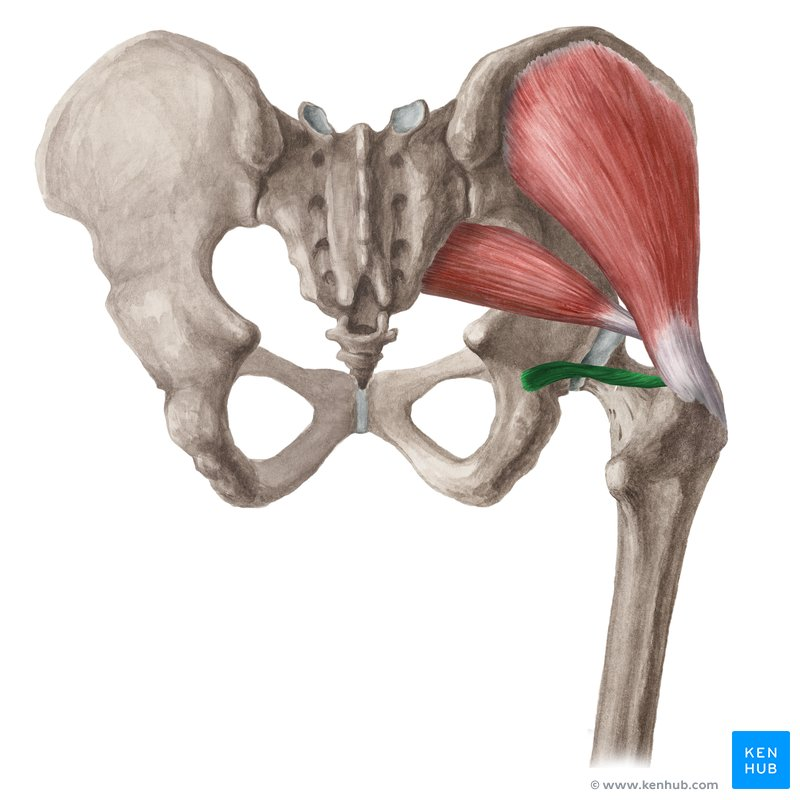
What Muscle?
Gemellus Inferior
O: Upper border of the ischial tuberosity
I: Medial surface of the greater trochanter of the femur, via the tendon of the obturator internus.
A: Laterally (externally) rotates the extended thigh at the hip joint and abducts the flexed thigh. It also helps stabilize the femoral head in the acetabulum.
In: Nerve to obturator internus (L5, S1) — note that it shares its nerve with the obturator internus muscle, whereas gemellus superior shares its nerve with quadratus femoris. [1, 2, 3, 4]
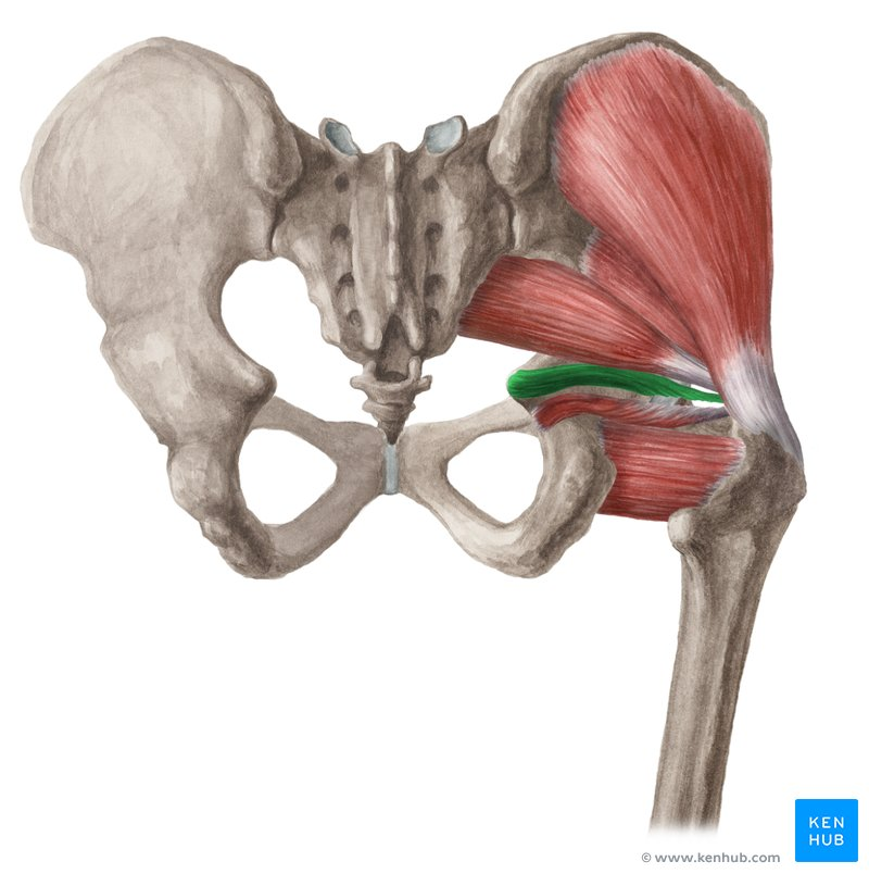
What Muscle?
Gemellus Superior
O: External surface of the ischial spine.
I: Medial surface of the greater trochanter of the femur, blending into the tendon of the obturator internus.
A: Laterally (externally) rotates the extended thigh at the hip joint and abducts the flexed thigh. It also stabilizes the femoral head within the acetabulum.
In: Nerve to obturator internus (L5, S1, S2).
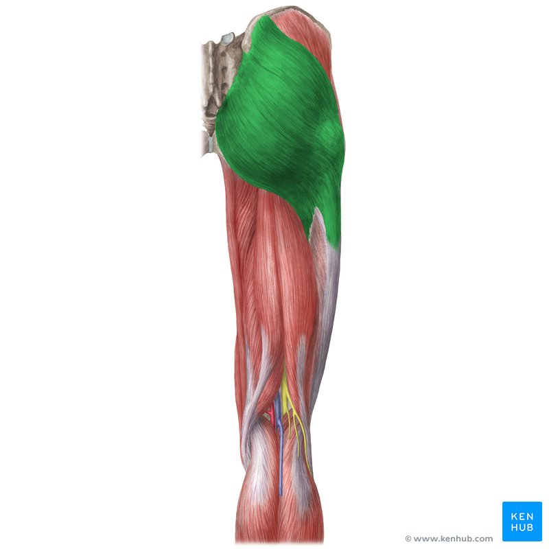
What Muscle?
Gluteal Maximus
O: Outer surface of the ilium (behind the posterior gluteal line), dorsal surface of the sacrum and coccyx, and the sacrotuberous ligament.
I: Upper/Superficial fibers: Iliotibial (IT) band of the fascia lata (three-quarters of the muscle).
Lower/Deep fibers: Gluteal tuberosity of the femur (one-quarter of the muscle). [1, 2, 3, 4, 5]
A: Major extensor of the thigh at the hip joint. It also laterally (externally) rotates the thigh, assists with steadying the trunk on the pelvis, and steadies the femur on the tibia via the IT band. [1, 2, 3, 4, 5]
In: Inferior gluteal nerve (L5, S1, S2)
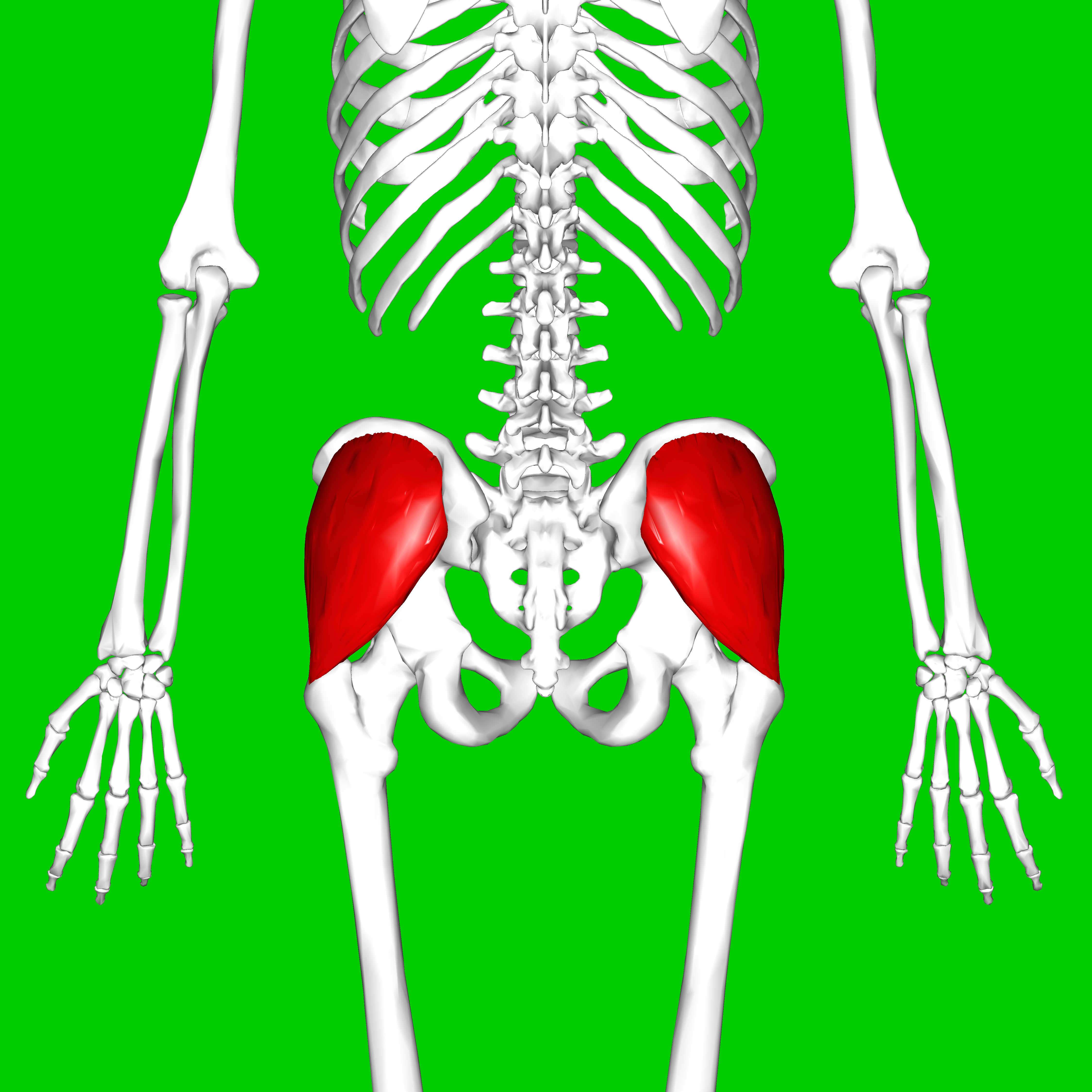
What Muscle?
Gluteus Medius
O: Outer surface of the ilium, between the anterior and posterior gluteal lines.
I: Lateral surface of the greater trochanter of the femur.
A: Abducts the thigh at the hip joint and stabilizes the pelvis during the stance phase of gait. Its anterior fibers also medially (internally) rotate the thigh.
In: Superior gluteal nerve (L4, L5, S1). [1, 2, 3, 4, 5]
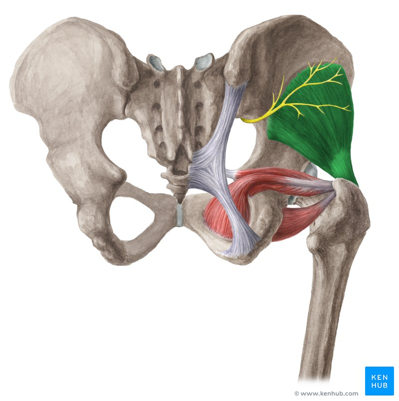
What Muscle?
Glutues Minimus
O: Outer surface of the ilium, between the anterior and inferior gluteal lines.
I: Anterior border of the greater trochanter of the femur.
A: Abducts the thigh at the hip joint and stabilizes the pelvis during gait. Its anterior fibers also medially (internally) rotate the thigh.
In: Superior gluteal nerve (L4, L5, S1). [1, 2, 3, 4, 5]
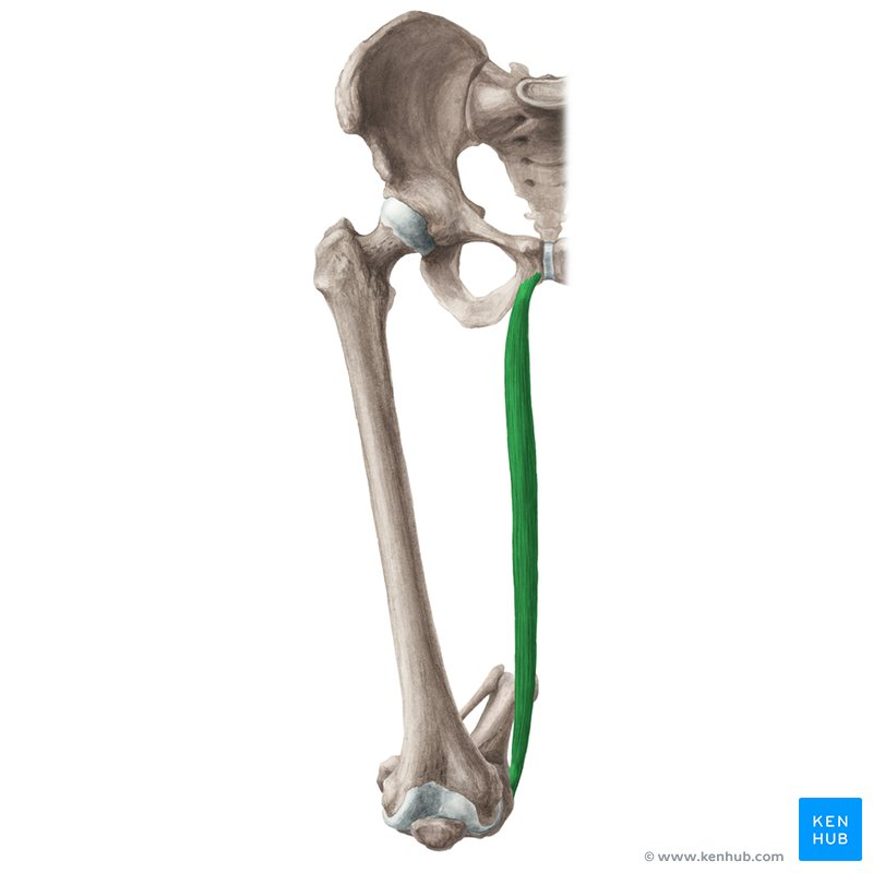
What Muscle?
Gracilis
O: Inferior ramus of the pubis, body of the pubis, and the adjacent ramus of the ischium.
I: Proximal medial surface of the shaft of the tibia, via the pes anserinus (goose's foot) tendon.
A: Adducts the thigh at the hip joint. It also flexes the leg at the knee joint and assists with medial (internal) rotation of the knee when it is flexed.
In: Obturator nerve (L2, L3). [1, 2, 3, 4, 5]
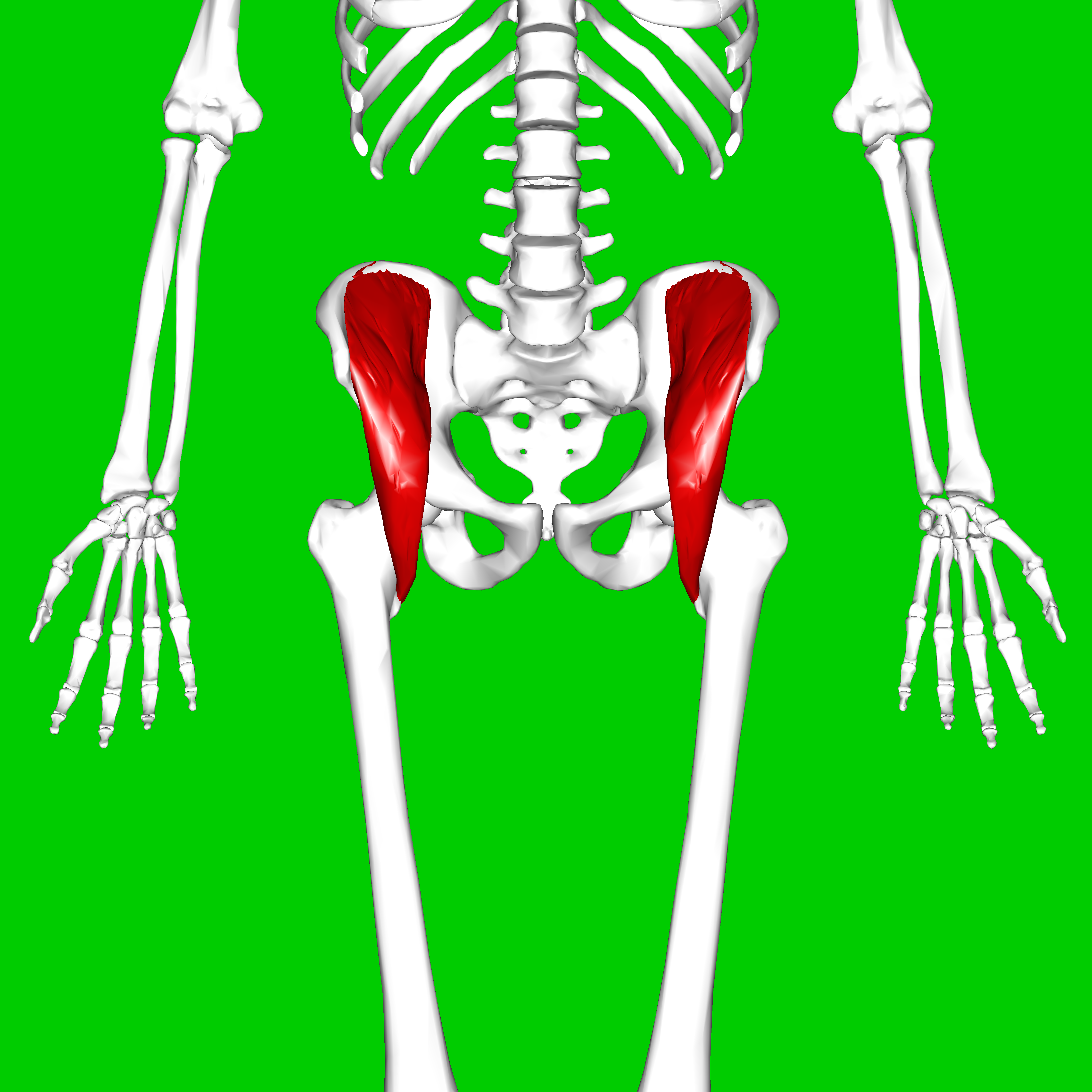
What Muscle?
Iliacus
O: Superior two-thirds of the iliac fossa, internal lip of the iliac crest, iliolumbar ligament, and the lateral ala of the sacrum.
I: Lesser trochanter of the femur, via the tendon common to it and the psoas major (iliopsoas tendon). [1, 2, 3, 4]
A: Flexes the thigh at the hip joint. When the thigh is fixed, it flexes the trunk forward (as in a sit-up) and stabilizes the hip joint. [1, 2, 3, 4]
In: Femoral nerve (L2, L3).
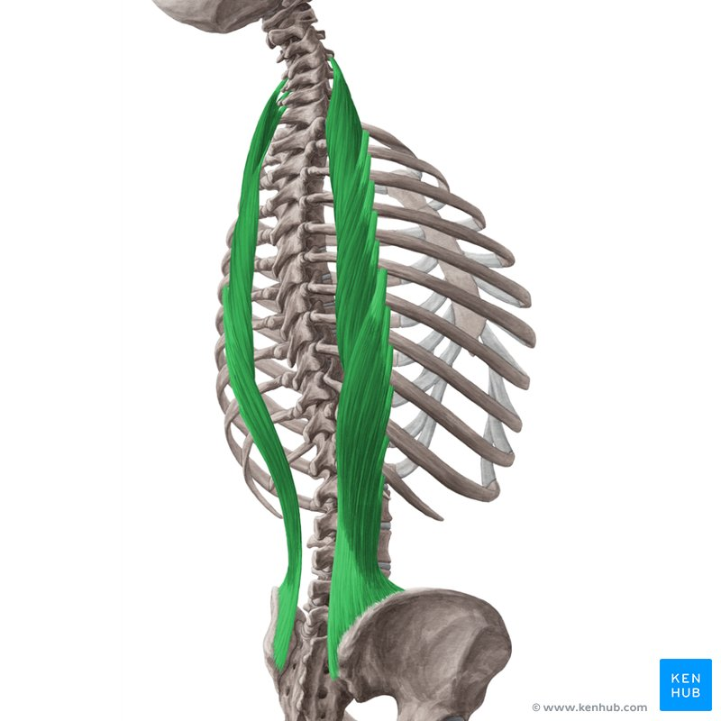
What Muscle?
Iliocostalis
O: Iliocostalis lumborum: Broad tendon from the posterior iliac crest, posterior sacrum, and lumbar spinous processes.
Iliocostalis thoracis: Angles of the lower 6 ribs (ribs 7–12).
Iliocostalis cervicis: Angles of ribs 3–6. [1, 2, 3, 4, 5]
I: Iliocostalis lumborum: Angles of the lower 6 or 7 ribs.
Iliocostalis thoracis: Angles of the upper 6 ribs and the transverse process of the C7 vertebra.
Iliocostalis cervicis: Transverse processes of vertebrae C4–C6.
A: Acting bilaterally, it extends the vertebral column and head. Acting unilaterally, it laterally flexes (side-bends) the vertebral column to the same side
In: Posterior rami (dorsal rami) of the respective spinal nerves in the lumbar, thoracic, and cervical regions. [1, 2, 3, 4, 5]
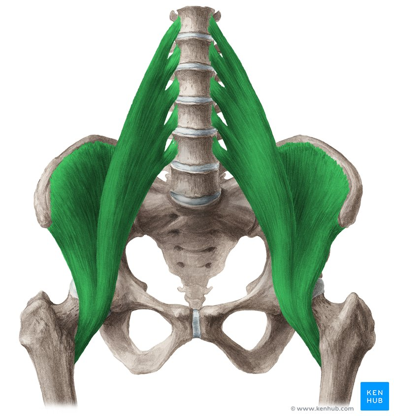
What Muscle?
Iliopsoas
O: Arises from two distinct anatomical regions corresponding to its component parts:
Iliacus part: Upper two-thirds of the iliac fossa, iliac crest, and lateral ala of the sacrum.
Psoas major part: Transverse processes, bodies, and intervertebral discs of T12–L5 vertebrae. [1, 2, 3, 4, 5]
I: Lesser trochanter of the femur, via a unified, shared tendon. [1, 2, 3]
A: Primary and powerful flexor of the thigh at the hip joint. When the femur is fixed, bilateral contraction flexes the trunk forward on the pelvis (such as raising the torso during a sit-up).
In: Dual nerve supply split between its components:
Iliacus part: Femoral nerve (L2, L3).
Psoas major part: Anterior (ventral) rami of lumbar spinal nerves (L1, L2, L3). [1, 2, 3, 4, 5]
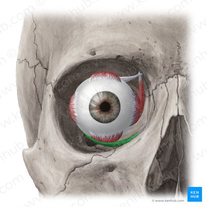
What Muscle?
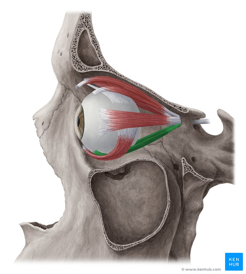
Inferior Oblique
O: Anterior part of the floor of the orbit, specifically on the orbital surface of the maxilla, just lateral to the lacrimal groove. [1, 2, 3, 4
I: Sclera of the eye in the posteroinferior quadrant, deep to the lateral rectus muscle.
A: Elevates, abducts (turns outward), and laterally rotates (extorts) the eyeball. Its primary clinical action is elevation when the eye is adducted (looking toward the nose).
In: Inferior division of the oculomotor nerve (CN III). [1, 2]
What Muscle?
Inferior Rectus
O: Lower part of the common tendinous ring (annulus of Zinn) at the apex of the orbit.
I: Anterior lower surface of the sclera of the eye, approximately 6.5 mm inferior to the corneal limbus.
A: Depresses (looks down), adducts (looks inward toward the nose), and laterally rotates / extorts (rolls outward) the eyeball. Its primary clinical action is depression when the eye is abducted.
In: Inferior division of the oculomotor nerve (CN III).
What Muscle?
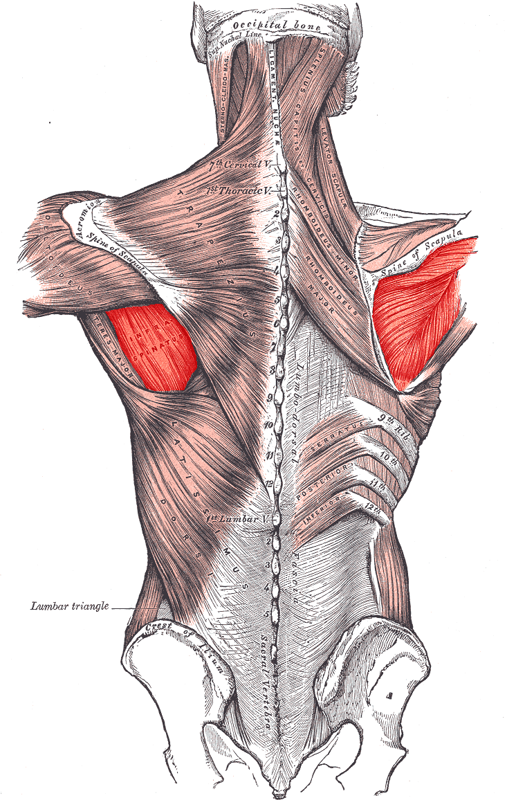
Infraspinatus
O: Infraspinous fossa of the scapula (shoulder blade) and the deep fascia covering the muscle.
I: Middle facet on the greater tubercle of the humerus.
A: Laterally (externally) rotates the arm at the glenohumeral (shoulder) joint and helps hold the head of the humerus securely within the glenoid cavity.
In: Suprascapular nerve (C5, C6). [1, 2, 3, 4, 5]
What Muscle?
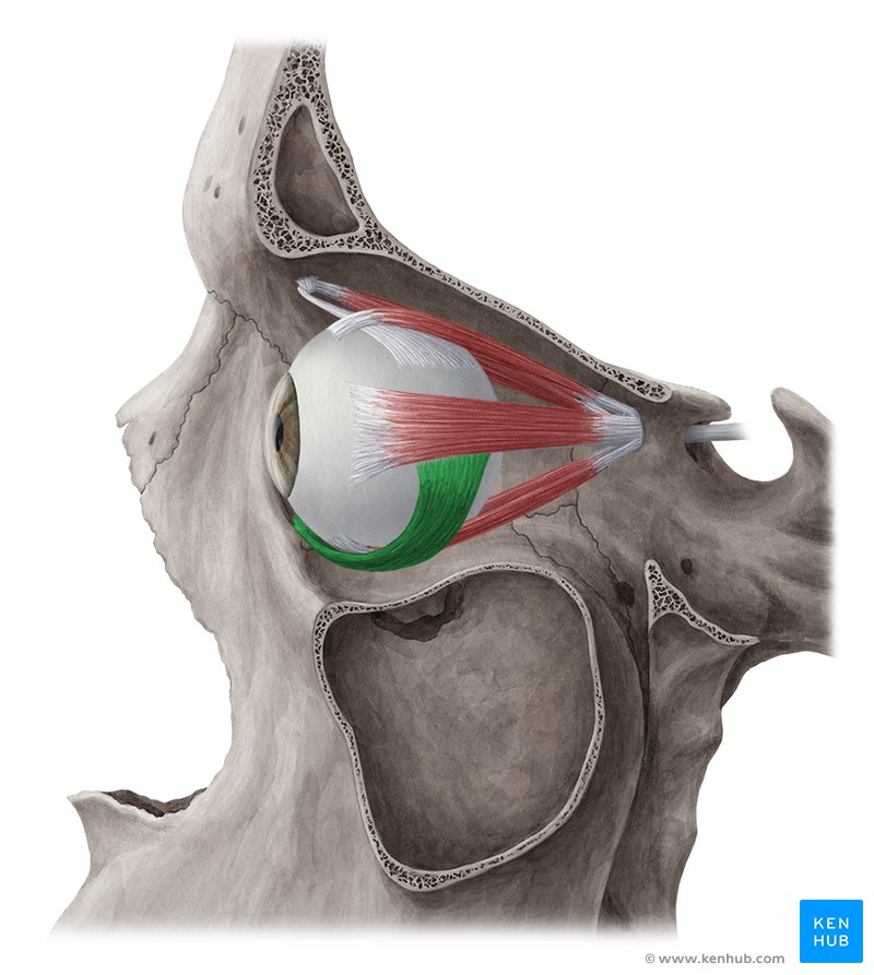
Inferior Oblique
O: Anterior floor of the orbit, on the orbital surface of the maxilla (just lateral to the lacrimal groove).
I: Sclera of the eye in the posteroinferior quadrant, deep to the lateral rectus muscle.
A: Elevates, abducts (turns outward), and extorts (laterally rotates) the eyeball.
In: Inferior division of the oculomotor nerve (CN III). [1, 2, 3, 4, 5]
What Muscle?
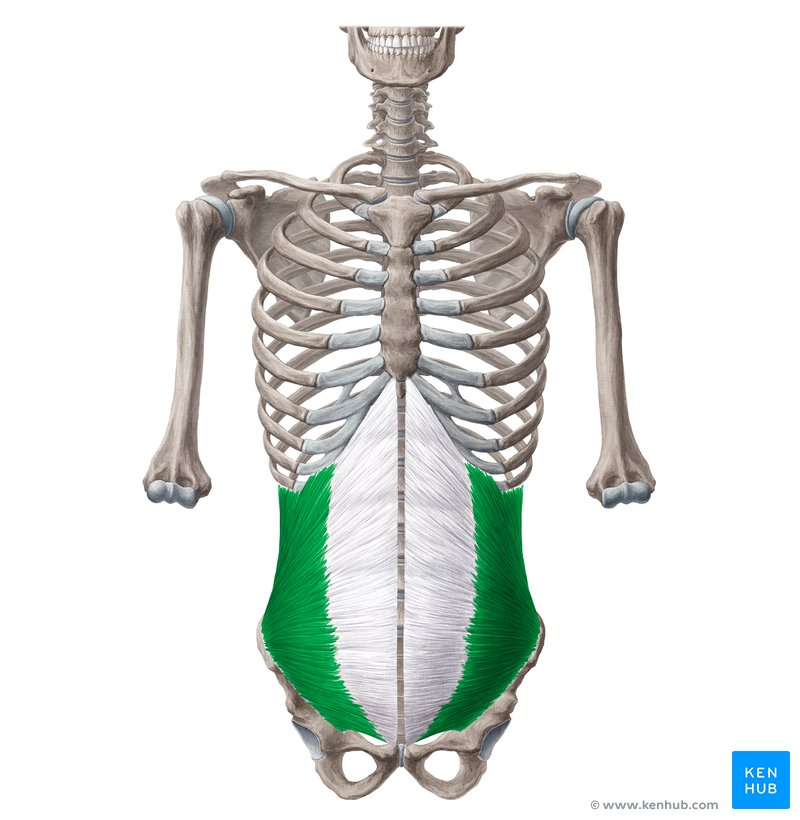
Internal Oblique
O: Thoracolumbar fascia, anterior two-thirds of the iliac crest, and the lateral half of the inguinal ligament.
I: Inferior borders of the lower three or four ribs (ribs 9–12), the linea alba, and the pubic crest (via the conjoint tendon).
A: Compresses and supports the abdominal viscera. Acting bilaterally, it flexes the trunk. Acting unilaterally, it rotates the trunk to the same side (ipsilateral rotation) and laterally flexes the trunk. [1, 2, 3, 4, 5]
In: Anterior rami of the lower six thoracic spinal nerves (T7–T12 intercostal and subcostal nerves), as well as the iliohypogastric and ilioinguinal nerves (L1). [1, 2, 3, 4, 5]
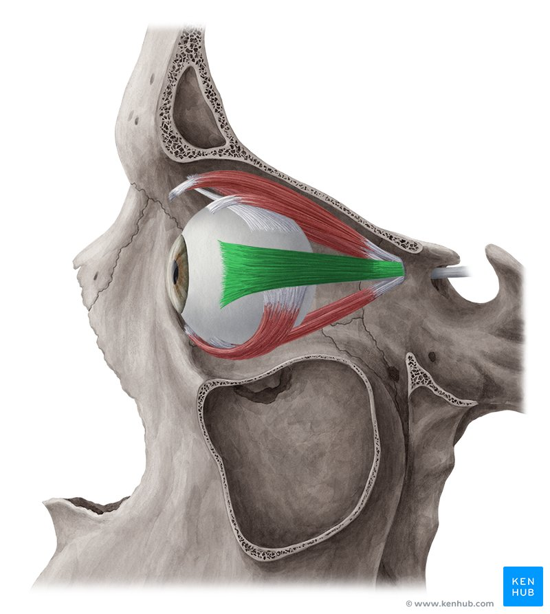
What Muscle?
Lateral Rectus
O: Lateral part of the common tendinous ring (annulus of Zinn) at the apex of the orbit, spanning across the superior orbital fissure.
I: Anterior lateral surface of the sclera of the eye, approximately 7 mm lateral to the corneal limbus.
A: Abducts the eyeball (turns the eye outward, away from the nose).
In: Abducens nerve (CN VI).
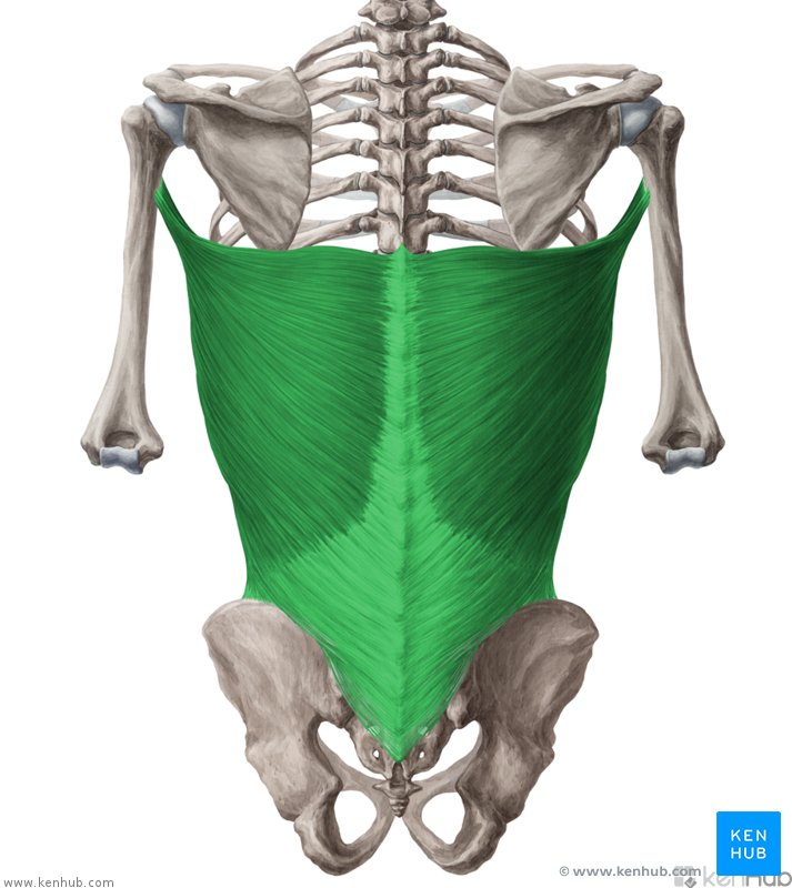
What Muscle?
Latissmimus Dorsi
O: Spinous processes of the lower 6 thoracic vertebrae (T7–T12), thoracolumbar fascia, iliac crest, and the lower 3 or 4 ribs. It also frequently has a small attachment at the inferior angle of the scapula. [1, 2, 3, 4, 5]
I: Floor of the intertubercular sulcus (bicipital groove) of the humerus.
A: Extends, adducts, and medially (internally) rotates the arm at the glenohumeral (shoulder) joint. It is a crucial muscle for pulling movements, climbing, and swimming.
In: Thoracodorsal nerve (C6, C7, C8).
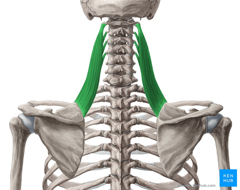
What Muscle?
Levator Scapulae
O: Transverse processes of the upper four cervical vertebrae (C1–C4).
I: Medial border of the scapula, specifically from the superior angle down to the root of the scapular spine.
A: Elevates the scapula and rotates its glenoid cavity downward. Acting unilaterally with the scapula fixed, it laterally flexes the neck to the same side and assists in neck rotation.
In: Anterior rami of cervical nerves C3 and C4, and the dorsal scapular nerve (C5). [1, 2, 3, 4, 5]
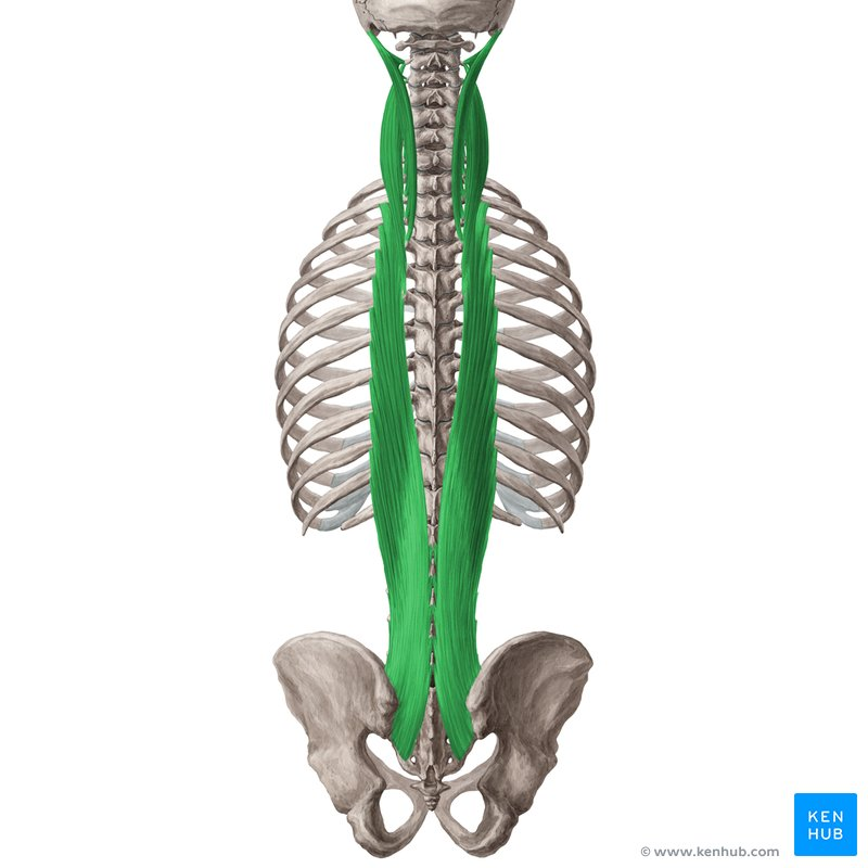
What Muscle?
Longissimus
O: Longissimus lumborum/thoracis: Common erector spinae tendon from the posterior sacrum, iliac crest, and lumbar spinous processes.
Longissimus cervicis: Transverse processes of the upper 4–6 thoracic vertebrae (T1–T6).
Longissimus capitis: Transverse processes of the upper 4–5 thoracic vertebrae (T1–T5) and articular processes of the lower 3–4 cervical vertebrae (C4–C7).
I: Longissimus thoracis: Transverse processes of all thoracic vertebrae and the lower 9 or 10 ribs (between the tubercles and angles).
Longissimus cervicis: Transverse processes of cervical vertebrae C2–C6.
Longissimus capitis: Posterior margin of the mastoid process of the temporal bone. [1, 2, 3, 4, 5]
A: Acting bilaterally, it extends the vertebral column; the capitis portion extends the head. Acting unilaterally, it laterally flexes (side-bends) the vertebral column and rotates the head to the same side (ipsilateral rotation). [1, 2, 3, 4, 5]
In: Posterior rami (dorsal rami) of the respective spinal nerves in each region. [1, 2]
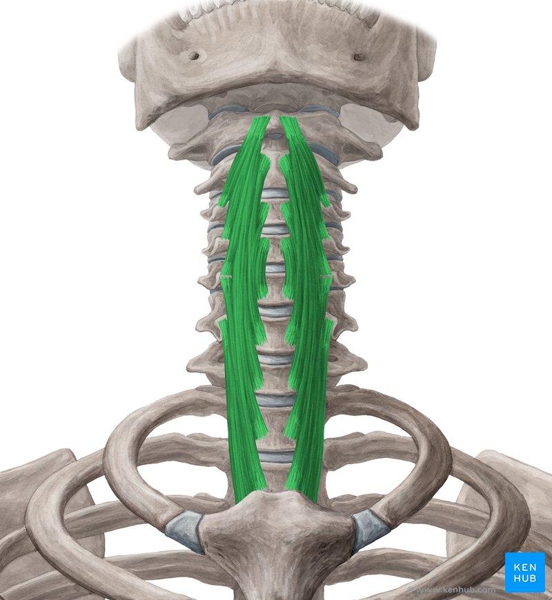
What Muscle?
Longus Colli
O: Superior oblique part: Anterior tubercles of the transverse processes of cervical vertebrae C3–C5.
Inferior oblique part: Anterior surfaces of the bodies of thoracic vertebrae T1–T3.
Vertical part: Anterior surfaces of the bodies of thoracic vertebrae T1–T3 and cervical vertebrae C5–C7.
I: Superior oblique part: Anterior tubercle of the atlas (C1 vertebra).
Inferior oblique part: Anterior tubercles of the transverse processes of cervical vertebrae C5–C6.
Vertical part: Anterior surfaces of the bodies of cervical vertebrae C2–C4. [1, 2, 3]
A: Action): Acting bilaterally, it flexes the neck anteriorly. Acting unilaterally, it laterally flexes (side-bends) the neck and assists in slight contralateral rotation of the head and neck.
In: Anterior (ventral) rami of cervical spinal nerves C2–C6
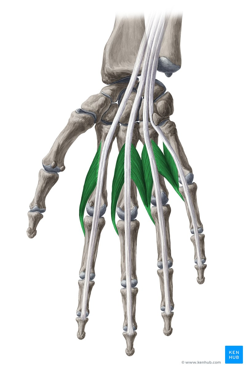
What Muscle?
Lumbricals
O: Tendons of the flexor digitorum profundus (FDP).
1 and 2 (Lateral): Unipennate from the lateral side of the tendons for digits 2 and 3.
3 and 4 (Medial): Bipennate from the adjacent sides of the tendons for digits 3, 4, and 5. [1, 2, 3, 4, 5]
I: Lateral sides of the extensor expansions of digits 2–5.
A: Flex the metacarpophalangeal (MCP) joints and extend the proximal (PIP) and distal (DIP) interphalangeal joints of digits 2–5. This combined action creates the "bye-bye" motion or writing posture. [1, 2]
In: Dual nerve supply:
1 and 2 (Lateral): Median nerve (C8, T1).
3 and 4 (Medial): Deep branch of the ulnar nerve (C8, T1). [1, 2, 3, 4]
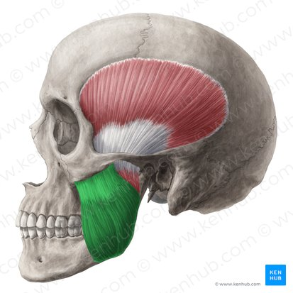
What Muscle?
Masseter
O: Superficial part: Anterior two-thirds of the lower border of the zygomatic arch and the maxillary process of the zygomatic bone.
Deep part: Posterior one-third of the lower border and the entire medial surface of the zygomatic arch
I: Lateral surface of the angle and ramus of the mandible. [1, 2]
A: Elevates the mandible (closes the jaw) and protrudes the jaw (primarily via the superficial fibers). It plays a key role in crushing and chewing food
In: Masseteric nerve, which is a branch of the mandibular division of the trigeminal nerve (CN V₃). [1, 2]
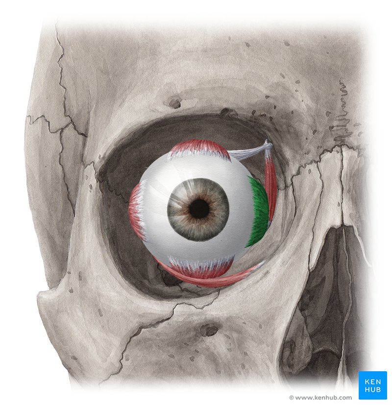
What Muscle?
Medial Rectus
O: Medial part of the common tendinous ring (annulus of Zinn) at the apex of the orbit.
I: Anterior medial surface of the sclera of the eye, approximately 5.5 mm medial to the corneal limbus.
A: Adducts the eyeball (turns the eye inward toward the nose).
In: Inferior division of the oculomotor nerve (CN III). [1, 2, 3, 4, 5]
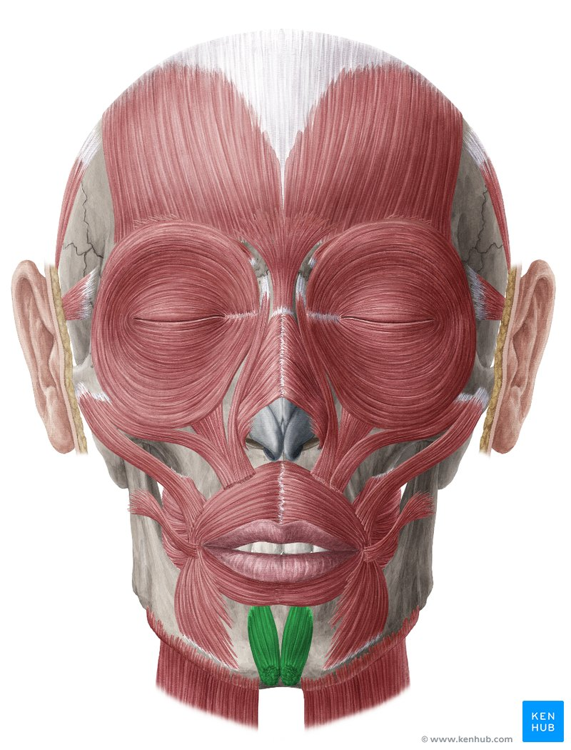
What Muscle?
Mentailis
O: Incisive fossa on the anterior surface of the mandible (just below the lower incisor teeth).
I: Skin of the chin (mentum).
A: Elevates and protrudes the lower lip (as in pouting). It also wrinkles the skin over the chin and helps express doubt or sadness.
In: Marginal mandibular branch of the facial nerve (CN VII). [1, 2, 3, 4, 5]
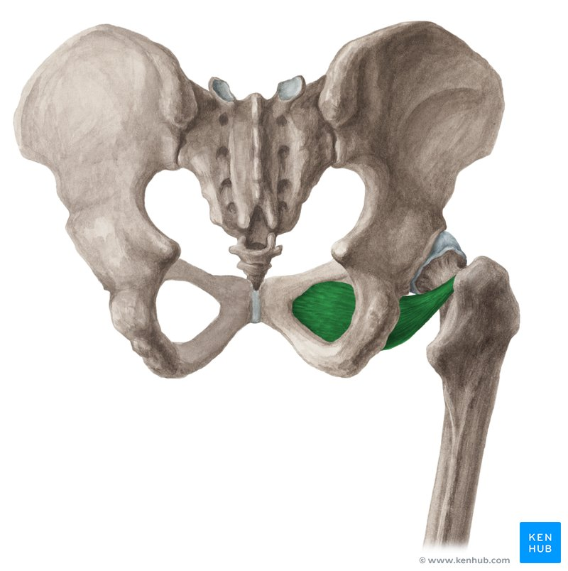
What Muscle?
Obturator Extwernus
O: External surface of the obturator membrane and the surrounding bony margins of the obturator foramen (specifically the rami of the pubis and ischium).
I: Trochanteric fossa of the femur (on the medial surface of the greater trochanter).
A: Laterally (externally) rotates the thigh at the hip joint. It also stabilizes the head of the femur inside the acetabulum when walking or standing.
In: Innervation): Posterior branch of the obturator nerve (L3, L4).
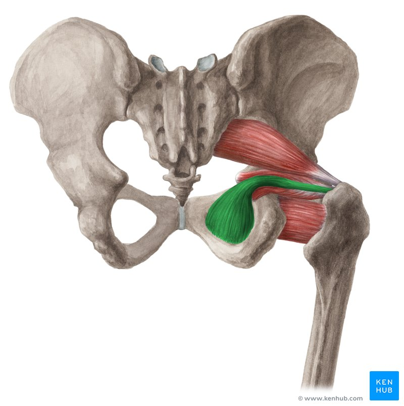
What Muscle?
Obturator Internus
O: Internal surface of the obturator membrane and the surrounding bony margins of the obturator foramen. [1, 2, 3]
I: Medial surface of the greater trochanter of the femur, superior to the trochanteric fossa (often sharing a common tendon with the superior and inferior gemelli). [1, 2, 3, 4]
A: Laterally (externally) rotates the extended thigh at the hip joint and abducts the flexed thigh. It also stabilizes the femoral head in the acetabulum. [1, 2, 3, 4]
In: Nerve to obturator internus (L5, S1, S2)
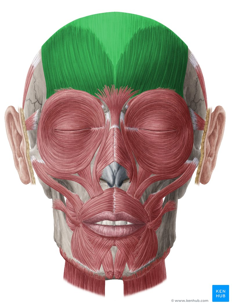
What Muscle?
Occiptofronatilis
Occipitalis
Frontialis
O: Lateral two-thirds of the superior nuchal line of the occipital bone and the mastoid process of the temporal bone.
I: Epicranial aponeurosis (galea aponeurotica).
A: Retracts (pulls back) the scalp, increasing the efficiency of the frontalis muscle.
In: Posterior auricular branch of the facial nerve (CN VII).
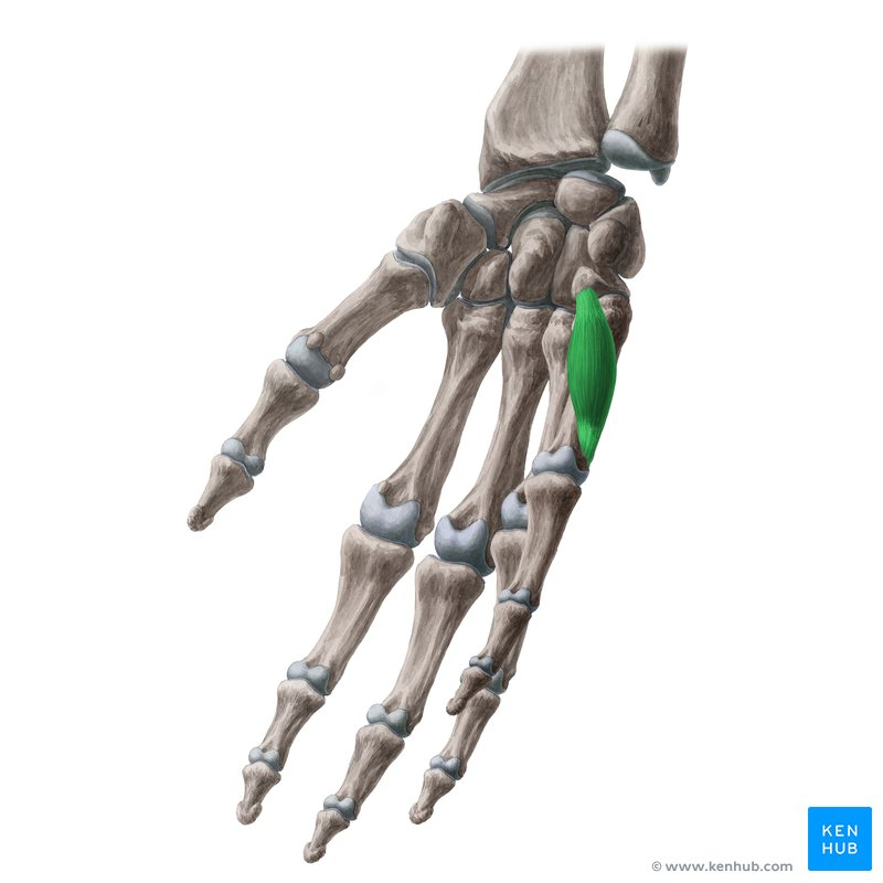
What Muscle?
Opponens Digiti Minimi
O: Hook of the hamate bone and the flexor retinaculum.
I: Medial (ulnar) margin of the entire shaft of the 5th metacarpal bone (little finger).
A: Flexes and laterally rotates the 5th metacarpal bone toward the palm, bringing the little finger into opposition with the thumb.
In: Deep branch of the ulnar nerve (C8, T1). [1, 2, 3, 4, 5]
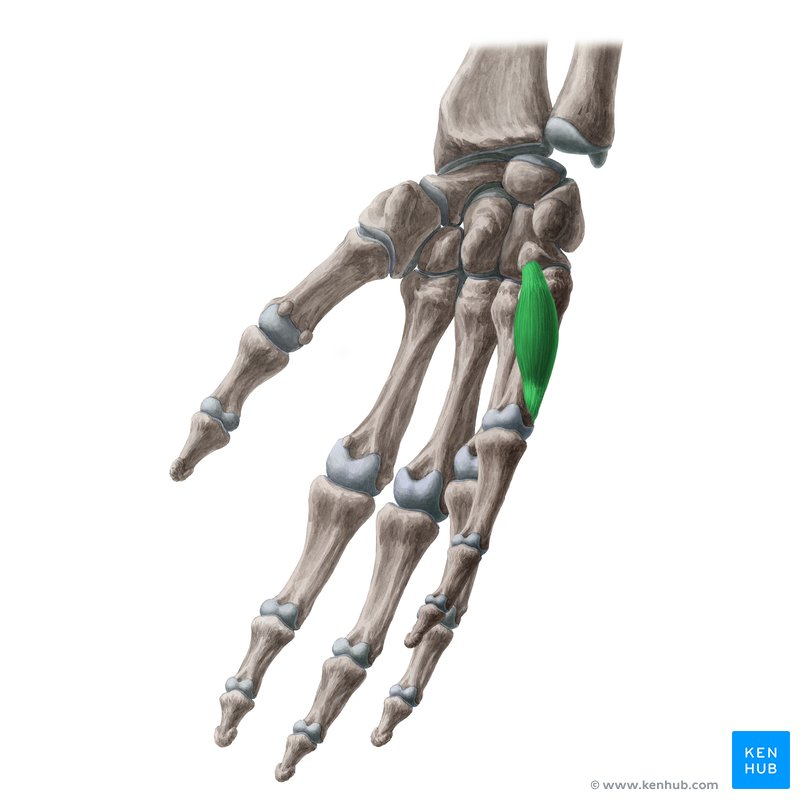
What Muscle?
Opponens Digiti Minimi
O: Hook of the hamate bone and the flexor retinaculum.
I: Medial (ulnar) margin of the entire shaft of the 5th metacarpal bone (little finger).
A: Flexes and laterally rotates the 5th metacarpal bone toward the palm, bringing the little finger into opposition with the thumb.
In: Deep branch of the ulnar nerve (C8, T1). [1, 2, 3, 4, 5]
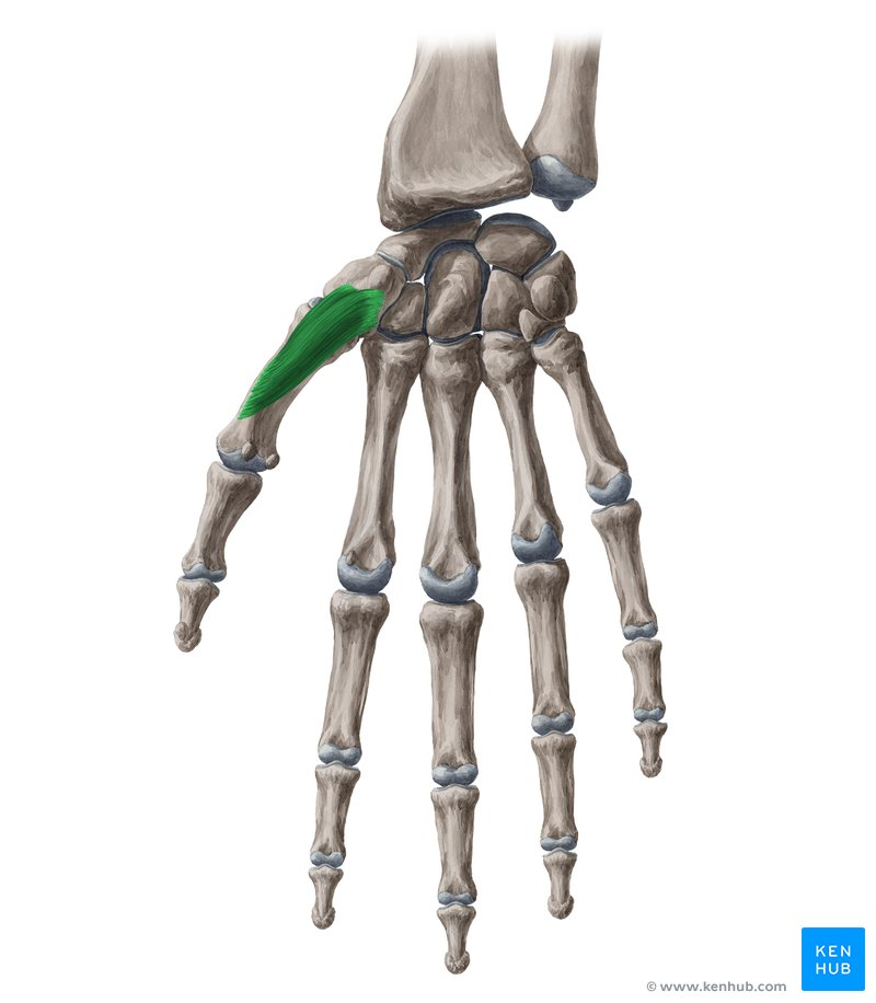
What Muscle?
Opponens Pollcis
O: Flexor retinaculum and the tubercle of the trapezium bone.
I: Lateral (radial) side of the entire shaft of the 1st metacarpal bone (thumb).
A: Opposes the thumb. It flexes and medially rotates the 1st metacarpal bone at the carpometacarpal (CMC) joint, bringing the pad of the thumb across the palm to face or touch the pads of the other digits.
In: Recurrent branch of the median nerve (C8, T1). [1, 2, 3, 4, 5]
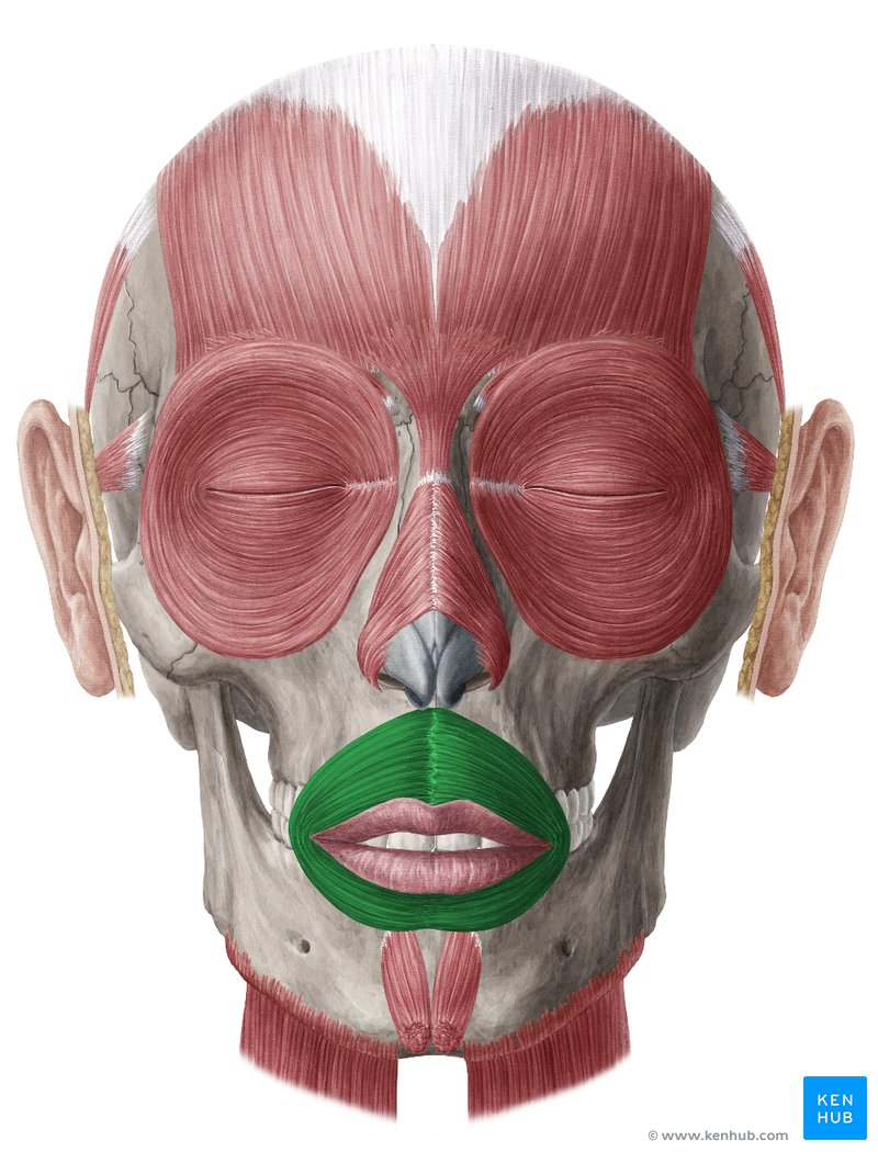
What Muscle?
Orbicualris Oris
O: Medial aspect of the maxilla and mandible, perioral muscles (such as the buccinator), and the deep surface of the skin around the lips.
I: Mucous membrane and skin of the lips, blending with fibers of other facial muscles at the corners of the mouth (the modiolus). [1, 2, 3, 4, 5]
A: Closes the oral fissure, compresses and protrudes the lips (as when kissing or whistling), and resists distension when blowing. [1, 2, 3]
In: Buccal and marginal mandibular branches of the facial nerve (CN VII). [1, 2]
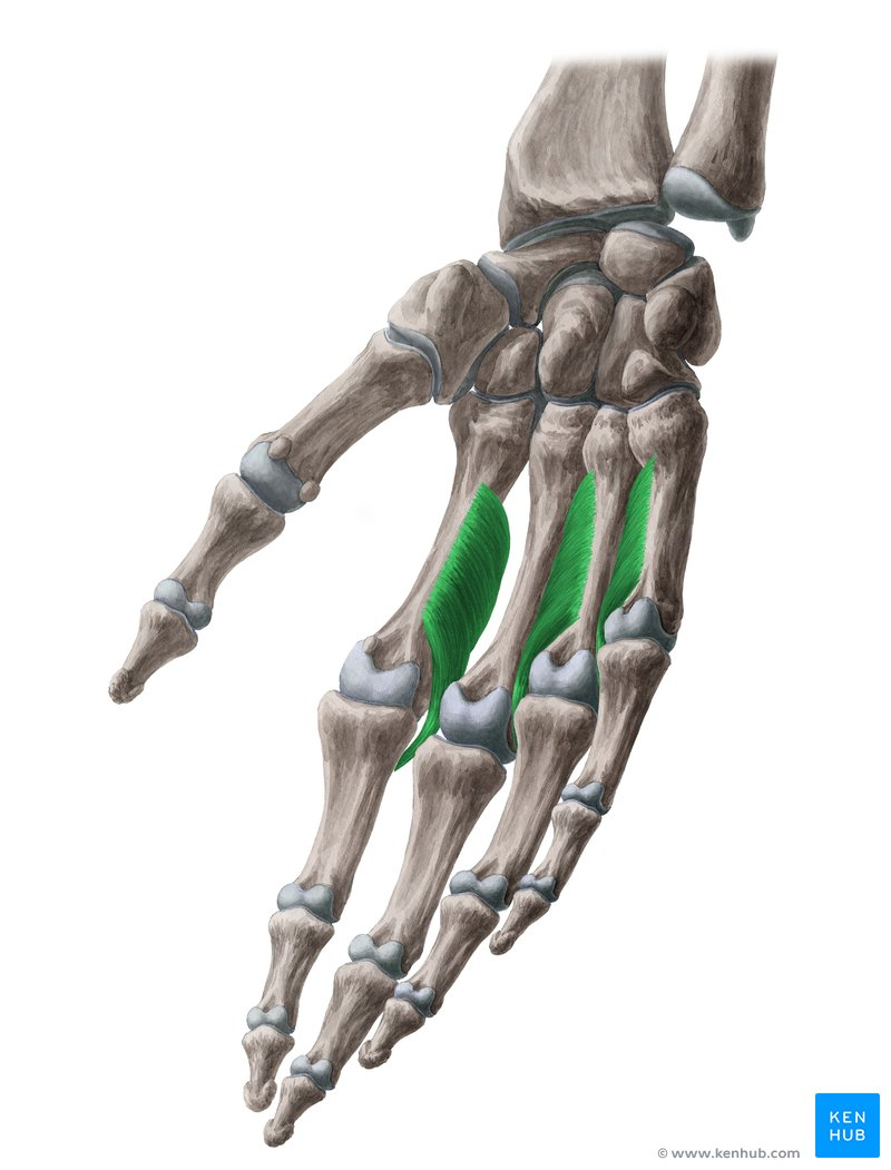
What Muscle?
Palmar Interossei
O: Palmar surfaces of the shafts of the 2nd, 4th, and 5th metacarpals. (If a 1st palmar interosseus is described, it arises from the base of the 1st metacarpal). [1, 2, 3, 4, 5]
I: Bases of the proximal phalanges and the extensor expansions of the respective digits (index, ring, and little fingers). [1, 2, 3, 4, 5]
A: Adducts the index, ring, and little fingers toward the middle finger (axial line of the hand). They also assist the lumbricals in flexing the metacarpophalangeal (MCP) joints and extending the interphalangeal (PIP and DIP) joints. [1, 2, 3, 4, 5]
In: Deep branch of the ulnar nerve (C8, T1). [1, 2]
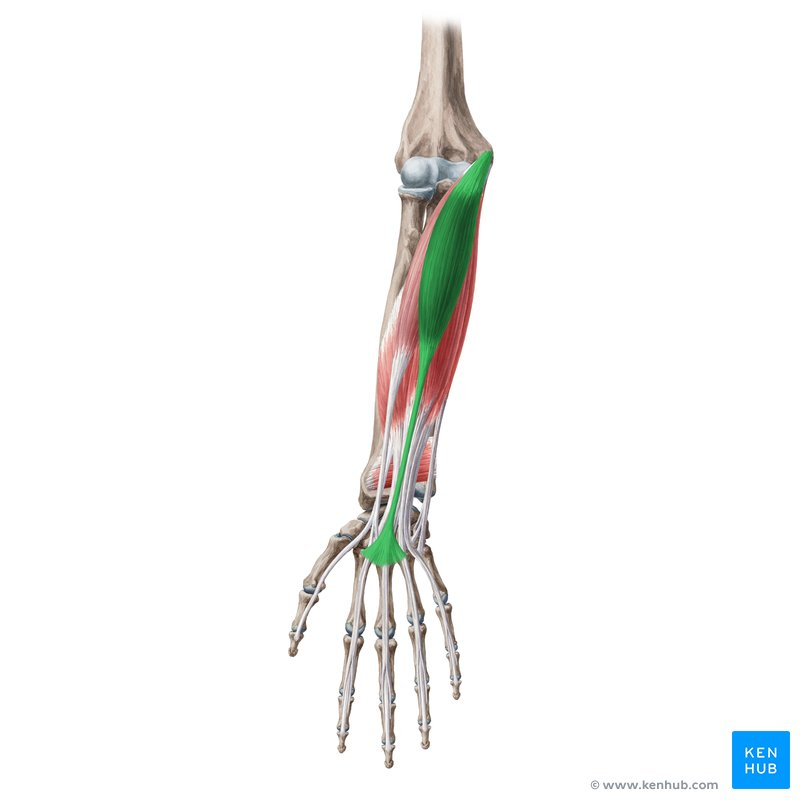
What Muscle?
Palmaris Longus
O: Medial epicondyle of the humerus via the common flexor tendon.
I: Flexor retinaculum and the palmar aponeurosis (the thick connective tissue layer of the palm).
A: Flexes the hand at the wrist joint and tenses the palmar aponeurosis.
In: Median nerve (C7, C8).
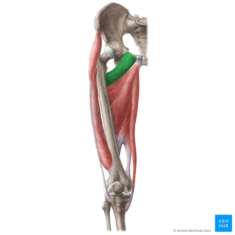
What Muscle?
Pectineus
O: Pectineal line (pecten pubis) on the superior ramus of the pubis. [1, 2]
I: Pectineal line of the femur, which runs from the lesser trochanter to the linea aspera. [1, 2, 3]
A: Adducts and flexes the thigh at the hip joint. It also assists with medial (internal) rotation of the thigh.
In: Dual nerve supply (primarily femoral, but can be mixed):
Primary: Femoral nerve (L2, L3).
Secondary/Variable: Obturator nerve (L2, L3) via its anterior branch in a portion of the population. [1, 2, 3, 4, 5]
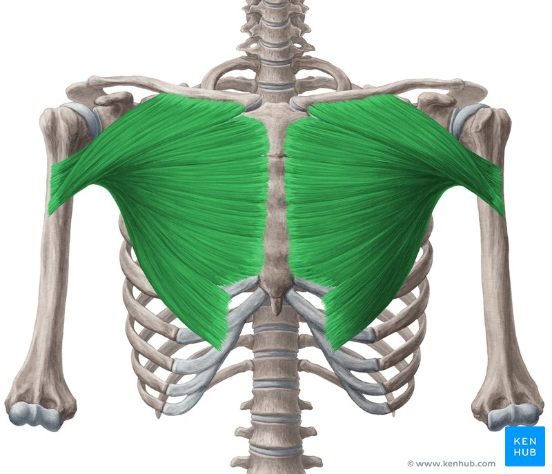
What Muscle?
Pectorialis Major
Clavicular+ Seternocostal Head
O: Clavicular head: Anterior surface of the medial half of the clavicle.
Sternocostal head: Anterior surface of the sternum, the superior six costal cartilages, and the aponeurosis of the external oblique muscle.
I: Lateral lip of the intertubercular sulcus (bicipital groove) of the humerus.
A: Unified actions: Adducts and medially (internally) rotates the arm at the glenohumeral (shoulder) joint.
Clavicular head alone: Flexes the extended arm.
Sternocostal head alone: Extends the flexed arm back down toward the trunk.
In:
Clavicular head: Lateral pectoral nerve (C5, C6, C7).
Sternocostal head: Medial pectoral nerve (C8, T1), along with some fibers from the lateral pectoral nerve.
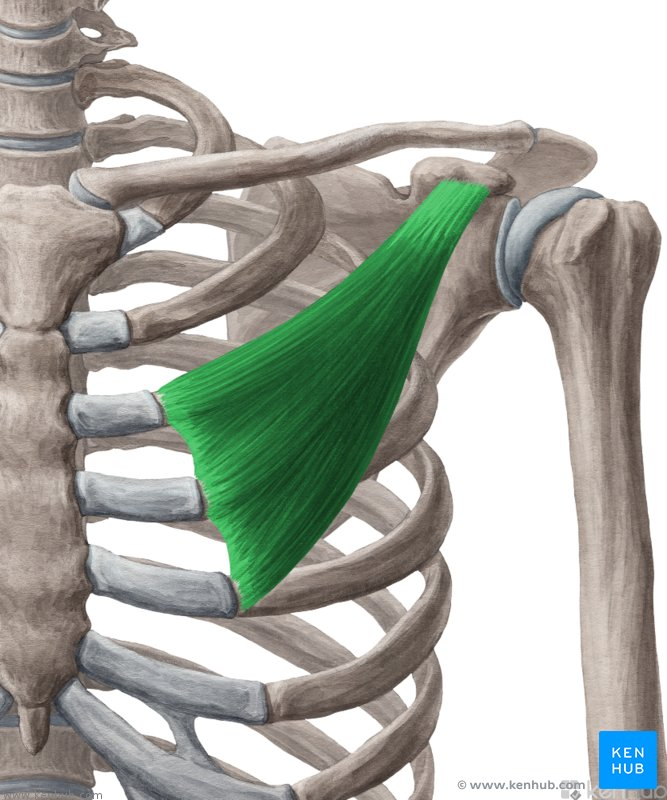
What Muscle?
Pectoralis Minor
O: Anterior surfaces of the 3rd, 4th, and 5th ribs near their costal cartilages.
I: Medial border and superior surface of the coracoid process of the scapula.
A: Stabilizes the scapula by drawing it inferiorly and anteriorly against the thoracic wall. It also rotates the scapula downward and can act as an accessory muscle of inspiration (elevating the ribs) when the scapula is fixed.
In: Medial pectoral nerve (C8, T1), which pierces through the muscle. [1, 2, 3, 4, 5]
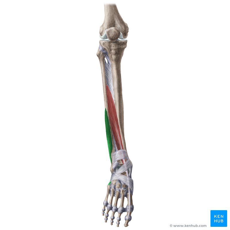
What Muscle?
Peroneus Brevis (Fibularis Brevis)
O: Inferior two-thirds of the lateral surface of the shaft of the fibula and the adjacent intermuscular septa.
I: Tuberosity on the lateral side of the base of the 5th metatarsal bone.
A: Everts the foot at the subtalar joint and weakly plantarflexes the foot at the ankle joint.
In: Superficial peroneal (fibularis) nerve (L5, S1). [1, 2, 3, 4, 5]
If you want, let me know:
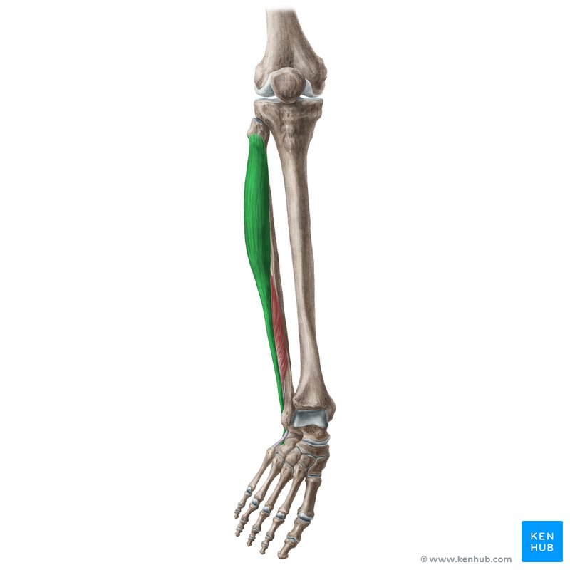
What Muscle?
Peroneus Longus (Fibularis Longus)
O: Head and superior two-thirds of the lateral surface of the fibula, as well as the adjacent intermuscular septa.
I: Lateral side of the base of the 1st metatarsal bone and the medial cuneiform bone.
A: Everts the foot at the subtalar joint and plantarflexes the foot at the ankle joint. It also plays a key role in supporting and stabilizing the transverse arch of the foot.
In: Superficial peroneal (fibularis) nerve (L5, S1, S2). [1, 2, 3, 4, 5]
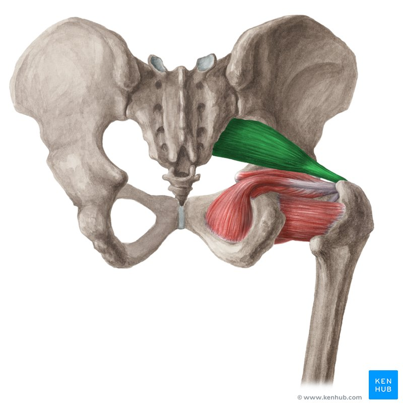
What Muscle?
Pririformis
O: Anterior surface of the sacrum (between the S2 and S4 anterior sacral foramina), gluteal surface of the ilium near the posterior inferior iliac spine, and the sacrotuberous ligament.
I: Superior border of the greater trochanter of the femur.
A: Laterally (externally) rotates the extended thigh at the hip joint and abducts the flexed thigh. It also helps stabilize the femoral head in the acetabulum.
In: Anterior rami of S1 and S2 spinal nerves (sometimes called the nerve to piriformis).
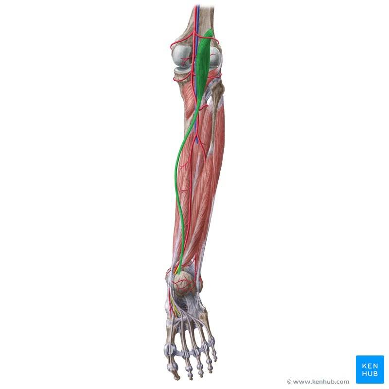
What Muscle?
Plantaris
O: Inferior end of the lateral supracondylar line of the femur and the oblique popliteal ligament.
I: Posterior surface of the calcaneus (heel bone), usually inserting just medial to the Achilles tendon.
A: Weakly assists the gastrocnemius in plantarflexing the foot at the ankle joint and flexing the leg at the knee joint.
In: Tibial nerve (S1, S2).
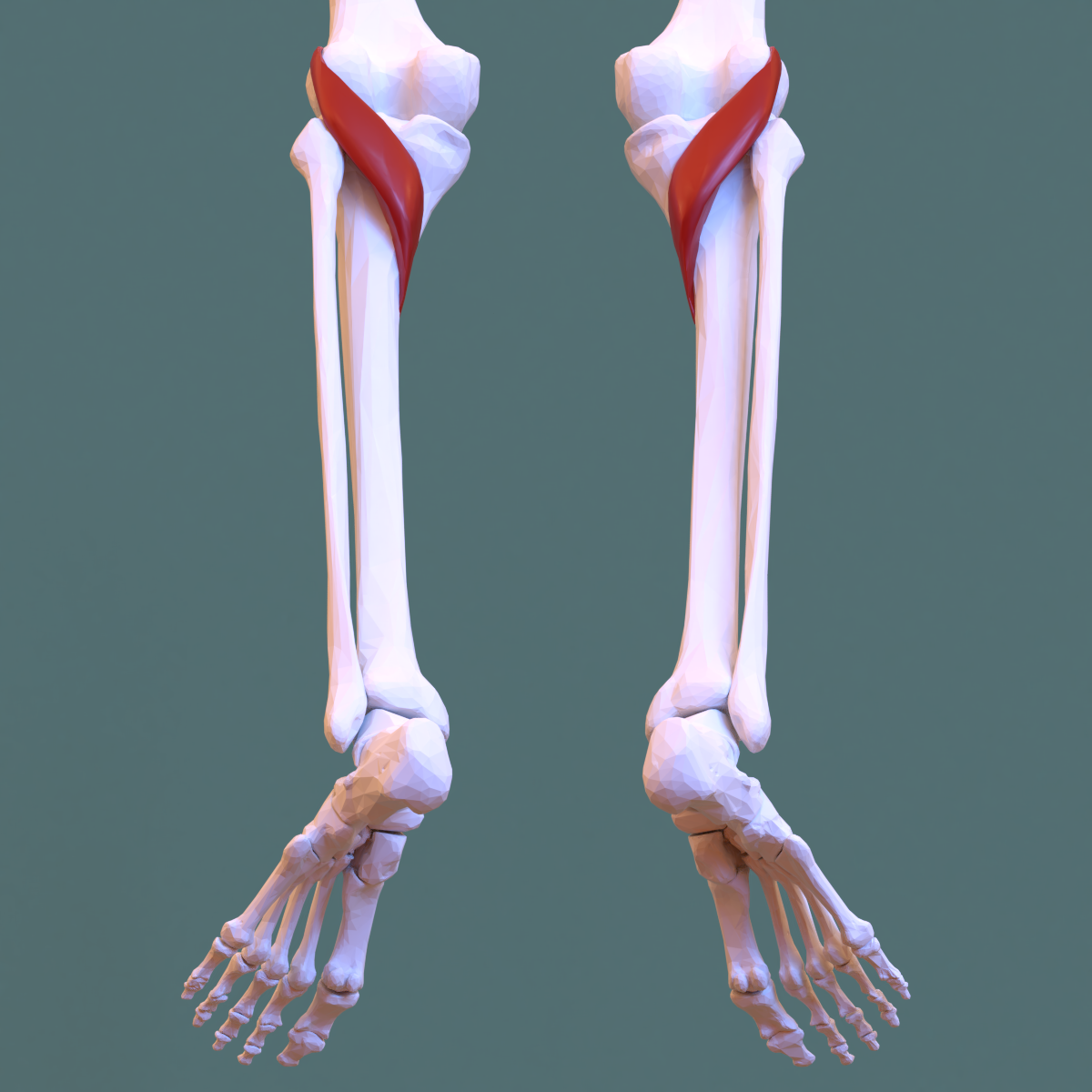
What Muscle?
Popliteus
O: Lateral surface of the lateral condyle of the femur and the posterior horn of the lateral meniscus.
I: Posterior surface of the shaft of the tibia, superior to the soleal line.
A: Flexes the leg at the knee joint and rotates it medially (internally). Crucially, when the leg is fully extended and bearing weight (screw-home mechanism), it laterally (externally) rotates the femur on the tibia to "unlock" the knee joint so flexion can begin.
In: Tibial nerve (L4, L5, S1). [1, 2, 3, 4, 5]
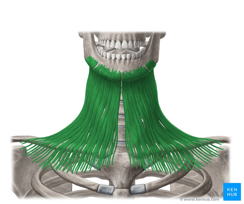
What Muscle?
Platysm
O: Subcutaneous tissue (fascia) covering the superior parts of the pectoralis major and deltoid muscles.
I: Base of the mandible, skin of the lower face, and the muscles around the corner of the mouth (such as the orbicularis oris and depressor anguli oris).
A: Depresses the mandible (against resistance) and tenses the skin of the lower face and neck (creating vertical wrinkles). It also draws the corners of the mouth inferiorly, expressing sadness, fright, or horror. [1, 2, 3, 4, 5]
In: Cervical branch of the facial nerve (CN VII)
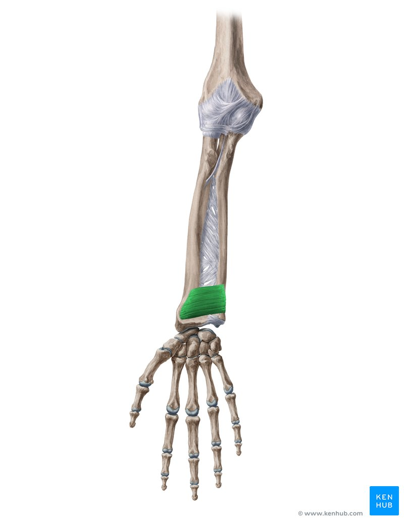
What Muscle?
Pronator Quadratus
O: Anterior surface of the distal fourth of the shaft of the ulna.
I: Anterior surface of the distal fourth of the shaft of the radius.
A: Pronates the forearm (rotates the radius medially over the ulna). It also helps bind the radius and ulna together to stabilize the distal radioulnar joint.
In: Anterior interosseous nerve, which is a branch of the median nerve (C8, T1). [1, 2, 3, 4, 5]
What Muscle?
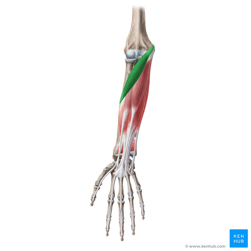
Pronator Teres
O: Humeral head: Medial epicondyle of the humerus via the common flexor tendon.
Ulnar head: Coronoid process of the ulna
I: Mid-lateral surface of the shaft of the radius (at the pronator tubercle). [1, 2]
A: Pronates the forearm (rotates the radius over the ulna) and weakly assists in flexing the forearm at the knee-like hinge of the elbow joint. [1, 2, 3]
In: Median nerve (C6, C7).
What Muscle?
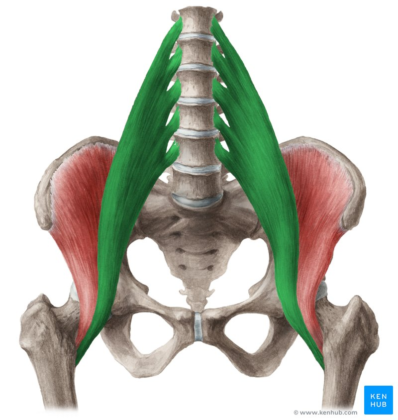
Psoas Major
O: Transverse processes of lumbar vertebrae (L1–L5), sides of the bodies of T12–L5 vertebrae, and their intervening intervertebral discs.
I: Lesser trochanter of the femur, via a unified tendon shared with the iliacus muscle (iliopsoas tendon)
A: Primary flexor of the thigh at the hip joint. When the femur is fixed, bilateral contraction flexes the trunk forward on the pelvis (as in a sit-up). It also assists with lateral flexion of the vertebral column when acting unilaterally.
In: Anterior (ventral) rami of lumbar spinal nerves (L1, L2, L3).
What Muscle?
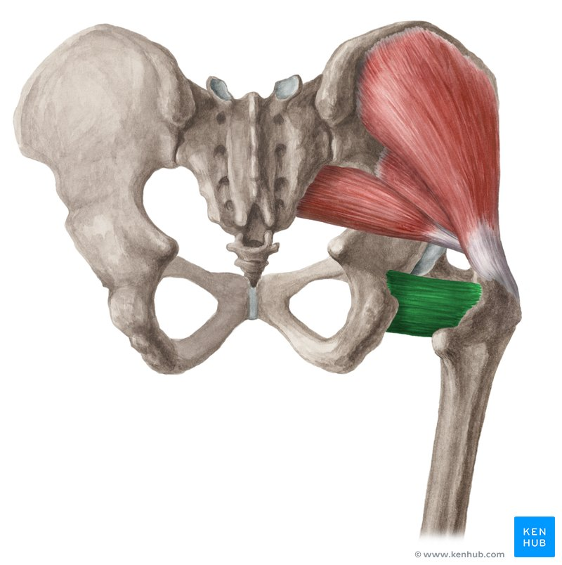
Quadratus Femoris
O: Lateral border of the ischial tuberosity.
I: Quadrate tubercle on the intertrochanteric crest of the femur.
A: Laterally (externally) rotates the thigh at the hip joint and assists with thigh adduction. It also stabilizes the head of the femur within the acetabulum.
In: Nerve to quadratus femoris (L5, S1). Note: This nerve also supplies the gemellus inferior muscle. [1, 2, 3, 4, 5]
What Muscle?
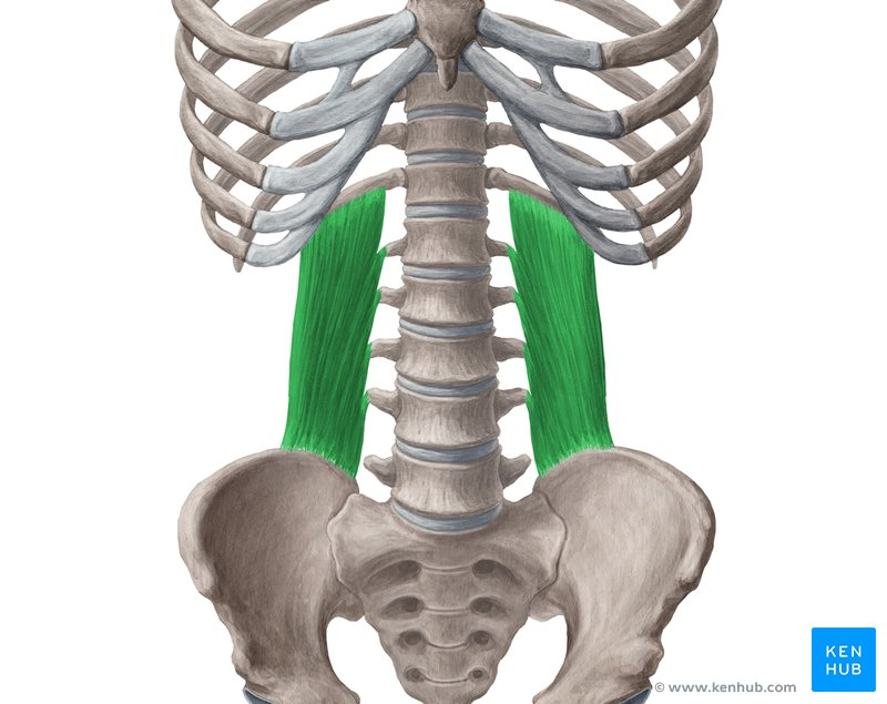
Quadratus Lumborum
O: Internal lip of the posterior iliac crest and the iliolumbar ligament.
I: Lower border of the 12th rib and the transverse processes of the upper four lumbar vertebrae (L1–L4).
A: Acting unilaterally, it laterally flexes (side-bends) the vertebral column to the same side. Acting bilaterally, it extends the lumbar spine and stabilizes the 12th rib during forced exhalation (fixing the rib for diaphragmatic contraction).
In: Anterior (ventral) rami of the T12 (subcostal) nerve and the upper three or four lumbar spinal nerves (L1–L4). [1, 2, 3, 4, 5]