(2) Cell injury, tissue adaptations and cell death
1/20
There's no tags or description
Looks like no tags are added yet.
Name | Mastery | Learn | Test | Matching | Spaced | Call with Kai |
|---|
No analytics yet
Send a link to your students to track their progress
21 Terms
Define homeostasis
maintaining balance and optimal environment in the body
What is a stressor?
Give examples.
(agent produce stress → disrupts homeostatis)
agent capable of producing stress → thereby disrupting homeostasis
1) change in pH
2) change in nutrient/O2 availablility, BF
3) increased/decreased hormone levels
4) change in ionic balance
5) accumulation of toxins
6) ionizing radiation
7) physical, thermal, chemical agents
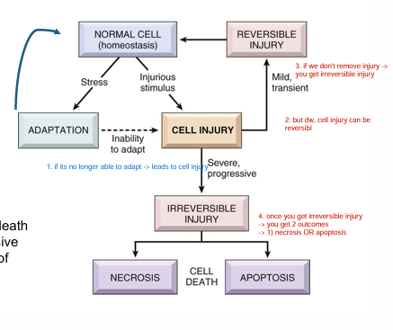
How may a body respond to stress?
TLDR: if you aren’t able to adapt
→ leads to irreversible injury/damage
→ when you hit irreversible damage
→ either get cell death (necrosis OR apoptosis)
Cellular adaptations are ___ in the __ of cells in response to changes in their environment
reversible changes
size, #, phenotype, metabolic activity, function
PNS MF
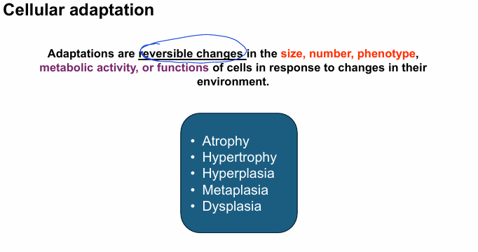
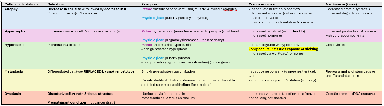
What are the different types of cellular adaptations?
endometrial hyperplasia = irregular thicker uterine lining (pathological; not normal)

Why might we say that metaplasia is a double edged sword?
Replacement of differentiated cell into another cell type
→ change in morphology
→ functional change
→ may be more prone to malignance/infection
Ex. Smoker
→ normal epithelium secrete mucus which traps chemicals
→ but as you smoke the tract gets irritated and body replaces with more resilient cells
→ these resilient cells don’t have cilia/mucous production so pathogens can easily break squamous cells
→ susceptible to infection.
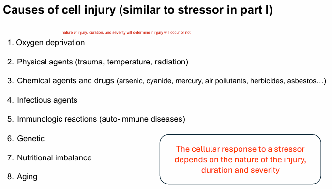
What are some causes of cell injury?
1) O2 deprivation
2) physical agents (temperature, trauma, radiation)
3) infectious agents
4) genetic
5) nutritional imbalance
6) aging

The cellular response to a stressor depends on __
nature of injury
duration
severity
(these 3 will determine if an injury will occur or not)
Differentiate b/w REVERSIBLE & IRREVERSIBLE injuries
Reversible = remove stressor → eliminate functional/morphological changes that occured
Irreversible = can’t adapt to stressor → leads to irreversible damage → cell death occurs
What are the hallmarks for REVERSIBLE injury?
1. Cell swelling/morphological changes
2. Intracellular accumulation
3. Decreased oxidative phosphorylation (LOW ATP)
4. Mitochondrial dysfunction (early)
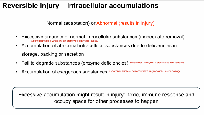
(LO) Explain intracellular accumulation.
How does it occur?
How does it occur:
1) excessive normal intracellular substance (inadequate removal) - were not removing it fast enough
2) accumulation of abnormal intracellular substance b/c of deficiencies in storage/packing/secretion
3) failing to degrade substances (enzyme deficiency)
4) accumulation of exogenous substance
ex. inhaling smoke → accumulate cytoplasm → cause damage
TLDR: so by removing these “stressors” (ex. too much intracellular substance), cell can revert
*normal = adaptation
*abnormal = results in injury
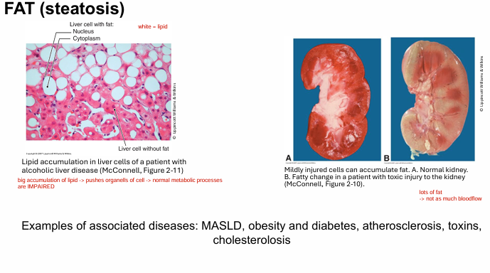
What’s an example of intracellular accumulation?
FAT (steatosis)
obsesity/diabetes
atheroscleorsis
Fat accumulation → pushes organelles → normal metabolic processes impaired
lots of fat, not a lot of blood flow to these organs (therefore no O2/nutrients)
Other substances that accumulate:
glycogen, proteins
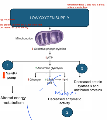
1) Hypoxia is the __.
2) Explain how hypoxia causes injury.
1) Most common cause of cell injury
2) Low O2
→ lack of aerobic resp
→ low ATP
→ 3 parts: (EXAM)
1) decreased Na/K+ pump
→ alters energy metabolism
2) Increased anaerobic glycolysis
→ increases lactic acid
→ decrease pH
→ decrease enzymatic activity
3) decreased protein synthesis & misfolded proteins
prolonged hypoxia = cell death
(LO)
1) What is ischemia? Why is it relevant?
2) How is it different from hypoxia?
*Note: hypoxia = loss of oxygen
1) Ischemia = less BF to area (so loss of O2 AND nutrients due to obstruction)
2) damage is more severe & faster than hypoxia
-b/c NO NUTRIENTS → not glucose → can’t do anaerobic glycolysis (so we get no #2 from #1-3)
How can reperfusion cause injury?
*Note: Reperfusion used to fix ischemia (remove blockage)
Abrupt sudden increase in O2 in area w/ low O2
→ large # of free radicals (extremely unstable reactive molecules) (ROS)
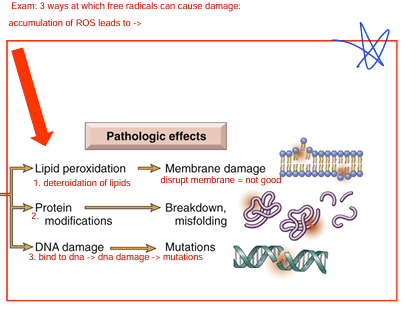
How free radicals cause damage (exam)
Accumulation of ROS leads to:
1) Lipid peroxidation (deteriorate lipids) = membrane damage
2) protein modification = breakdown/misfolding
3) DNA damage (ROS bind to dna) = mutations
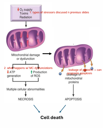
Discuss MC dysfunction
Once exposure to stressors become chronic, __ becomes _ and cell death occurs
1) depletion of ATP → alters cellular metabolism → tissue necrosis
2) ROS production → tissue necrosis
→ lipid peroxi
→ protein modification
→ DNA damage
3) leakage of proteins
→ proteins leaking → programs apoptosis
MC damage, irreversible
Once exposure to stressors become chronic
mitochondria damage becomes irreversible, and
cell death occurs

(LO)
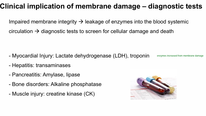
Identify the events leading to the impairments of cell membrane (lipid membrane) integrity
Main causes:
1) Ischemia/hypoxia
- no ATP
- activate phospholipase (breakdown lipids) (affects the lipid membrane)
- activate protease (breakdown proteins) (no proteins mean no rebuilding)
2) bacteria/viral toxins
3) ROS (lipid peroxidation)
- deteriorating lipids
- too much ROS accumulation → damage lysosome → release enzymes for cell death
4) decreased phospholipid synthesis (MC dysfunction, lipid peroxidation)
- not producing enough lipids to reproduce lipid membrane
Dunno if you need to know..
Compare and contrast cell death (apoptosis vs. necrosis)
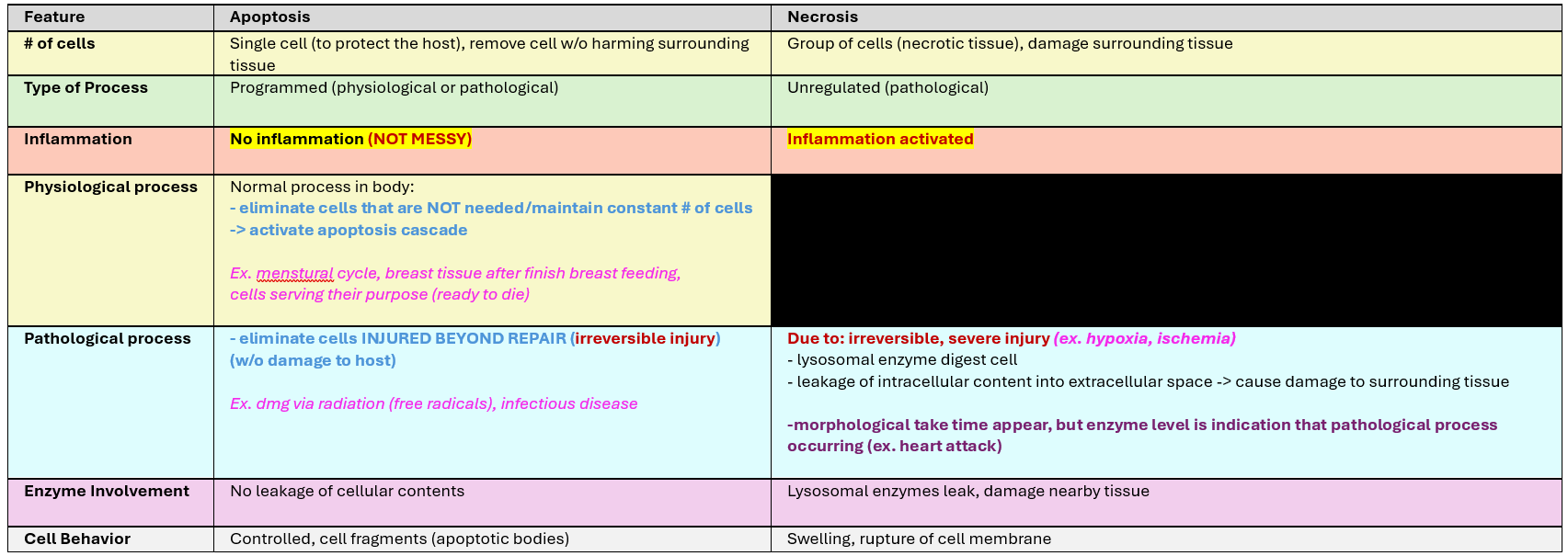
What are the different types of necrosis
1) Coagulative = architecture of dead tissue preserved for few days
- most common necrosis
- hypoxic injury → lack of ATP
- ischemia (lack of blood flow)
- seen in tissue infract (brain exception)
2) Liquefactive
- enzymes digest tissue in brain
3) Caseous (combination of liquefactvie and coagulative)
- crumbled cheese
- seen in TB
- associated w/ chornic inflammation + granulomas
- lung
4) Fat necrosis
- fat cells digested
- pancreas
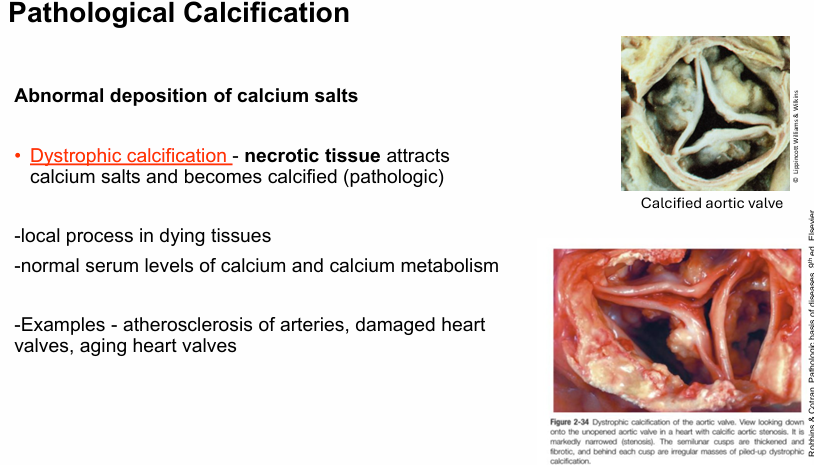
What is pathological calcification?
abnormal deposition of calcium salts
dystrophic calcification: necrotic tissue attracts calcium salts and become calcified
(Jerry, an 80-year-old, has large calcium deposits in his lung tissue secondary to Tuberculosis and caseous necrosis. Jerry’s serum calcium levels remain normal. This form of substance accumulation in the tissues is best described as)
Ex. atherosclerosis, damaged heart valves, aging heart valves