Exam 2: Postsynaptic Potentials
1/16
There's no tags or description
Looks like no tags are added yet.
Name | Mastery | Learn | Test | Matching | Spaced | Call with Kai |
|---|
No analytics yet
Send a link to your students to track their progress
17 Terms
How does the membrane get depolarized?
When a particular neurotransmitter binds to postsynaptic receptors, the ion channels open up to a specific type of ion and let them through which changes the membrane potential.
What happens when glutamate binds to receptors on the postsynaptic membrane?
An excitatory neurotransmitter (glutamate) opens up Na+ channels, resulting Na+ influx into the cell, which depolarizes the cell (not that sodium is positive, so its influx decreases the cell’s negative resting potential, making the inside a bit more positive). The cell is depolarized and is more likely to fire (i.e. when it reaches a threshold of, say, -55mV).
What ions can enter through the membrane?
Na+ (sodium)
K+ (potassium)
Ca2+ (calcium)
Cl- (chloride)
What are EPSPs?
Excitatory postsynaptic potentials; a temporary change in a neuron’s electrical potential that make it more likely to fire an action potential
What is the approximate voltage of a depolarized patch of the membrane?
About -55mV
What happens to the cell membrane voltage?
Hyperpolarization - IPSPs (inhibitory postsynaptic potentials) are generated when an inhibitory neurotransmitter is released into the synapse (such as GABA). GABA opens chloride channels, which results in the influx of Cl-. The membrane potential becomes more damaging than the resting potential and the cell is not hyperpolarized and is less likely to fire.
What are the main characteristics of postsynaptic potentials? (aka? location? dependent on? types of summation? speed? initiates or impedes?)
Postsynaptic potentials are known as graded potentials (because they vary in amplitude: they reflect the strength of the input; they can be depolarizing and hyperpolarizing).
They reflect the summation of postsynaptic potentials on the dendrites and the soma; they are local, decrease with distance from the source, and weaken with time.
They depend on the strength of the stimulus (i.e. more neurotransmitters will cause stronger postsynaptic potentials, or a louder sound will cause stronger postsynaptic potentials).
The summation of postsynaptic potentials can be: temporal - if they overlap in time; and spatial - if they are close in space. And both of these increase the chance of firing (reaching the firing threshold)
Important: Postsynaptic potentials caused by a single input is weak, so the summation is needed to reach the threshold: if these postsynaptic potentials overlap in time and space, if they are primarily excitatory, they will depolarize the membrane, build on each other and can trigger an action potential at the axon hillock that will travel down the axon (ie the neuron will fire)
Postsynaptic potentials (PSPs) are relatively SLOW (tens of milliseconds), which makes it possible for them to overlap in time and summate
Can be excitatory or inhibitory
What is an action potential (ie “cell firing”)?
A rapid, all-or-nothing electrical impulse that travels down a neuron’s axon to transmit signals
How do action potentials happen? What is the necessary criterion? Where is it generated? How fast does it move down the axon?
If the cell is sufficiently depolarized (i.e. if the summated EPSP reaches the threshold, the negative potential is decreased sufficiently), it fires. If the threshold is not reached, there is no action potential.
An action potential is generated at the axon hillock.
The action potential moves down the axon very FAST (1-2 ms). Myelinated axons with regions without myelin (nodes of Ranvier), the action potential is regenerated at each node.

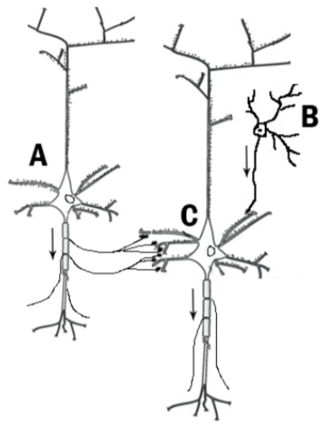
Can you recognize all the cell types? How are they connected? Which cells receive excitatory vs inhibitory input? Which cell(s) are more likely to fire?
Can you recognize all the cell types? A and C are pyramidal cells. B is an inhibitory cell
How are they “connected”? Through complex circuits that include feedback inhibition, direct excitation, and reciprocal connections
Which cells receive excitatory vs inhibitory input? Both pyramidal and inhibitory cells receive both types of input. Pyramidal cells are the main excitatory neurons. Inhibitory cells primarily receive excitatory input from other neurons.
Which cell(s) are more likely to fire? Pyramidal cells (A and C)
What type of potentials do we record as EEG?
Summated postsynaptic potentials
Note that currents flow through the brain tissue, the cerebrospinal fluid, the skull, and the skin and are recorded with EEG electrodes. So, what are these currents like outside of the cell?
EPSP (Na+ influx into the cell) cause positive ions to enter the cell, which leaves a deficit of positive charges and a relatively negative potential outside the cell
What is the current sink and the current source?
Current sink - area with negative charges (where Na+ flows into the cell); when glutamate is released at the synapse, it binds with Na+ channels, which open and let Na+ ions flow into the cell; results in negative external potential; this is an ACTIVE process caused by synaptic events
Current source - area with positive charges (currents flow from source to sink) (in the electric field, positive charges always flow toward negative charges); as a result of the sink, there is relative negativity (a deficit of positive ions) outside the cell, which attracts other positive ions in the vicinity; represents the source of positive ions
What happens outside the cell during EPSPs, and where do these currents flow?
From the source toward the sink; this flow of charged ions between the sink and the source creates an electrical current
What happens during IPSPs outside the cell?
IPSPs (at GABA synapses) cause an influx of Cl- ions into the cell (which are negative), resulting in an area of positive charges outside the cell membrane
How can we model these currents with dipoles?
The sink and the source are 2 poles of a DIPOLE (their positive/negative imbalance cases ionic charges to move within the extracellular and intracellular fluid)
Especially relevant are these extracellular currents that propagate through the head volume in all directions (in 3D); they are conducted by the brain tissue, especially by the cerebrospinal fluid, less well by the bone, but they reach the scalp, where they are measured with EEG electrodes
These ionic currents are generated by summated postsynaptic potentials along the apical dendrites of numerous highly synchronized pyramidal cells. A dipole model can approximate them at a distance (meaning away from the individual cells)
The current flow through the extracellular space is measured as voltage (EEG) on the scalp. Important: voltage is a relative measure (you cannot measure EEG with a single electrode)
The voltage will be positive or negative depending on the dipole orientation. This means that if the sink is closer to the surface of the head, the measured voltage will be negative.
Why is EEG generated by postsynaptic potentials and not action potentials?
Action potentials are very fast (1-2ms). They are not synchronized in time or space (not aligned). In contrast, graded potentials are slow-ish (tens of ms, and they can summate).