Clinical Monitoring Cardiac - Exam 1
 Call Kai
 Learn
Learn Practice Test
 Spaced Repetition
 Match
Match Flashcards
 Knowt Play
1/73

There's no tags or description
Looks like no tags are added yet.
Name | Mastery | Learn | Test | Matching | Spaced | Call with Kai | Chat |
|---|
No analytics yet
Send a link to your students to track their progress
74 Terms
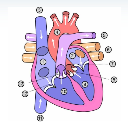
Label 1-13
1- Right atrium
2- Left atrium
3- Superior Vena Cava
4- Aorta
5- Pulmonary artery
6- Pulmonary veins
7- Mitral valve
8- Aortic Valve
9- Left ventricle
10- Right ventricle
11- Inferior vena cava
12- Tricuspid valve
13- Pulmonary valve
What are the chambers of the heart
Right and left atrium
Right and left ventricle
Which are semilunar valves
pulmonary and aortic valves
Which are AV valves
Atrioventricular valves
tricuspid
mitral
Structure of tricuspid valve
3 leaflets
normal size 4-6cm²
Mitral valve structure
2 leaflets
normal size= 4-5 cm²
Chordae Tendineae
fibrous cords that connect edges of valve to papillary muscle
Papillary muscle
muscular projections that arise from the inner surface of the ventricles
semilunar valves structure
three half-moon shaped leaflets attached to a fibrous ring
normal size: 3-4 cm²
what constitutes aortic stenosis
<2.0 cm²
Coronary Ostia
small opening of coronary artery
Fibrous Pericardium
outermost, tough, dense connective tissue
Anchors the heart to the great vessels, diaphragm, and sternum
Helps to limit excessive cardiac distention
Serous Pericardium
thin, smooth membrane - made up of two layers
Parietal layer
lines and is fused to the inner surface of the fibrous pericardium
Visceral layer
also called the epicardium; directly covers the heart
pericardial cavity/sac
lies between the parietal and visceral layers; contains a small amount of fluid, reduces friction
Myocardium
muscular, middle layer of the heart → has both contractile and conduction properties
Endocardium
serous membrane that lines the inner surface of the heart and extends out to form the heart valves → similar to endothelial cells that line blood vessels
What is a sacromere
basic functional contractile unit of cardiac muscle
contains actin and myosin filaments
during contraction actin and myosin slide past one another → shortening the sarcomere and generating tension
Why is calcium important for contraction
Increase in intracellular Ca2+ initiates contraction
Binds to troponin which causes shape change and moves tropomyosin
Myosin binding site open for Actin-Myosin crossbridge
More intracellular calcium = longer/stronger contraction
What is the pulmonary capillary wedge pressure?
It is an estimate of left atrial pressure
Obtained by inflating a small balloon at the tip of a catheter placed in a branch of the pulmonary artery
Right atrium pressure
0-4 mmHg
Right ventricle pressure
25 systolic; 4 diastolic
Normal Pulmonary artery pressure
25 systolic, 10 diastolic
Left atrium pressure
4-12 mmHg
Left ventricle pressure
120 systolic, 10 diastolic
Aortic pressure
120 systolic, 80 diastolic
What are the 5 major steps of the cardiac cycle
isovolumetric relaxation
ventricular filling
arterial systole
isovolumetric ventricular contraction
ventricular ejection
Identify the nodes of the cardiac conduction system
a. “Some Believe In Acting Badly Before Performing”
i. Sinoatrial (SA) Node → upper part of RA, pacemaker
ii. Bachmann Bundles → inner wall of LA, atrial conduction system
iii. Intermodal Pathway → atrial wall, SA node – AV node
iv. Atrioventricular Node → AV node delay, allows atria to empty/finish contraction before stimulating ventricles
v. Bundle of His → passes through ventricular septum, transmits signals from AV node to ventricles
vi. Right and Left bundle branches
vii. Purkinje Fibers → provide synchronized contractions of the ventricles
How to find Cardiac Output + normal value
heart rate x stroke volume; normal value = 4-8 L/min
stroke volume
= End-diastolic volume - end-systolic volume
determined by
preload - ventricular stretch
contractility - force of myocardial contraction
afterload - resistance/pressure must overcome
Frank Starling Law
as stroke volume increases as the volume of ventricular filling (EDV) increases
stretch of the myocardium, lining up of sarcomere, must be optimal alignment/overlap for proper contraction and stroke volume
Centric Hypertrophy
Thickening of the myocardial wall; increase in the number of muscle layers
occurs because of increased workload → need to push/contract against persistently high resistance
Eccentric (ventricular) hypertrophy
Dilation of heart chamber → widens/lengthens
Increased blood volume - stretch → increased wall tension
Atrial Natriuretic Peptide
released from atria; stimulated by atrial stretch or increased volume
promotes natriuresis and diuresis → offload of excess volume
vasodilation
Decreases blood volume, preload, and vascular resistance
B-type Natriuretic Peptide
released by the ventricular myocardium, stimulated by ventricular wall stretch and increased filling pressure
BNP and NT-proBNP are biomarkers for cardiac wall stress
Decreases blood volume, preload, and vascular resistance
Nervous system control of myocardium
Sympathetic NS = flight or fight
norepinephrine and epinephrin stimulate Beta 1 adrenergic receptors
increase HR, AV conduction, myocardial contractility
increased CO to meet metabolic need
Parasympathetic NS = rest and digest
medicated by vagus nerve
Acetylcholine stimulates M2 muscarinic receptors
decreased SA node firing → decrease HR
decrease AV nodal conduction
Arterial Pressure Waveform
a. As the arterial pressure wave transmits further away from the heart → systolic pressure increases and diastolic pressure decreases
i. Increases pulse pressure
ii. MAP declines
b.Systolic upstroke = sharp, rapid rise in pressure caused by left ventricular ejection
c. Dicrotic Notch = downward deflection/notch on the downstroke signifying aortic valve closure and the end of systole
How is CO to each organ determined
Demand, function, and metabolic needs of organs
Resistance across organs → individual vascular resistance makes it easier/harder to perfuse and exchange oxygen
What percentage of C.O. goes to the lungs
100% of CO from right heart
What percentage of CO goes to the digestive system
21%
What percentage of CO goes to the Kidneys
20%
What percentage of CO goes to the Skeletal muscle
15%
What percentage of CO goes to the brain
13%
What percentage of CO goes to the skin
9%
What percentage of CO goes to the liver
6%
What percentage of CO goes to the bone
5%
What percentage of CO goes to the heart muscle (coronary circulation)
3%
Right Coronary Artery pathway
travels between right atrium and ventricle
Acute Marginal branches = supply the right ventricular wall
Posterior descending artery, if dominant
Left main coronary artery
Short vessel from the left coronary sinus and ascending aorta
left anterior descending
Diagonal branches - anterolateral LV
Septal perforators - anterior interventricular septum
Left Circumflex
Obtuse marginal branches
Ramous
Coronary dominance
Determined by which coronary artery gives rise to the posterior descending artery
85% right
8-10% left
5-10% codominant
Average diameter of coronary artery
3-5 mm
Law of Laplace
larger the vessel radius, larger wall tension required to withstand a given internal fluid pressure
as radius increases vessels must generate more force to create the same internal pressure
Pascal’s Principle
pressure is transmitted equally in an enclosed static fluid
push on a fluid anywhere in a enclosed system → pressure spreads evenly throughout system
Poiseuille’s principle
Flow is determined by the change in pressure, resistance, and the radius of the vessel to the fourth power
The radius has a very strong influence on the flow
small changes to the radius has huge change to flow
double radius = increase flow by 16x
What governs fluid movement across a capillary membrane
Hydrostatic pressure - physical pressure exerted by capillaries → pushes fluid out of the capillary into the interstitium
- going in high, 25-30 mmHg
- going out low, 10-15 mmHgColloid Osmotic Pressure - attraction of protein molecules for water across membrane
- proteins (75% albumin) act like a magnet pulling water back in
At the atrial end, high hydrostatic pressure pushes fluid out → venous end hydrostatic pressure drops COP allowed to pull more in
Syncope
Brief loss of consciousness caused by temporary cerebral hypoperfusion
low CO, pulmonary embolism, arrhythmias, acute myocardial ischemia
Presyncope
lightheadedness or feeling faint
lower O2 delivery - decreased energy
Palpitations
conscious awareness of one’s heartbeat
Bradycardia
slowed heart rate, below 60 BPM
tachycardia
fast heart rate, higher than 100 BPM
Stage 1 hypertension
systolic 130-139; diastolic 80-90
Stage 2 hypertension
systolic >140; diastolic >90
Stage 3 hypertension
systolic >180; diastolic >120
Arterial Pressure Measure Sites
adult cardiac surgery: radial or femoral preferred
pediatric cardiac surgery: dorsalis pedis
carotid, branchial
Pulsus Alternans
regular alternation between strong and weak pulse amplitudes
alternating stroke volume, may occur with LV dysfunction
Pulsus Paradoxus
Abnormal drop in systolic blood pressure during inspiration
drop >10 mmHg
may occur with cardiac tamponade, severe obstructive lung disease
Auscultation
Listening to heart sounds, closure of heart valves
S1 = closure of mitral and tricuspid valves → isovolumetric contraction
S2 - closure of aortic and pulmonic valves → isovolumetric relaxation
Systolic murmur
when heart muscle contracts between S1 and S2
Diastolic murmur
when heart muscle relaxes between beats, after S2 and before S1
Cardiac Stress test: how, purpose
uses a treadmill/exercise to put the heart under stress and measures heart rate/rhythm via EKG or ECHO
can be used to determine workload, blood flow, oxygenation, etc
Cardiac stress test for non-ambulatory patients
Give patients drugs that make the heart behave as if it’s been exercising
regadenoson/lexiscan
dobutamine
adenosine
Why wouldn’t we perform a stress test
if patient is unstable
acute MI
severe hypertension
unstable angina
severe aortic stenosis
heart failure
Coronary angiography/cardiac catheterization
invasive procedure that allows direct visualization of coronary anatomy
presence and severity of coronary stenosis
coronary blood flow
intracardiac pressure
hemodynamic
possible interventions