BIOS242 Microbiology - Exam 1 Flashcards
 Call Kai
 Learn
Learn Practice Test
 Spaced Repetition
 Match
Match Flashcards
 Knowt Play
1/166

There's no tags or description
Looks like no tags are added yet.
Name | Mastery | Learn | Test | Matching | Spaced | Call with Kai | Chat |
|---|
No analytics yet
Send a link to your students to track their progress
167 Terms
Medical Microbiology
Deals with microbes that cause diseases in humans and animals
Example: Looking at pathogens, their transmission, diagnosis, and treatment.
Agricultural Microbiology
Relationships between microbes, farm animals, and crops
Example: what microbes can help grow or what is harmful to crops of farm animals
Public Health Microbiology and Epidemiology
Monitor and control the spread of diseases in communities
Example: the flu season, and whether it has spread through a local community, a county, a state, or multiple states (epidemic, pandemic, etc)
Immunology Microbiology
Investigates protective substances and cells produced in response to infection
Example: looking at the human body response to pathogens
Industrial Microbiology
Looks at safeguarding our food and water supplies from microbial contamination and ensuring safety standards in production
Example: taking samples of beef and investigating for unacceptable E. coli levels, along with looking at wastewater treatment plants, whether it’s safe to release into the community or back into rivers/lakes
Environmental Microbiology
Studies the effects of microbes on the Earth’s diverse habitats (aquatic, soil, geomicrobiology, astrobiology)
Example: Microorganisms that could be in space
Medical Microbiology Notes and Example
Focuses on microorganisms responsible for illnesses in humans and animals
Scientists investigate the elements contributing to the virulence of these microorganisms and explore mechanisms to counteract their harmful effects
Example: A laboratory scientist examines a sample in a biotech lab focused on the creation of new vaccines.
Public Health Microbiology and Epidemiology Notes and Example
Oversees and manages the dissemination of diseases within societies
Entities engaged in these efforts include the CDC at the national level and the WHO at the international level
Example: A doctor tests a patient for a virus during a pandemic
Immunology Notes and Example
Delves into the intricate network of defensive elements and cells generated as a reaction to infections
Encompasses various domains, including immunization, blood, analysis, and allergic reactions
Immunologists explore the immune system’s involvement in conditions like cancer and autoimmune disorders
Example: A laboratory assistant prepares a blood sample for research
Industrial Microbiology Notes and Example
Ensures the safety of the food and water supply
Uses biotechnology, which harnesses microbial metabolism to produce a wide array of desired products, such as bread, biofuels, and gene therapy
Microorganisms are harnessed to yield significant quantities of various substances like amino acids, alcohol, medications, enzymes, and vitamins
Example: Biologists at a wastewater treatment plant check for microbes present in the water supply
Agricultural Microbiology Notes and Example
Revolves around the interactions between microorganisms and cultivated plants, and domesticated animals
Within the realm of plants, experts concentrate on issues like plant diseases, soil fertility, and nutritional dynamics
Specialists in animal-related aspects delve into infectious diseases and other symbiotic relationships animals establish with microorganisms
Example: Veterinarians conduct a routine check on livestock
Environmental Microbiology Notes and Example
Investigates how microorganisms inhabit the varied ecosystems of our planet
Various subdisciplines focus on specific environments and even outer space
Example: Biologists collect water samples to take back to the lab for further investigation
Louie Pasteur
Invented pasteurization
Developed the germ theory of disease (microorganisms are responsible for diseases)
Eventually developed a vaccine for rabies
Disproved the theory of spontaneous generation
Discovered yeast cells produce alcohol
Robert Hooke
Notable for his work in microbiology, particularly in microscopy
First to describe cells in cork using a microscope
First to observe microbes
Known as one of the pioneers of microscopy, first to use the term "cell" for the fundamental unit of life.
Antonie van Leeuwenhoek
Made a small crude microscope by fixing pieces of glass together to examine fabric and saw small molecules floating around in the air that were moving, calling them “animalcules”
Drew “animalcules” from rainwater and scrapes from his teeth
Robert Koch
Developed postulates to link a specific organism to a specific disease
Developed an aseptic technique
Identified the causative agent for anthrax
Robert Koch’s Postulates:
The suspected causative agent must be found in every case of the disease and be absent from healthy hosts
The agent must be isolated and grown outside the host
When the agent is introduced into a healthy susceptible host, the host must get the disease
The same agent must be re-isolated from the diseased experimental host
John Needham:
Attempted to disprove spontaneous generation but failed
Did not realize that boiling did not kill all organisms
John Tyndall
Demonstrated that some microbes in the dust and air have very high heat resistance
Something stronger than boiling was needed to destroy these microbes
Joseph Lister
First to utilize handwashing and misting operating rooms with antiseptic chemicals
Advanced the idea of antisepsis in healthcare settings with the use of phenol
Credited with developing ASEPTIC technique
Oliver Wendell Holmes
Observed that mothers who gave birth at home experienced fewer infections than mothers who gave birth at the hospital
Dr. Ignaz Semmelweis
Showed that women became infected in the maternity ward after examinations by physicians who had been working in the autopsy rooms without washing their hands
Required medical students to wash their hands in chlorinated lime water before attending births to protect against puerperal fever
Carl von Linne (known as Linnaeus
Established rules for classification
Developed a taxonomic system for naming living organisms and grouping similar organisms together between the years of 1735-1760
Nomenclature still used today
Binomial Naming System
The scientific name is the genus name followed by the species name
Example: Staphylococcus aureus
Most general to most specific classifications of taxonomy
Domain → Kingdom → Phylum/Division → Class
→ Order → Family → Genus → Species
Mnemonic: Dear King Philip Came Over For Good Soup (D.K.P.C.O.F.G.S)
Objective Lens (4)
Lens closest to the specimen and forms the real image that is magnified and projected into the eyepiece. It typically comes in various magnifications, such as 4x, 10x, 40x, or 100x.
Arm
Used to carry the microscope and support the optical components.
Mechanical Stage
Platform that holds the slide in place for observation.
Base with light source
Is the bottom part of the microscope that supports the entire structure and contains the light source for illuminating the specimen.
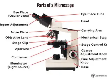
Aperture diaphragm control
Regulates the amount of light that passes through the stage and specimen for optimal visibility.
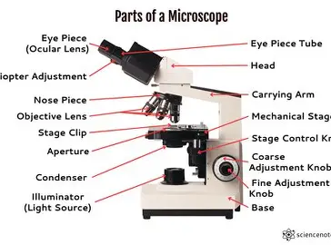
Substage condenser
A lens system located beneath the stage that focuses light onto the specimen, enhancing image clarity and contrast.
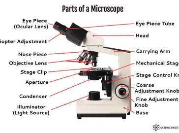
Nosepiece
The rotating part of the microscope that holds multiple objective lenses and allows the user to switch between them for varying levels of magnification.
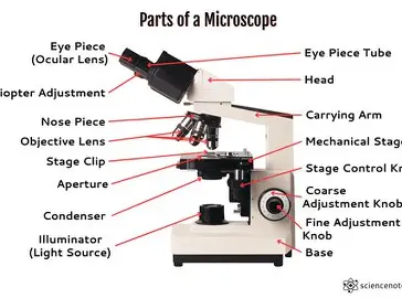
Eyepieces (ocular lens)
Lenses located at the top of the microscope that magnify the image from the objective lenses, allowing the user to view the specimen.
Coarse focus adjustment knob
A large knob on the microscope used for making broad adjustments to the focus of the specimen by moving the stage up and down.
Fine focus adjustment knob
A smaller knob on the microscope that allows for precise adjustments to the focus of the specimen, enabling clearer viewing of details.
Field diaphragm lever
A component that controls the aperture of light entering the microscope, allowing for regulation of illumination and contrast when viewing a specimen.
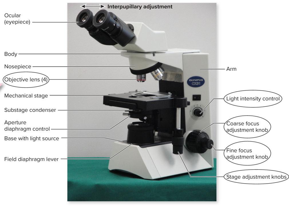
Stage adjustment knob
A knob on the microscope that controls the horizontal and vertical positioning of the stage, allowing for precise movement of the specimen under observation.
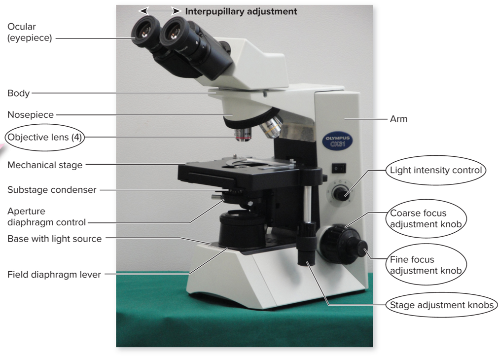
Light intensity control
A feature on the microscope that adjusts the brightness of the light source, enhancing visibility of the specimen.
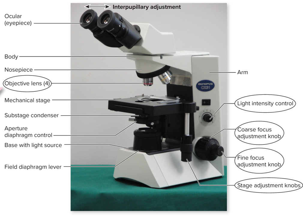
Light microscopes: bright-field
A type of optical microscope that transmits light through the specimen, producing a bright image against a darker background, commonly used for viewing stained biological samples. The most widely used microscope.
Light microscopes: dark-field
A type of optical microscope that directs light at an angle to the specimen, creating a bright image of the specimen against a dark background. This method is particularly useful for observing live, hard-to-stain, or fragile samples.
Phase-contrast microscope
A type of optical microscope that enhances contrasts in unstained specimens by amplifying the differences in refractive index, making it particularly useful for viewing living cells and their internal structures.
Differential interference microscope
A type of optical microscope that uses polarized light and special optics to enhance contrast in transparent specimens, allowing for a three-dimensional appearance. It is especially effective for examining live cells in detail.
Fluorescent microscope
A type of optical microscope that utilizes ultraviolet rays to excite special dyes that emit visible light, allowing for the visualization of specific cellular components and structures. This method is widely used in biological and medical research to study complex cellular processes.
Confocal microscope
A type of optical microscope that uses a laser to scan samples in multiple layers (thicknesses) within a cell, producing high-resolution, 3D images of complex structures within cells. It allows for enhanced imaging of specific areas while minimizing background noise.
Transmission electron microscope
A type of electron microscope that transmits electrons through a specimen to form an image. This allows for extremely high-resolution imaging at the nanometer scale, revealing internal structures of cells and tissues. Typically used for viewing the detailed structure of cells and their organelles and viruses.
Scanning electron microscope
A type of electron microscope that scans the surface of a specimen with a focused beam of electrons, producing detailed 3D images of the sample's surface topography. It provides high-resolution images and is widely used in materials science, biology, and semiconductor research.
Simple Stains
Stains involve using a single dye to highlight specific features of microbial cells, enhancing visibility under a microscope. This technique is commonly used to determine cell shape, arrangement, and size.
Differential stain
A staining technique that uses two or more dyes to differentiate between types of cells or structures. This method allows for the identification of specific cellular components and is crucial in microbiology for distinguishing between Gram-positive and Gram-negative bacteria.
Special stain
A staining technique used to identify specific bacterial structures, such as spores, capsules, or flagella, which are not visible with basic stains. Special stains provide essential information on the morphology and characteristics of different microorganisms.
Negative stain
A staining technique that creates a contrast between the organism and the background by staining the background instead of the cells themselves. This method allows for better visualization of cell morphology and size without altering the cells.
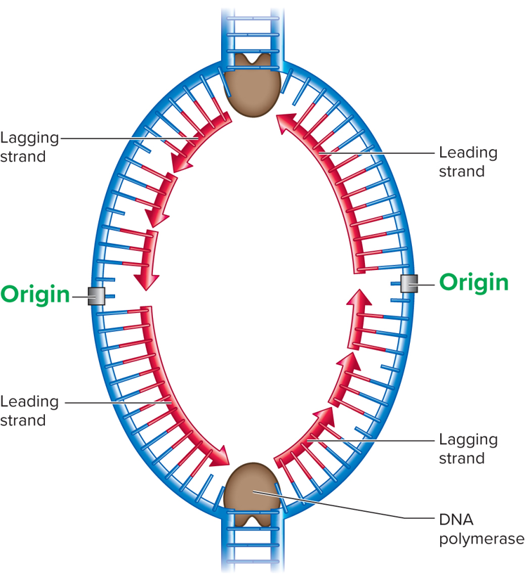
DNA replication pt 1
The process by which a cell duplicates its DNA, creating two identical copies.
Each daughter molecule is identical to the parent in composition
The template strand is an original parental DNA strand
The newly synthesized DNA is made in a discontinuous fashion
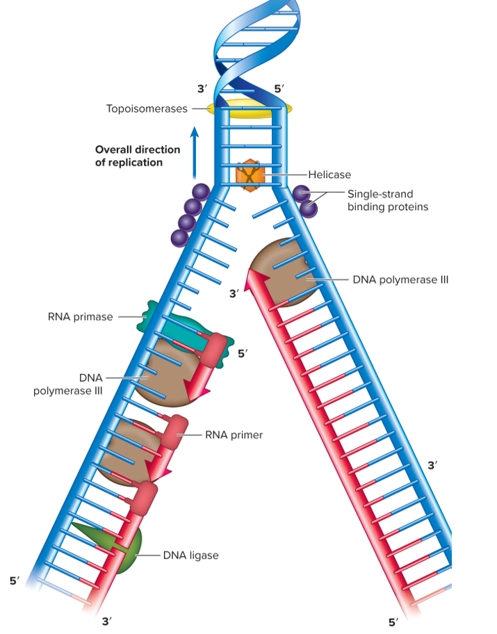
DNA replication pt 2
It involves unwinding the double helix and synthesizing new complementary strands using the original strands as templates.
Primase makes an RNA primer
DNA polymerase III adds new nucleotides to the growing DNA chain
DNA ligase fills in any resulting gaps in the newly formed DNA
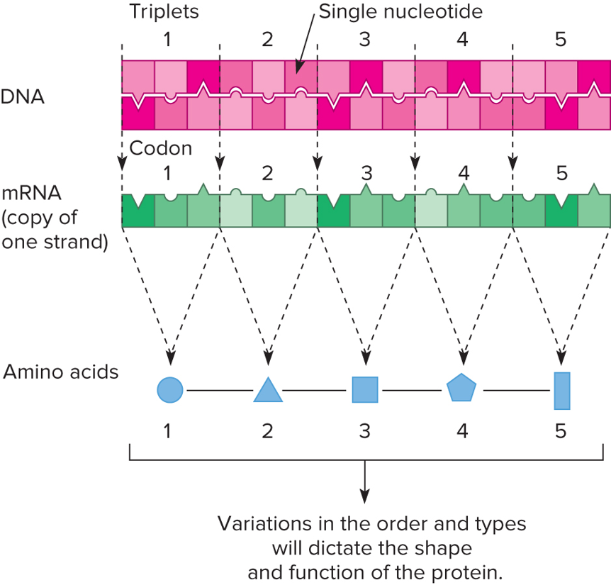
Central Dogma of Microbiology
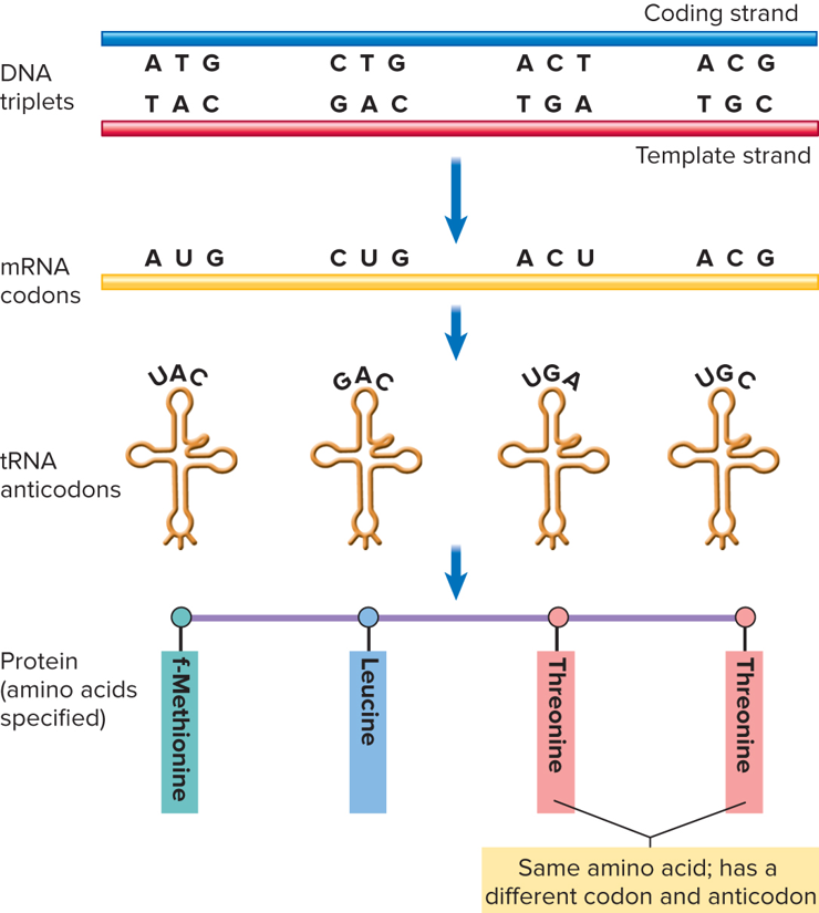
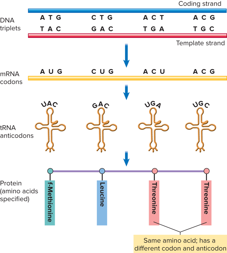
DNA is transcribed into mRNA and mRNA is translated into protein
DNA → RNA → Protein
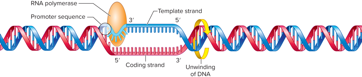
Transcription
The process by which the genetic information in DNA is copied into messenger RNA (mRNA). This occurs in the nucleus and is the first step in gene expression.
DNA → RNA (mRNA)
Translation
The process of synthesizing proteins from mRNA sequences. This involves ribosomes reading the mRNA and linking amino acids together in the appropriate order to form proteins.
RNA → Protein
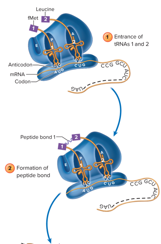
Initiation
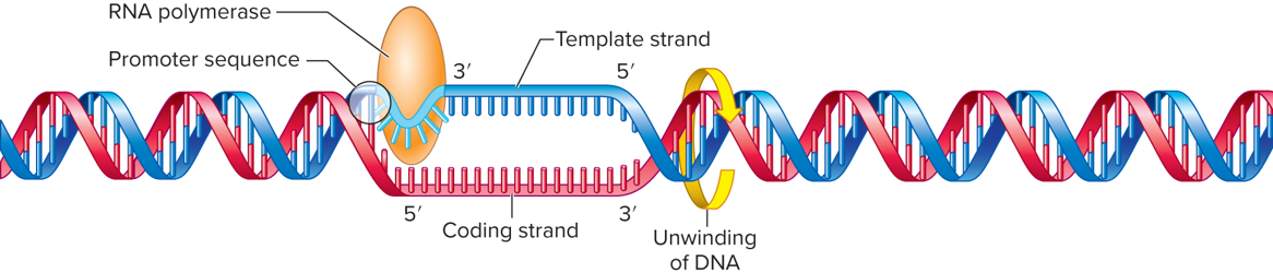
The first stage of transcription or translation, where the necessary components assemble at the start site of the gene to begin the process of RNA synthesis or protein production.
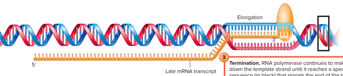
Elongation
The stage during transcription or translation where the RNA strand or polypeptide chain is extended by the addition of nucleotides or amino acids, respectively.
Termination
The final stage of transcription or translation, where the synthesis of RNA or protein is completed and the newly formed molecule is released from the ribosome or RNA polymerase.
mRNA
A type of RNA that conveys genetic information from DNA to the ribosome, where it serves as a template for protein synthesis.
tRNA
A type of RNA that transfers specific amino acids to the ribosome during protein synthesis, matching its anticodon with the corresponding codon on the mRNA.
Ribosomes
Cellular structures that facilitate protein synthesis by reading mRNA and assembling amino acids into polypeptides.
Contains two subunits, a small subunit and a large subunit, directing the translation process by determining where on the mRNA to begin translation
Genome
Includes:
DNA in chromosomes
DNA in the mitochondria
DNA in the chloroplasts
DNA in the floating plasmids
DNA replication characteristics
Double helix, each strand of the DNA can serve as a template to make new DNA molecules
Self-replicating
Each DNA base must hydrogen bond with each other in a specific pairing
During replication, the DNA double helix is unwound by enzymes producing two single DNA strands
Transcription of mRNA characteristics
A complementary nucleotide is inserted opposite to the nucleotide in the DNA template
The strand that RNA polymerase reads to make the mRNA is the template strand
Adenine pairs with uracil, not thymine
DNA replication to transcription to translation
The central dogma of molecular biology describing the flow of genetic information from DNA to RNA and then to protein.
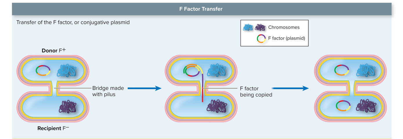
Conjugation
A process of horizontal gene transfer in bacteria where one bacterium transfers genetic material to another through direct contact, often via a plasmid.
This process typically involves the formation of a conjugative pilus that connects the two bacteria, allowing for the transfer of plasmid DNA, which can carry beneficial traits such as antibiotic resistance.
Example: Drug resistance; resistance to metals; toxin production; enzymes; adherence molecules; degradation of toxic substances; uptake of iron
Transformation
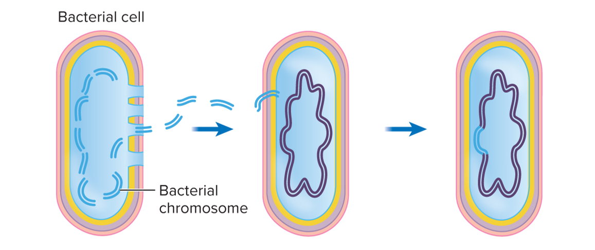
The process by which bacteria take up foreign genetic material from their surroundings and incorporate it into their own genome, leading to genetic changes.
An indirect transfer technique that does not require physical interaction between two cells; one cell (recipient) is alive, while the other (donor) is dead
After a cell dies, the cell wall and membrane are broken down, exposing the dead cell’s DNA
The donor DNA (now located in the environment) will also be broken into smaller pieces
Once inside the cell, the pieces of DNA containing genes are incorporated into the bacterial genome (chromosome)
All new cells derived from the competent cell will now contain these new genes
Example: Polysaccharide capsule: unlimited with cloning technique
Transduction
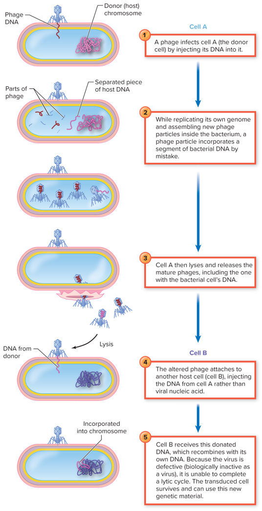
The process of horizontal gene transfer in bacteria, where bacteriophages (viruses that infect bacteria) transfer genetic material from one bacterium to another. During infection, the phage incorporates bacterial DNA into its own genome, which is then introduced into a new host bacterium during subsequent infections.
The bacteriophage infects a bacterial cell, the viral genome is replicated, and then the viral proteins and nucleic acid are assembled into a mature bacteriophage
The subsequent release of mature bacteriophage kills the donor bacterial cell; the newly released bacteriophage is genetically altered and defective
The defective bacteriophage can then inject its genome into a new recipient bacterial cell, due to the genome being altered, the bacteriophage cannot replicate in the recipient cell; however, the recipient bacterial cell can incorporate the genes from the original donor bacterial cell
Example: Toxins; enzymes for sugar fermentation; drug resistance
Transposons
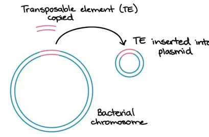
Transposable elements, or "jumping genes," can move within and between genomes, causing mutations and altering gene expression.
Widespread among cells and viruses
Can replicate and move to multiple regions in the chromosome
Can move from a plasmid to a chromosome, from a chromosome to a plasmid, and from one chromosome to another chromosome
Known to be involved with changes in traits such as colony morphology, pigmentation, antigenic characteristics, replacement of damaged DNA, and the transfer of drug resistance between bacteria
First proposed by Barbara McClintock in 1951
Spontaneous mutation
A permanent change in the DNA sequence that occurs without external influence, typically arising during cellular processes such as DNA replication.
Result of errors during DNA replication
Induced mutations
Changes in the DNA sequence that occur due to external factors, such as exposure to radiation, chemicals, or biological agents, which increase the frequency of mutations beyond the normal rate.
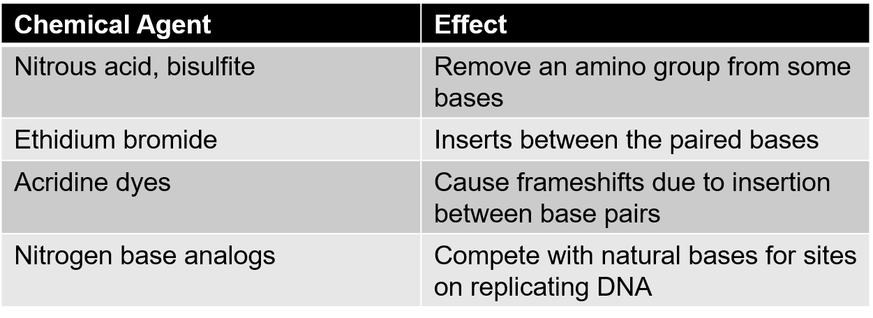
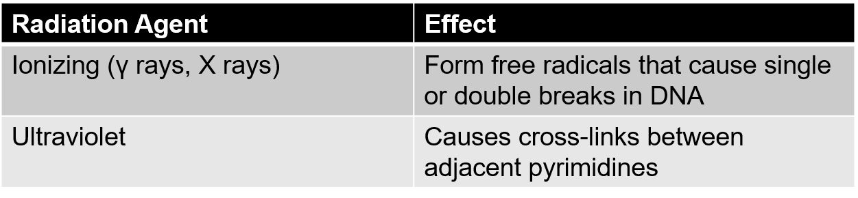
Can be induced by exposure to mutagens
Mutagens are chemical or physical agents that interact with the DNA and alter the DNA sequence
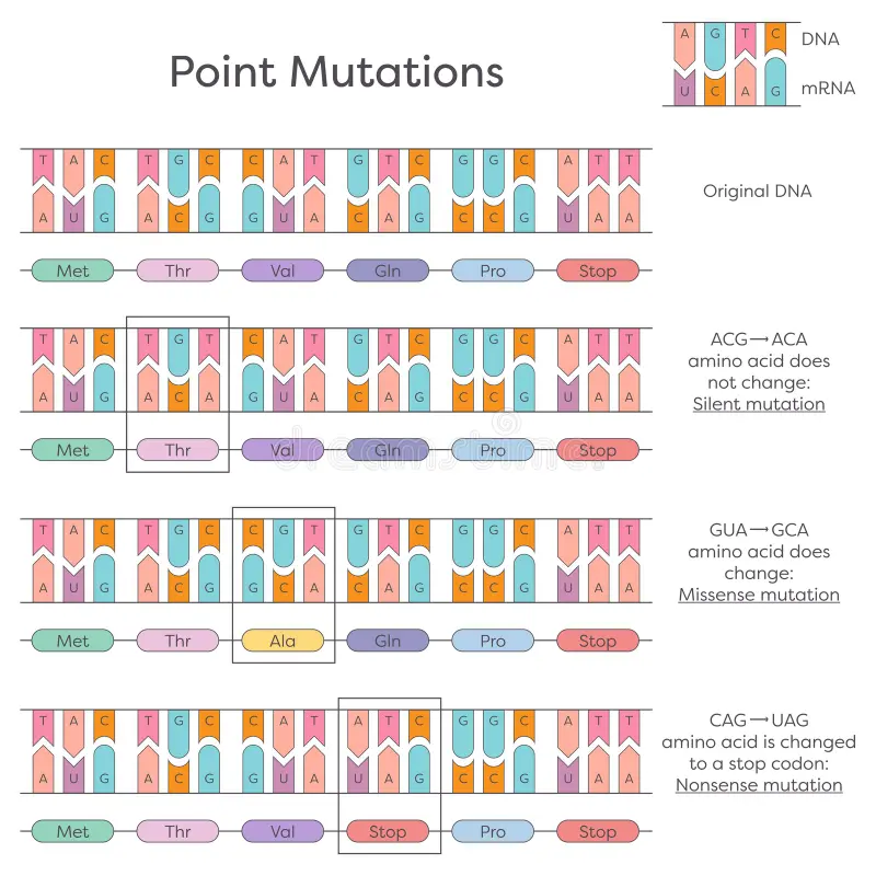
Point mutations
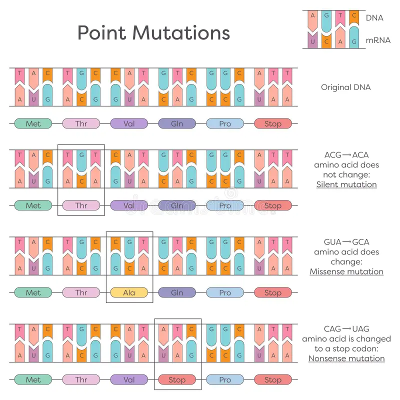
A type of genetic mutation where a single nucleotide change results in a different amino acid in the protein sequence, potentially altering its function. They can be classified as silent, missense, or nonsense mutations depending on the effect on the resulting protein.
Small mutations that affect only a single base on a gene
Involve addition, deletion, or substitution of single bases
Lethal mutation
A type of mutation that causes the death of the organism or cell that carries it, usually disrupting essential biological functions. Lethal mutations can result from significant changes in key genes critical for survival.
Missense mutation
A type of point mutation where a single nucleotide change results in the coding of a different amino acid, potentially altering protein function.
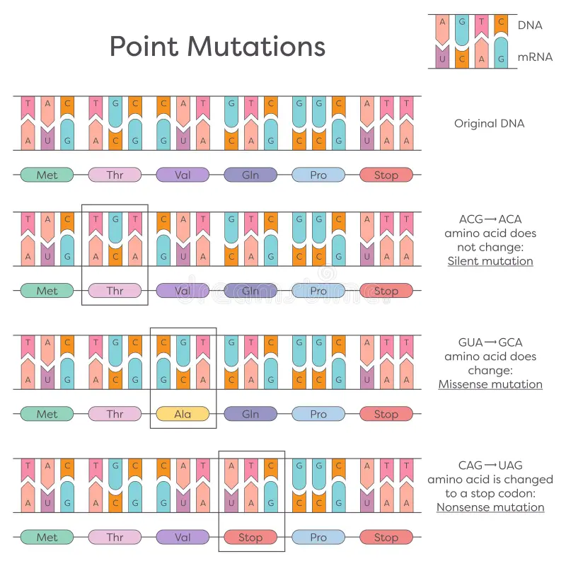
Nonsense mutation
A type of point mutation in which a single nucleotide change creates a premature stop codon, leading to a truncated protein that is usually nonfunctional.
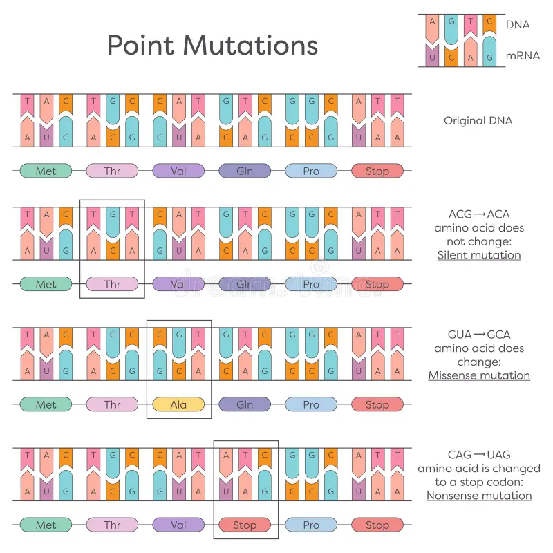
Silent mutation
A type of point mutation where a single nucleotide change does not alter the amino acid sequence of the protein, resulting in no functional change.
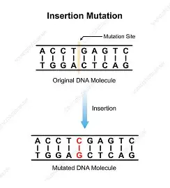
Insertion mutation
A type of mutation where one or more nucleotides are added into the DNA sequence, which can disrupt the reading frame and alter protein function.
Results in all amino acids downstream (to the right) of that insertion being changed
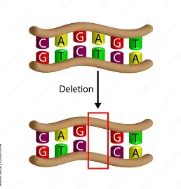
Deletion mutation
A type of mutation where one or more nucleotides are removed from the DNA sequence, potentially changing the reading frame and affecting protein function.
Results in all amino acids downstream (to the right) of the deletion being changed
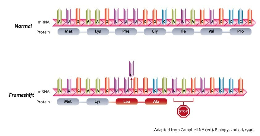
Frameshift mutation
A type of mutation caused by insertion or deletion of nucleotides that shifts the reading frame of the DNA sequence, leading to widespread changes in the amino acid sequence of the protein.
Nearly always results in a nonfunctional protein
Every amino acid after the mutation is different from what is coded for in the original DNA
Insertion of bases in multiples of three does not disturb the reading frame
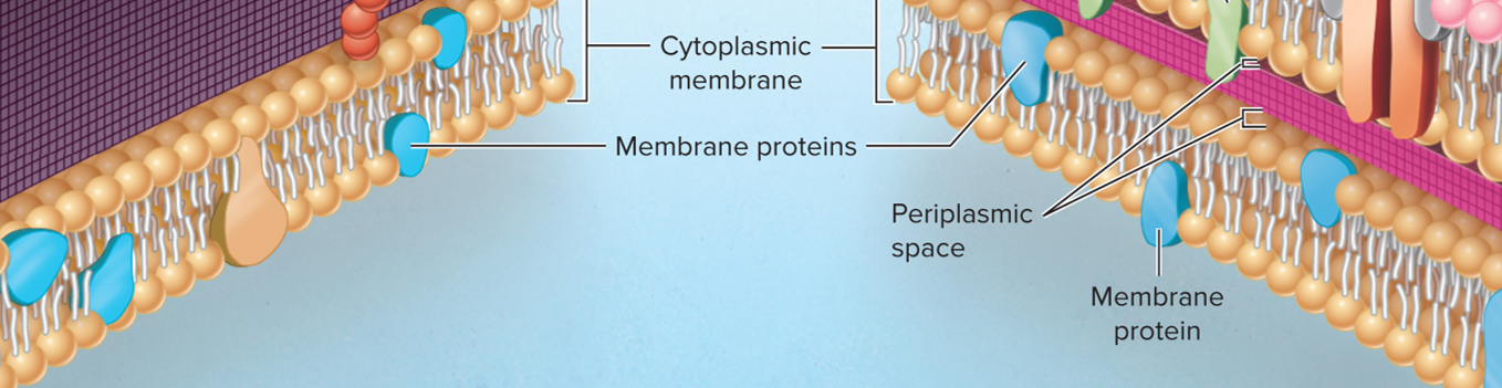
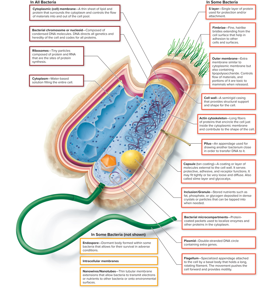
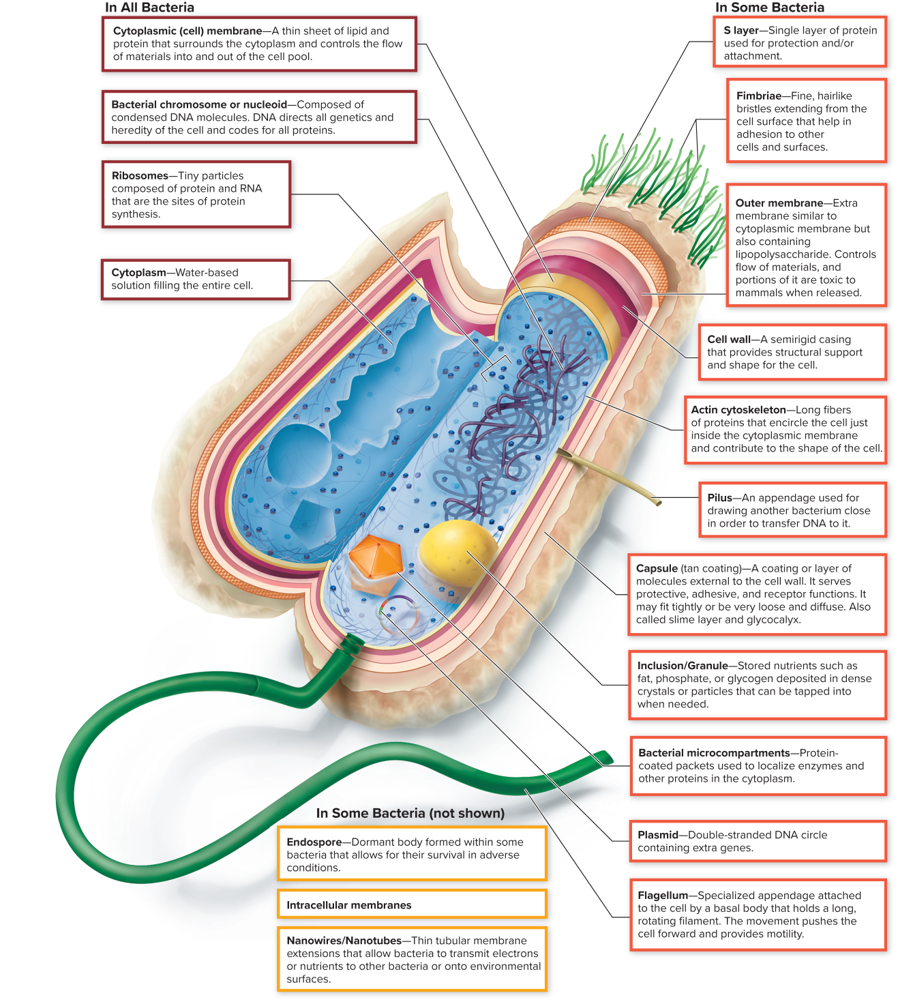
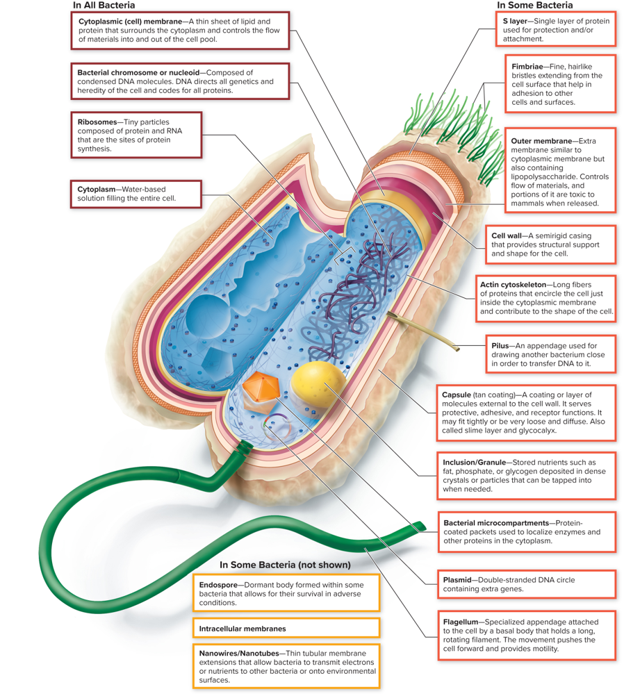
In ALL Cytoplasmic (cell) membrane
A thin sheet of lipid and protein that surrounds the cytoplasm and controls the flow of material into and out of the cell pool
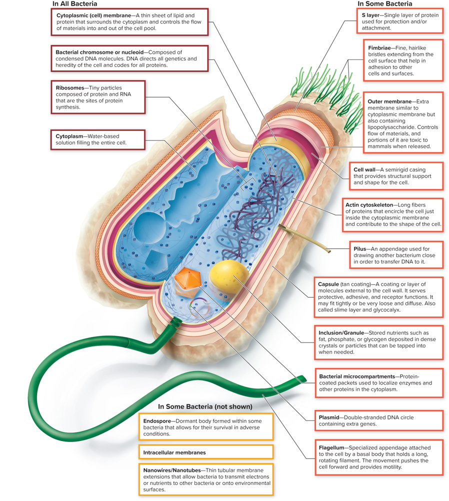
In ALL Bacteria: Bacterial chromosome (aka Nucleoid)
Composed of condensed DNA molecules. DNA directs all genetics and heredity of the cell and codes for all proteins.
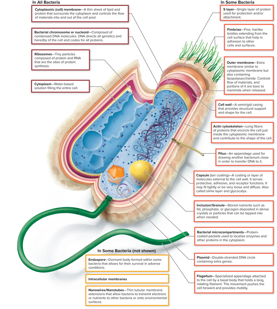
In ALL Bacteria: Cytoplasm
A water-based/gel-like substance filling the entire cell and contains dissolved ions, proteins, and other molecules, and is the site of various cellular processes.
Houses organelles and cellular components
In SOME Bacteria: S layer
Single layer of protein used for protection and/or attachment.
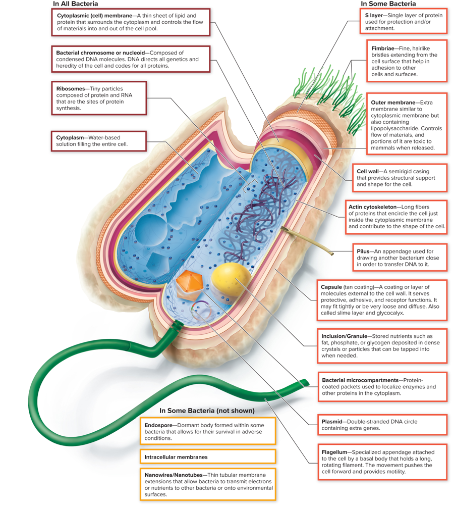
In SOME Bacteria: Fimbriae
Fine, hairlike bristles extending from the cell surface that help in adhesion to other cells and surfaces.
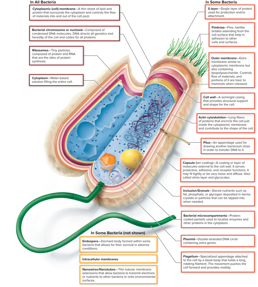
In SOME Bacteria: Outer membrane
An extra membrane similar to the cytoplasmic membrane but also containing lipopolysaccharide. Controls flow of materials, and portions of it are toxic to mammals when released.
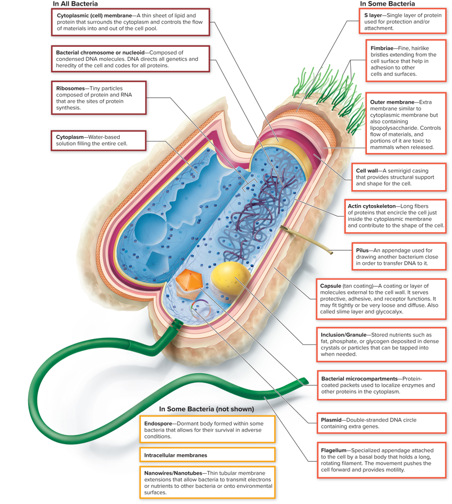
In SOME Bacteria: Cell Wall
A semi-grid casing that provides structural support and shape for the cell.
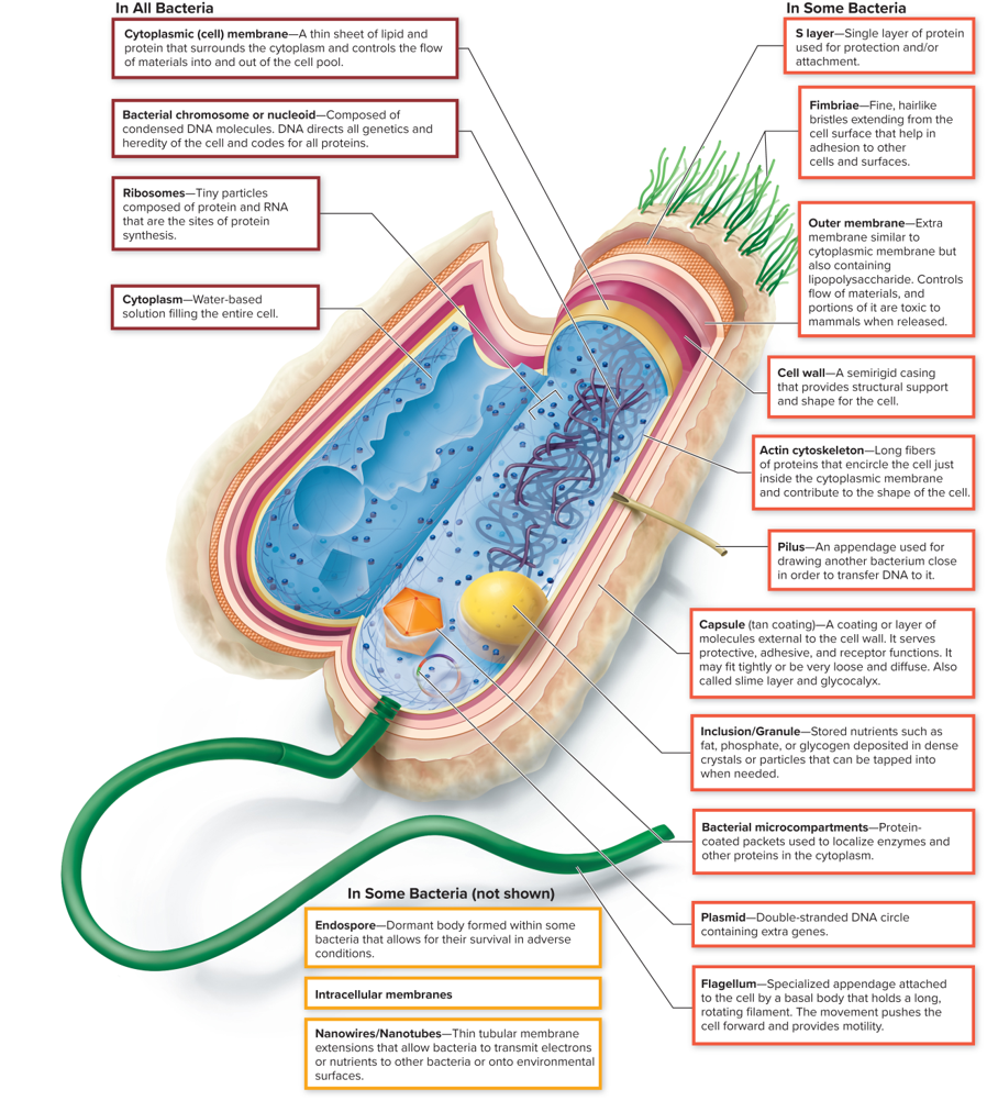
In SOME Bacteria: Actin cytoskeleton
Long fibers of proteins that encircle the cell just inside the cytoplasmic membrane and contribute to the shape of the cell.
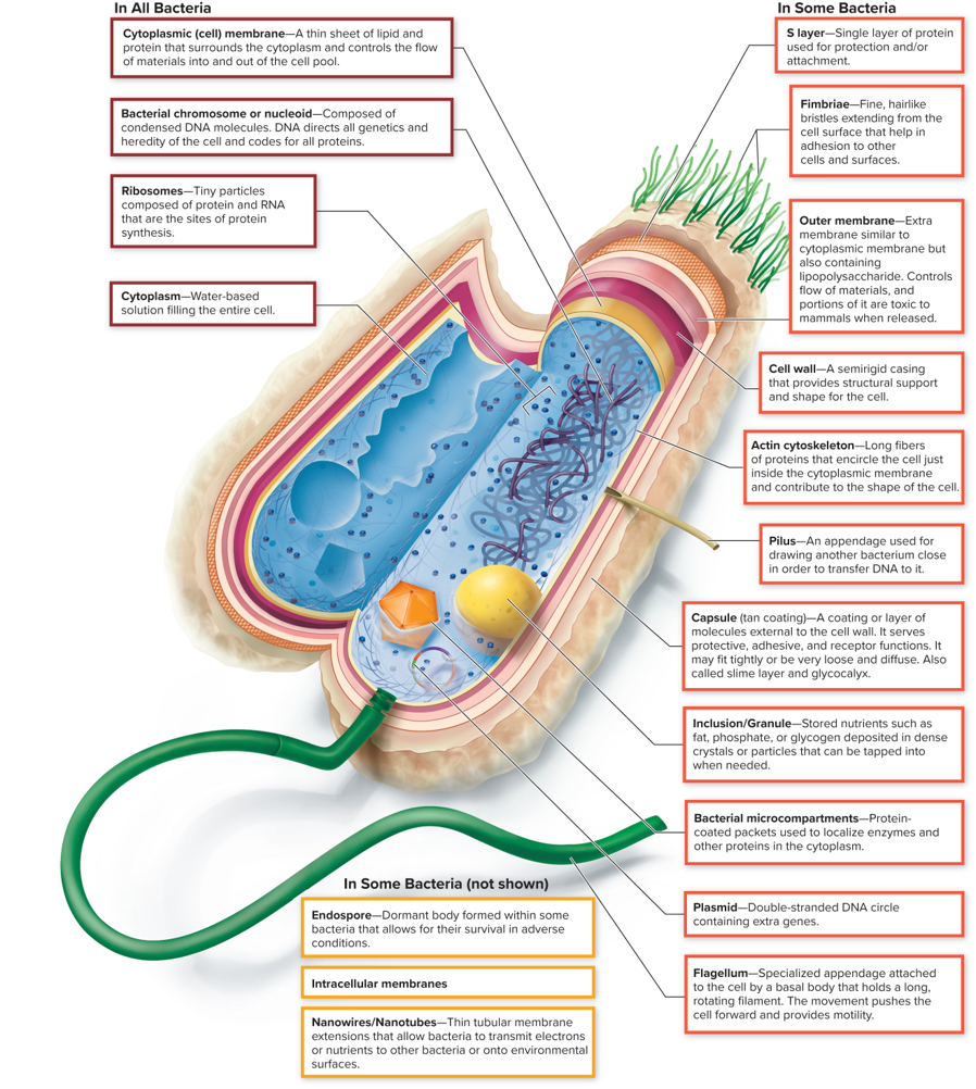
In SOME Bacteria: Pilus
An appendage used for drawing another bacterium close in order to transfer DNA to it.
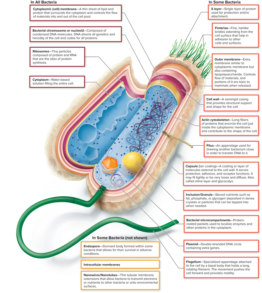
In SOME Bacteria: Capsule (tan coating)
A coating or layer of molecules external to the cell wall. It serves protective, adhesive, and receptor functions. It may fit tightly or be loose and diffuse. Also called the slime layer and glycocalyx.
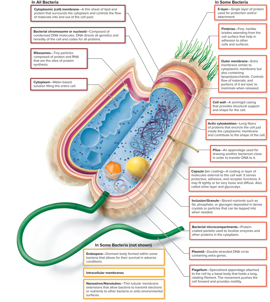
In SOME Bacteria: Inclusion/Granule
Stored nutrients such as fat, phosphate, or glycogen deposited in dense crystals or particles that can be tapped into when needed.
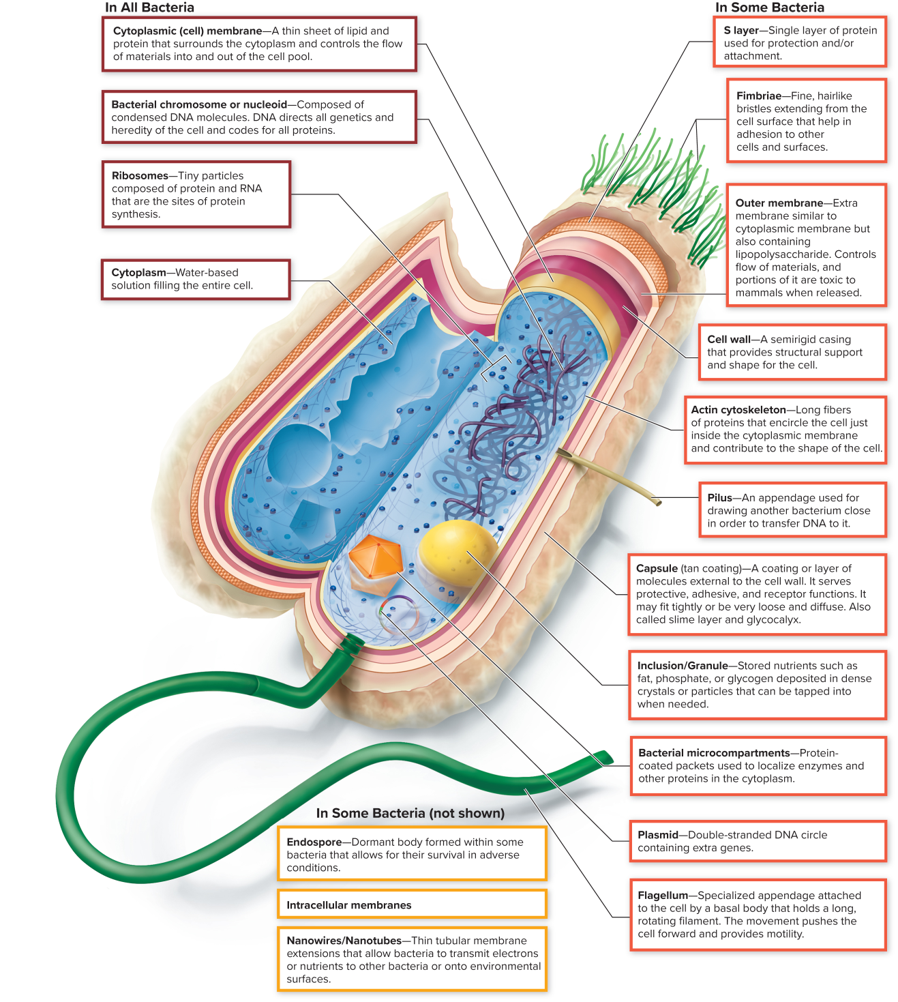
In SOME Bacteria: Bacterial microcompartments
Protein-coated packets used to localize enzymes and other proteins in the cytoplasm.
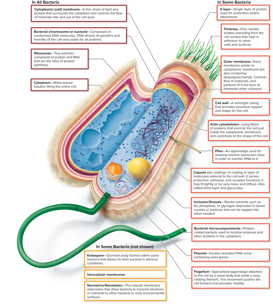
In SOME Bacteria: Plasmid
Double-stranded DNA circle containing extra genes that provide advantages such as antibiotic resistance or metabolic capabilities.
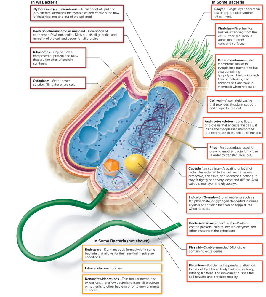
In SOME Bacteria: Flagellum
A long, whip-like structure that aids in bacterial motility by rotating and propelling the cell through liquid environments.
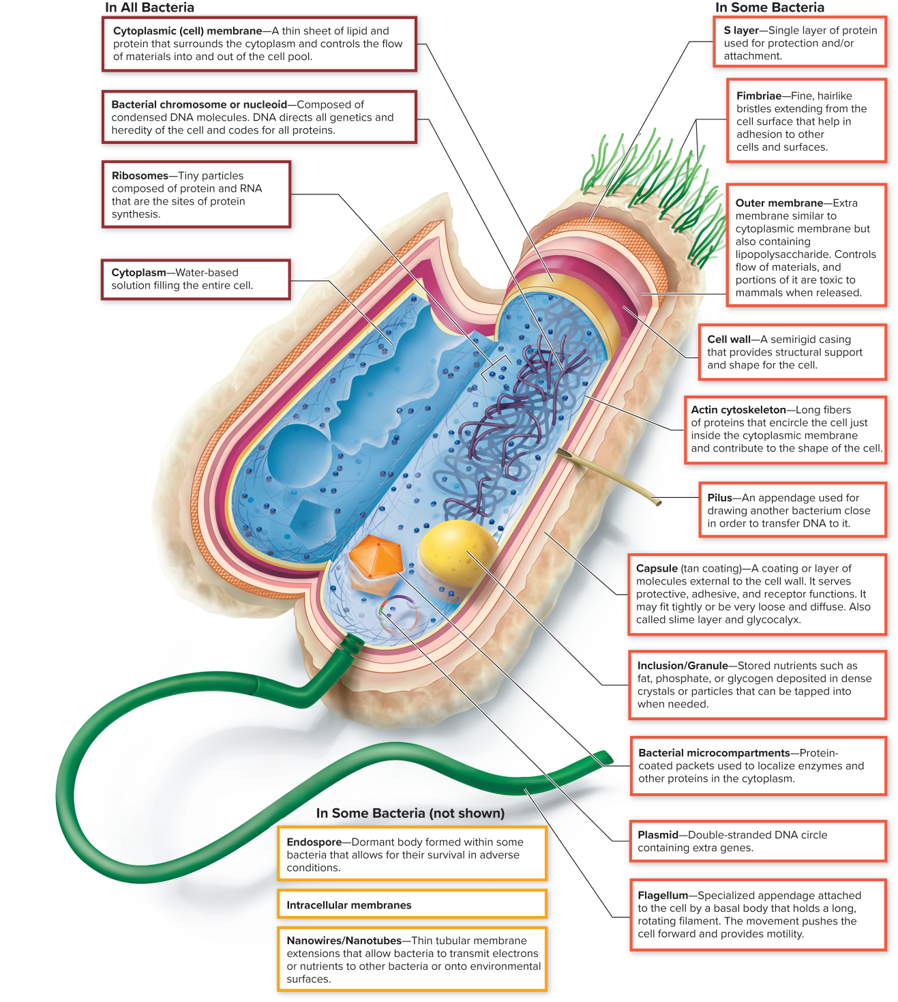
In SOME Bacteria: Endospore
A dormant body formed within some bacteria that allows for their survival in adverse conditions.
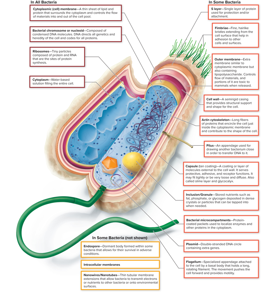
In SOME Bacteria: Intracellular membranes
Membrane-bound structures within the cytoplasm that compartmentalize various cellular processes, allowing for specialized functions.
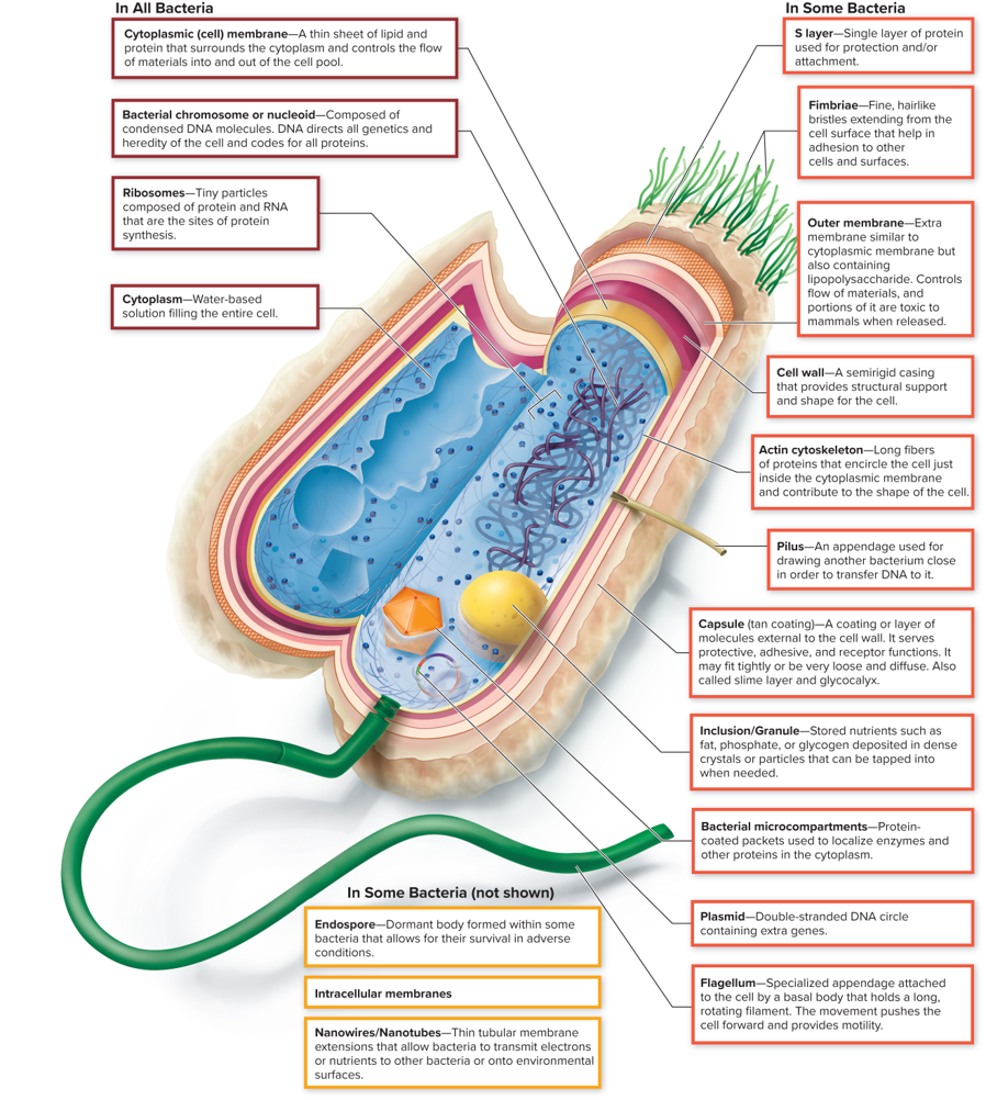
In SOME Bacteria: Nanowires/Nanotubes
Thin tubular membrane extensions that allow bacteria to transmit electrons or nutrients to other bacteria or onto environmental surfaces.
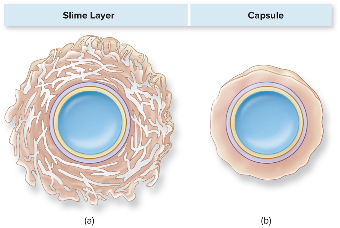
Slime Layer
A layer of extracellular polysaccharides that helps bacteria adhere to surfaces and provides protection against environmental factors.
It’s a loose covering which helps in the formation of biofilms
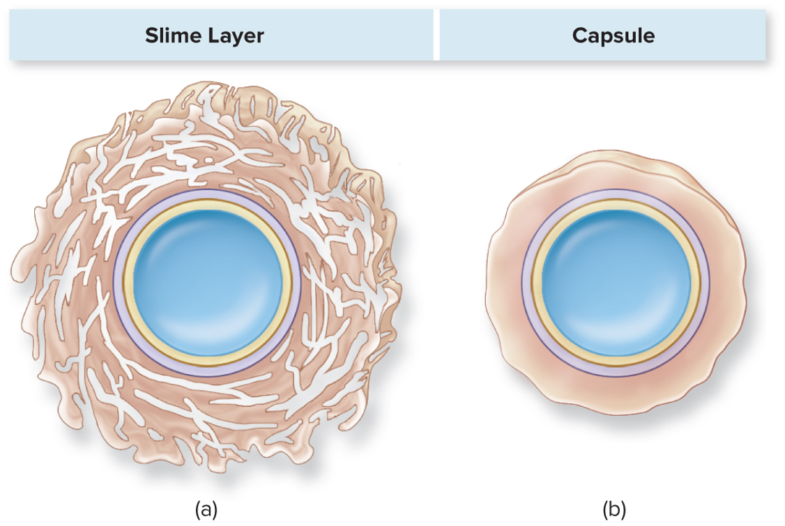
Capsule
A well-organized layer of polysaccharides or proteins that protects bacteria from phagocytosis and desiccation, aiding in adherence and biofilm formation.
Helps cells hide from the immune system
Cell Envelope: Cell Membrane
The protective outer layer of a bacterial cell, composed of the cell membrane, peptidoglycan layer, and sometimes an outer membrane. It maintains cellular integrity and regulates the transport of substances in and out of the cell.
Help classify bacteria into Gram-positive (thick peptidoglycan layer) and Gram-negative (thin peptidoglycan layer)
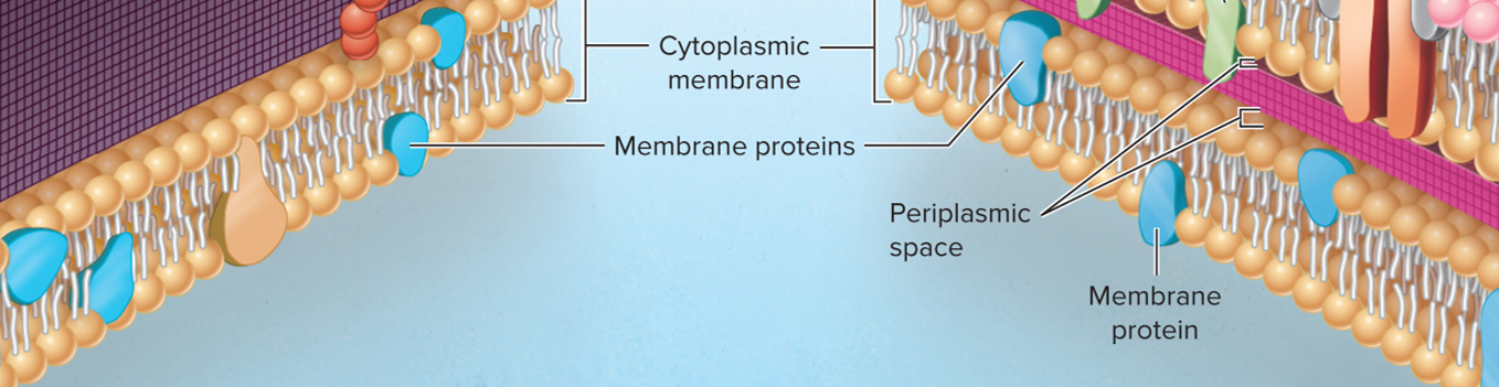
Membrane proteins
Integral and peripheral proteins that assist in various cellular functions, such as transport, signaling, and maintaining structure.