Brain Anatomy I
1/70
Earn XP
Description and Tags
Brain Anatomy Part I Quiz I from MRIQuiz
Name | Mastery | Learn | Test | Matching | Spaced |
|---|
No study sessions yet.
71 Terms
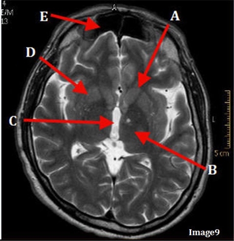
Image 9 is an example of a _____ weighted sequence acquired in the _______ scan plane.
T2; axial

Letter A in Image 9 is pointing to:
Caudate nucleus

Letter B in Image 9 is pointing to:
Thalamus

Letter C in Image 9 is pointing to:
Third ventricle

Letter D in Image 9 is pointing to:
lentiform nucleus

Letter E in image 9 is pointing to
frontal sinus

Letter A in Image 10 is pointing to:
Basal ganglia

Letter B in Image 10 is pointing to:
Posterior horn lateral ventricle
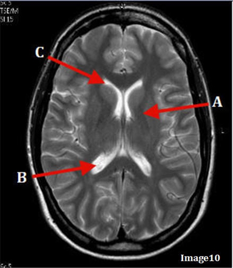
Letter C in Image 10 is pointing to:
Anterior horn lateral ventricle

Letter A in Image 13 is pointing to:
Genu of the corpus callosum

Letter B in Image 13 is pointing to:
Caudate nucleus

Letter C in Image 13 is pointing to:
Internal capsule
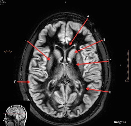
Letter D in Image 13 is pointing to:
White matter

Letter E im Image 13 is pointing to:
Grey matter

Letter F in image 13 is pointing to
lentiform nucleus
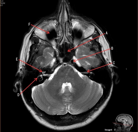
Image 6 is an example of a ___ weighted sequence acquired in the axial imaging plane
T2

Letter A in image 6 is pointing to
sphenoid sinus

Letter B in image 6 is pointing to:
internal carotid artery

Letter C in image 6 is pointing to
7th cranial nerve

Letter D in image 6 is pointing to:
Semicircular canal

Letter E in Image 6 is pointing to:
Cochlea

Letter F in Image 6 is pointing to
maxillary sinus

Letter A in Image 12 is pointing to
medial rectus muscle
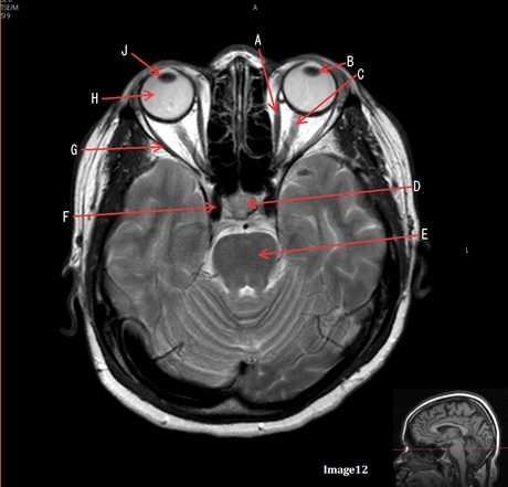
Letter B in Image 12 is pointing to:
lens

Letter C in image 12 is pointing to:
Left optic nerve

Letter D in Image 12 is pointing to
Pituitary gland

Letter E in Image 12 is pointing to:
Pons

Letter F in Image 12 is pointing to:
Internal carotid artery

Letter G in Image 12 is pointing to:
Lateral rectus muscle

Letter H in image 12 is pointing to:
globe

Letter J in Image 12 is pointing to:
right lens

Letter A in Image 8 is pointing to:
Lateral ventricle

Letter B in image 8 is pointint to:
corpus callsoum

Letter C in image 8 is pointing to
thalamus
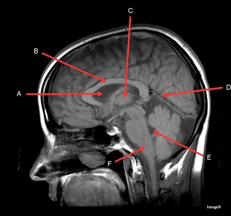
Letter D in image 8 is pointing to
tentorium

Letter E in Image 8 is pointing to:
4th ventricle

Letter F in Image 8 is pointing to:
Medulla oblongata

Image 7 is an example of a ___ weighted sequence acquired in the ____ scan plane
T2 FLAIR; Sagittal

Letter A in Image 7 is pointing to:
Genu to the corpus callosum
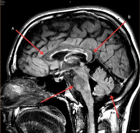
Letter B in Image 7 is pointing to:
Splenium of the corpus callosum

Letter C in Image 7 is pointing to
pons

Letter D in Image 7 is pointing to:
cerebellum

Image 5 is an example of an:
MRA Circle of Willis

Letter A in image 5 is pointing to:
Anterior cerebral artery

Letter B in image 5 is pointing to
Internal carotid artery

Letter C in image 5 is pointing to
vertebral artery

Letter D in Image 5 is pointing to:
basilar artery

Letter E in image 5 is pointing to
posterior cerebral artery
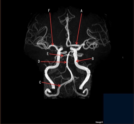
Letter F in Image 5 is pointing to:
Middle cerebral artery

Letter D in Image 14 is pointing to:
Sagittal sinus
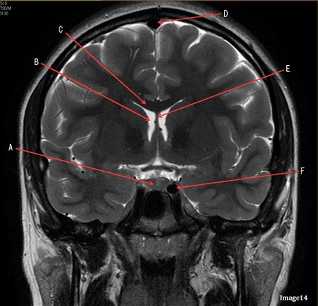
Letter E in Image 14 is pointing to:
Fornix
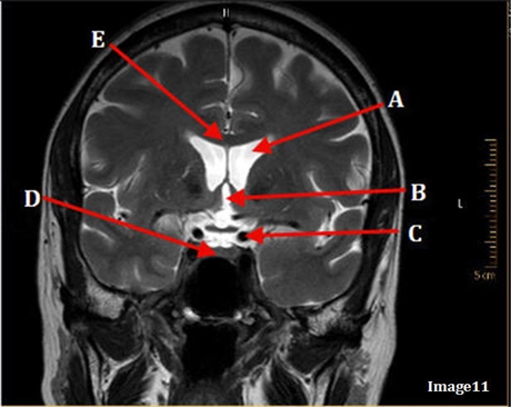
Image 11 is an example of a T2 weighted sequence acquired in the ___ scan plane
Coronal

Image 16 is an example of what type of MR image?
MRA extracranial circulartion
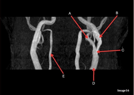
Letter A in Image 16 is pointing to:
External carotid artery

Letter A in Image 16 is responsible for blood supply in the:
face

Letter B in Image 16 is pointing to:
Internal carotid artery

Letter B in Image 16 is responsible for blood supply for the
anterior brain

Letter C in Image 16 is pointing to:
common carotid bifurcation

Letter D in Image 16 is pointing to:
common cartoid artery

Letter E in image 16 is pointing to:
vertebral artery

Letter E in image 16 is responsible for blood supply to the:
posterior brain

Letter F in Image 14 is pointing to:
Internal carotid artery

Letter C in image 14 is pointing to
corpus callosum

Letter A in Image 14 is pointing to:
Pituitary gland

Letter B in Image 14 is pointing to:
lateral ventricle
The pituitary stalk is also known as:
infundibulum
Which intracranial artery passes through the sylvian fissure?
middle cerebral
The medial and lateral rectus muscles are located in the:
eyes
Image 8 is an example of a _______ weighted sequence acquired in the _______ scan plane.
T1; sagittal
Which arteries join together to form the basilar artery?
vertebral arteries
the right and left optic nerve join at the
optic chiasm