14 Pathologic Conditions of the Temporomandibular Joint and Restorative Dentistry
 Call Kai
 Learn
Learn Practice Test
 Spaced Repetition
 Match
Match Flashcards
 Knowt Play
1/73

There's no tags or description
Looks like no tags are added yet.
Name | Mastery | Learn | Test | Matching | Spaced | Call with Kai | Chat |
|---|
No analytics yet
Send a link to your students to track their progress
74 Terms
Mandibular head on the condylar process of the mandible, mandibular fossa on the squamous part of the temporal bone
What are the two parts that make up the temporomandibular joint?
xx
Reminder of where the coronoid VERSUS condylar process is
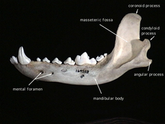
Temporomandibular joint
This part of the body is the lateral aspect of the mandibular head that moves in a rostral and ventral arc during mouth opening
Masseter, temporalis, and pterygoid
What muscles hold the condylar process in tight congruity with the fossa in the temporomandibular joint?
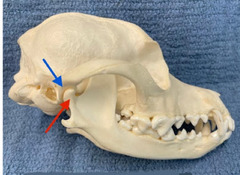
Blue: mandibular fossa of temporal bone
Red: condylar process of mandible
Name the parts of the temporomandibular joint
Dysplasia
This refers to abnormal growth or development of tissue
TMJ dysplasia
This problem results in excessive joint laxity with subluxation or luxation, often a rostral shifting causing ventrolateral displacement and abnormal contact of the coronoid on the zygomatic arch
Open mouth locking
What is a clinical finding of TMJ dysplasia, where there are intermittent episodes of inability to close the mouth due to inappropriate relationship of the coronoid to the zygoma?
Ramus of mandible sits behind (medial to) zygomatic arch in health
The ramus of the mandible sits WHERE in comparison to the zygomatic arch in health?
Shallow mandibular fossa, flattened mandibular head, abnormal obliquity of mandibular head/fossa, joint capsule/ligament laxity
What changes to the TMJ result in joint dysplasia?
TMJ dysplasia
A Cavalier King Charles Spaniel presents with intermittent inability to close the mouth, a few times a day. When you see the dog with this problem, you find that opening the mouth wider will allow you to disengage it. The dog now shows no abnormalities. What is happening?
CT/CBCT
Which should you use to diagnose TMJ dysplasia? *radiographs or CT/CBCT*?
Prevent further locking NOT stabilize
Surgery is the only way you can treat TMJ dysplasia. Is the goal of this surgery to *prevent further locking* or *stabilize the TMJ*?
Zygomatic arch resection
This treatment for TMJ dysplasia involves a semilunar rim ostectomy of 1/2 the rostral arch *length* and 1/2-2/3 the arch *height*
Reduction of coronoid process
This treatment for TMJ dysplasia involves blunt dissection of the deep masseter and fascia of temporal muscle incised parallel to the coronoid crest in order to remove 1/2-3/4 the coronoid *height*
Zygomatic arch resection and Reduction of coronoid process
What two treatments are performed at the same time for TMJ dysplasia?
TMJ dysplasia surgery: zygomatic arch resection and reduction of coronoid process
Which surgical procedure may have complications like facial asymmetry or recurrence due to inadequate removal/regrowth of bone?
Dislocation
This is the displacement of any part of the joint away from its normal position where the joint components stay within the capsule
Luxation
This is the complete dislocation where the articular surfaces are completely separated
Subluxation
This is partial or incomplete dislocation
Condylar process fracture and mandibular head fracture
Fractures of what two areas affect the TMJ?
TOWARDS the affected side
Where does mandibular shift typically occur with TMJ dysplasia?
FRACTURE of TMJ
An animal presents with its jaw back and TOWARDS the affected side. Is this a *fracture or luxation*
Luxation of TMJ
An animal presents with an inability to close or open the mouth with or without malocclusion. There is shift of the mandible to the CONTRALATERAL side (side that is not affected). Is this a *fracture or luxation*?
1: no fragmentation
3: major fragmentation
What is the difference between level 1 and level 3 fragmentation in fractures?
0: complete full surface contact remains
2: loss of contact
What is the difference between level 0 and level 2 displacement in fractures?
Rostrodorsal displacement of condylar process from mandibular fossa
What is the most common displacement position for luxation of the TMJ?
CLOSED (for subluxation. use fulcrum and push mandibles caudally with counterclockwise rotation, then do muzzle)
OPEN (condylectomy only in dire situations then do maxillomandibular fixation aka gluing the canines)
How do you treat TMJ luxation?
Nonsurgical = MMF (gluing canines together) for 7-14 days if young and 2-3 weeks in adults
UNLESS bone fragments are within joint space
What is the treatment of choice for TMJ fractures? With what exception?
Fractures and luxations
Complications of WHICH TMJ problem include ankylosis, fibrous healing, and DJD?
TMJ ankylosis
This is boney or fibrous fusion of the mandibular head to the mandibular fossa (often intraarticular from the destruction of the disc and narrowing of the joint space)
TLDR 2 parts of articulation are fused together
TMJ Pseudoankylosis
This is extra-articular, fibrous or bony encapsulation of the joint or structures remote to the joint with minimal intra-articular involvement
TLDR stuff around the joint are forcing the joint not to go thru its normal range of motion
Ankylosis or pseudoankylosis
This problem is characterized by extensive accumulation of fibrous or boney tissue in the TMJ
TMJ ankylosis or pseudoankylosis
An animal presents with gradual reduction in the ability to open the mouth, drifting towards the affected side, complete inability to open the mouth with tongue entrapment, discomfort on TMJ palpation, and reduced mandibular range of motion. Thoughts?
Bridging of the coronoid to the zygomatic arch
What is the most common form of pseudoankylosis of TMJ?
TMJ ankylosis and pseudoankylosis
In young patients with this condition you can do a gap arthroplasty instead of a condylectomy due to rapid bone growth and liklihood to re fuse
Condylectomy, gap arthroplasty, and salvage mandibulectomy
What 3 surgical procedures can you do to correct ankylosis and pseudoankylosis of TMJ?
Condylectomy
This procedure to correct TMJ ankylosis/pseudoankylosis involves cutting from the mandibular notch in a caudal and ventral direction with a slightly semilunar configuration
Gap arthroplasty
This procedure to correct TMJ ankylosis/pseudoankylosis involves zygomectomy, coronoidectomy, AND fossectomy, often used in young patients
Salvage mandibulectomy
This procedure to correct TMJ ankylosis/pseudoankylosis is used in EXTREME cases of bony proliferation where the base of the cranium is involved
Hard food should be reintroduced at 3-5 days post op to encourage movement
Complications of TMJ ankylosis/pseudoankylosis is FURTHER ankylosis. How do you prevent this?
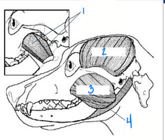
Medial and lateral pterygoid
What is 1?
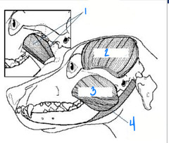
Muscles of mastication: pterygoid, temporalis, masseter, digastricus
What muscles are affected with masticatory muscle myositis?
Temporalis
What is 2?
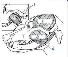
Masseter
What is 3?
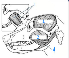
Digastricus
What is 4?
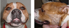
Masticatory muscle myositis
This condition is an inflammatory, focal myopathy with autoimmune origin. It exclusively effects 2M fibers of the masticatory muscles
Masticatory muscle myositis
What is this?
Masticatory muscle myositis
note: in image B, on the right side of the image in the temporalis muscle, there is brightness called "hyperattenuation" which is pathognomonic for MMM
What is this?
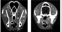
Masticatory muscle myositis = 2M fiber antibody test (may also do muscle biopsy or CT)
A dog comes in presenting with pain upon opening the mouth, swelling and atrophy of the masticatory muscles, inability to open the mouth, exophthalmos (bc of temporalis swelling), lethargy and fever. What do you think it happening and what can you do to diagnose it for sure?
Prednisone = generally good to excellent if they respond positively to prednisone within 1-3 days of treatment
What is the treatment and prognosis like for masticatory muscle myositis?
Dentin
What makes up the bulk of the tooth, comprised of tubules and made by odontoblasts throughout the life of the tooth?
Acute loss of enamel exposing dentin tubules
Which causes the tooth to become sensitive, *acute loss of enamel exposing dentin tubules* or *chronic wear and tertiary dentin*
Restoration
This is the process of sealing exposed dentin tubules
Restoration
The purpose of this process is to eliminate or reduce tooth sensitivity in areas of exposed dentin and help prevent pulpitis or pulp necrosis
SHALLOW: use a bonding agent
DEEPER: use composite restoration
What is the difference in restoration between shallow defects and deeper defects?
1. UNCOMPLICATED crown fracture (no pulp exposure)
2. dental caries (cavities) with NO pulp exposure on occlusal maxillary molar 1
3. restoration of root canal access
4. enamel defects from hypoplasia or hypocalcification
When would you do restorative therapy for teeth?
1. crown fractures with pulp exposure
2. crown fractures with NEAR pulp exposure
3. teeth that have endodontic disease
4. teeth that have periodontitis
5. *cats with tooth resorption*
When would you NOT DO restorative therapy for teeth?
b. enamel defects from hypoplasia or hypocalcification
d. restoration of root canal access
f. dental caries without pulp exposure on maxillary molar 1
Select the indications for doing restorative therapy for teeth:
a. complicated crown fracture with pulp exposure
b. enamel defects from hypoplasia or hypocalcification
c. cats with tooth resorption
d. restoration of root canal access
e. teeth with endodontic disease
f. dental caries without pulp exposure on maxillary molar 1
a. complicated crown fracture with pulp exposure
c. cats with tooth resorption
e. teeth with endodontic disease
Select the indications for NOT doing restorative therapy for teeth:
a. complicated crown fracture with pulp exposure
b. enamel defects from hypoplasia or hypocalcification
c. cats with tooth resorption
d. restoration of root canal access
e. teeth with endodontic disease
f. dental caries without pulp exposure on maxillary molar 1
Etchant, primer, adhesive, fillers, solvent
Name the components of dentin bonding agents?
Etchant
This dentin bonding agent is typically 35-37% phosphoric acid that prepares the enamel and dentin to receive the primer
Primer
This dentin bonding agent is a hydrophilic monomer carried in a water soluble solvent that promotes good flow into the hydrophilic enamel and dentin
Adhesive
This dentin bonding agent promotes bonding between enamel or dentin and the resin composite restorative material or cement that acts as a link between hydrophilic resin primer and hydrophobic resin composite
Fillers
This dentin bonding agent includes sub micron glass particles and nanofillers, whose purpose is to control handling and improve strength
Solvent
This dentin bonding agent includes acetone, ethanol, and water, whose purpose is to control evaporation rate
5th
What is the most commonly used generation of bonding agents?
37% phosphoric acid etch (separate) and primer + adhesive in one bottle
What are the components of the 5th generation bonding agents?
Mechanical retention to etched enamel
What is the mechanism of bonding to enamel?
Results from penetration of dentin tubules and formation of resin-collagen hybrid layer
What is the mechanism of bonding to dentin?
1. probe gingival sulcus
2. radiograph tooth (no bone loss, compare root canal to other teeth, no apical lucency)
3. transilluminate to ensure it is vital
What do you have to do to determine if something is an uncomplicated crown fracture to determine whether or not it will be bonded?
Use dental explorer/fine endodontic file/fine needle tip = extract or refer for endodontic therapy is pulp is found
How can you probe a crown fracture for tooth exposure?
Restoration of uncomplicated crown fracture
The following are steps for WHAT procedure:
1. clean tooth
2. probe for pulp exposure
3. remove sharp enamel edges (arkansas white stone)
4. smoothen fracture surface
5. polish with NON fluoride polish
6. isolate tooth w gauze
7. 37% acid etchant
8. bonding agent
9. air dry
10. light cure
6-12 months (of affected tooth and contralateral tooth + compare width of root canals)
When should you do followup radiographs after a restorative procedure of an uncomplicated crown fracture?