unit 1 glossary
1/103
There's no tags or description
Looks like no tags are added yet.
Name | Mastery | Learn | Test | Matching | Spaced | Call with Kai |
|---|
No analytics yet
Send a link to your students to track their progress
104 Terms
gyrus
a “ridge” of cerebral cortex
sulcus
a “valley” of cerebral cortex
cortical sheet
the outer surface covering of cerebral cortex, composed of gray matter (neuron cell bodies)
cerebellum
latin for “little brain”; the small, highly ridged portion of brain that sits inferior to the cerebral cortex and posterior to the brainstem
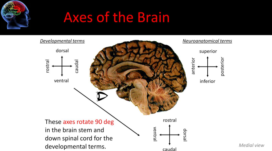
developmental and neuroanatomical terms
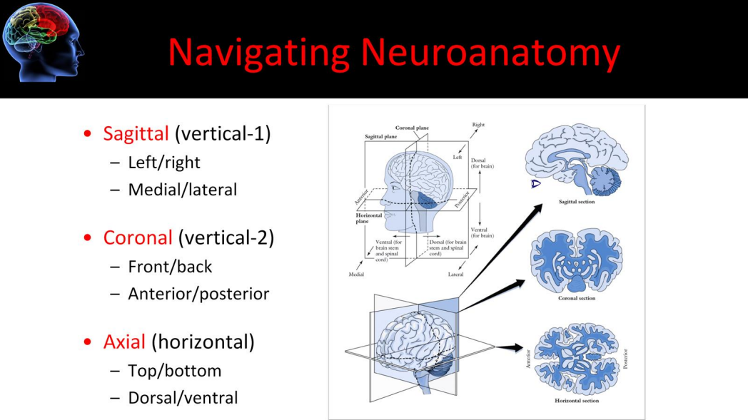
anatomical planes/slices
central nervous system (CNS)
composed of the brain and spinal cord
peripheral nervous system (PNS)
composed of peripheral nerves that connect the CNS to the limbs, trunk, and internal organs
autonomous nervous system (ANS)
a subdivision of the PNS that controls visceral functions
includes parasympathetic and sympathetic nervous systems
cranial nerves
a set of 12 specialized nerves that act as the PNS (motor control and sensory info) to the head and neck
meninges
the three protective layers of tissue between the brain and the skull
dura mater
“hard mother”
the durable, leathery outer protective layer of the meninges
arachnoid mater
“spiderlike mother”
the spiderweb-like middle protective layer of the meninges that is filled with cerebral spinal fluid
pia mater
“tender mother”
the thin, shiny, inner protective layer of the meninges that “shrink-wraps” the brain
lobes of the brain
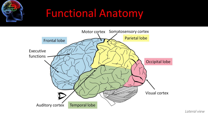
central sulcus
the sulcus dividing the frontal and parietal lobes, surrounded on each side by motor and sensory cortex
lateral fissure
the gap that divides the temporal from the frontal and parietal lobes
a fissure is another name for a large sulcus
parieto-occipital sulcus
the sulcus that divides the parietal and occipital lobes
pre-occipital notch
the notch that serves as the bottom point of the imaginary dividing line between the temporal and occipital lobes
the top of the parieto-occipital sulcus is the top point
brodmann’s areas
~50 cytoarchitectural areas defined by neuroanatomist Brodmann according to cell size, cell density, number and thickness of cortical layers, and density of myelinated axons; numbering system is becoming less common as neuroimaging measurements take over tissue histology
gray matter
outer “bark” of the cerebral cortex composed of neuronal cell bodies
this is where computations happen
much of cortex consists of six layers
white matter
inner region of cerebral cortex composed of the axons of the neurons with cell bodies in the gray matter
can be thought of as the “wiring” connecting different regions of gray matter
neuron
the basic cell in the brain that processes and transmits information in the form of electrical and chemical signals
dendrite
the branched portion of a neuron which receives inputs from synapses with other cells and sends small depolarizations towards the cell body
cell body
the “main” portion of a cell that contains the nucleus, mitochondria, and other organelles necessary for the cell to survive
axon hillock
the base of the axon, where it meets the cell body of the neuron
action potentials are initiated here
axon
the long cell structure that carries depolarizations (action potentials) away from the cell body of a neuron to the synapse
node of ranvier
the small gaps between myelin sheaths in myelinated axons involved in fast action potential propagation down the axon
synapse
the region/space which information flows across from one neuron to another neuron
space between neurons can be called the synaptic cleft
axon terminal
the very end of a branch of a neuron’s axon, specialized to release neurotransmitters from vesicles into the synapse in response to an action potential
glial cell
surround neurons in CNS and PNS and provide myelination as well as other support for them
most abundant cell types in CNS; types differ between CNS and PNS
CNS: oligodendrocytes, astrocytes, ependymal cells, microglia
PNS: satellite cells, Schwann cells
myelin sheath
a layer of protective tissue wrapped around axons of neurons to hasten the transmission of action potentials
neuronal communication
includes electrical conduction (action potential) along the axon and chemical transmission via neurotransmitter release at the synapse
corpus callosum
the main connection of white matter that is integral for communication between the two cerebral hemispheres
cerebral spinal fluid (CSF)
the fluid surrounding the brain and spinal cord that cushions the nervous system
fluid is similar to blood plasma
ventricle
CSF-filled cavities in the brain, four total (left, right, third, and fourth)
choroid plexus
the specialized cells lining the ventricles responsible for the creation of CSF
lumbar puncture
a method to withdraw CSF for testing from a low part of the spinal column just below the spinal cord
arachnoid granulations
the bubble-like portions of the arachnoid mater (middle layer of meninges) into the draining venous sinus system that are responsible for the removal of CSF from around the brain
CSF is “recycled” into the blood stream
hydrocephalus
“water on the brain”
disorder of CSF causing problems with CSF flow or reuptake
leads to enlargement, developmental problems, changes in eye gaze, and if left untreated, death
may be developmental or acquired
primarily treated with a shunt to siphon CSF away from the brain into the abdomen
hydrocephalus ex vacuo
large spaces develop inside cortex due to loss of cortical tissue - “cortical atrophy”
seen in dementia
NOT really hydrocephalus
non-communicating hydrocephalus
hydrocephalus caused by something obstructing the normal flow of CSF
CSF behind obstruction (between production in choroid plexus and obstruction) would increase in pressure
blockages can be from things like a tumor/mass or a clot of blood/infection
communicating hydrocephalus
hydrocephalus caused by a problem with the normal uptake of CSF through the arachnoid granulations
whole CSF system would have increased pressure
shunt
in brain disorders: a tube placed inside the skull to drain off extra fluid, as seen in hydrocephalus (ventriculoperitoneal) and strokes (intraventricular)
blood vessels
the part of the circulatory system that transports blood throughout the human body
three major types: the arteries which carry oxygenated blood away from the heart, the capillaries which enable the blood and the tissues, and the veins which carry de-oxygenated blood from the capillaries back toward the heart
the arteries carry high pressure blood pumping from the heart
this high pressure decreases as larger arteries branch into progressively smaller arteries
by the capillary bed, the system is at very low pressure. this low pressure continues into the venous system and back to the heart
note that the arteries have the largest smooth muscle layer. this layer provides the mechanical strength to withstand a persons blood pressure
circle of willis
a circle of arteries that supply blood to the brain
this arrangement of blood vessels allows for collateral blood flow to the brain
carotid artery
a blood vessel that supplies the head and neck with oxygenated blood
there are two, one on each side. they supply blood to the anterior part of the brain
divides in the neck into the internal (supplies circle of willis) and external (the artery you take a pulse from)
vertebral artery
a blood vessel that runs up the back of the neck
there are two one on each side, that join at the base of the skull to form the basilar artery
these vessels supply the posterior part of the brain
basilar artery
the artery that supplies the pons, cerebellum, posterior cerebellum, and inner ear
this vessel is formed by the merging of the vertebral arteries
middle cerebral artery
the artery that supplies lateral cerebral cortex and anterior temporal lobes
strokes here can affect face, arm and language use
anterior cerebral artery
the arteries that supply oxygen to most medial portions of frontal lobes and superior medial parietal lobes
strokes here can affect leg use
stroke
rapid loss of brain tissue and function as a result of disruption of the blood supply to the brain
transient ischemic attack (TIA)
“mini stroke”
same wide-range of possible symptoms as a stroke, but symptoms are only temporary
ischemia
lack of oxygen arising from a restriction in blood supply
ischemic stroke
a stroke resulting from restriction of blood flow into a region of brain tissue
thrombus
a clot, or atherosclerotic plaque, that forms in place within a blood vessel obstructing blood flow
can close off blood flow at the place it forms or may break apart to form an embolus
embolus
a moving clot that then lodges in a small vessel
carotid stenosis
abnormal narrowing of the carotid artery, often caused by atherosclerotic plaque formation
hemorrhage
bleeding, the loss of blood from the circulatory system
hemorrhagic stroke
a stroke resulting from blood bleeding into the brain, damaging tissue
aneurysm
a localized, blood-filled bulge of blood vessel
subarachnoid hemorrhage
bleeding that occurs between the arachnoid and the pial meningeal layers
this is the space where the CSF flows around the brain and the spinal cord, the space that is also filled with spiderweb-like protrusions of the arachnoid mater
intracerebral hemorrhage
bleeding that occurs within the brain tissue itself
this would be below the pia mater
intraventricular hemorrhage
bleeding that occurs from vessels along the ventricles
the bleeding in this case would be directed into the ventricles
tPA
drug made from tissue plasminogen activator, a protein involved in the breakdown of blood clots
used to treat ischemic stroke if within 3 hours of symptom onset
venous malformation
a general term for congenital vascular anomalies
the anomalies- unusual formation- can involve only veins or veins and arteries
often increase the patient’s risk of bleeding in the brain and/or abnormal oxygen delivery to the brain tissue
arteriovenous malformation (AVM)
a vascular malformation that is a tangle of abnormal blood vessels connecting arteries and veins in the brain
has increased risk of bleeding and decreases normal oxygen flow to local tissue (no capillary bed for gas exchange)
venous thrombosis
a blood clot that forms within a vein
in the brain, this may occur in the large venous sinuses and block off blood flow out of the brain, which frequently is fatal
bridging veins
veins that drain the neural tissue and puncture (“bridge”) through the dura mater to drain into the venous sinuses
they may tear with trauma and bleed to cause a subdural hematoma
have a higher risk of rupturing from trauma from a fall in patients who suffer from alcoholism
long term alcoholism both weakens the vein vessel walls throughout the body and shrinks the brain, which puts greater stress on them specifically
hematoma
a solid swelling of clotted blood within the tissues, which can be located anywhere in the body
also called a bruise when in/under the skin
in the brain: causes local mass effects, such as compressing and injuring surrounding brain tissue like a brain tumor can
subdural hematoma
bleeding that occurs below the dura- between the dura and the brain (or spinal cord), resulting in a build up of blood that compresses the brain (or spinal cord)
this type usually comes from trauma, like a fall that tears the bridging veins
compared to epidural hematomas, these usually have a slower onset, as lower pressure venous blood is usually involved
epidural hematoma
bleeding that occurs between the skull and the dura (or spinal column and dura), resulting in a build-up of blood that compresses the brain (or spinal cord)
this type usually develops from trauma, especially in conjunction with a skull fracture
compared to subdural hematomas, these tend to have a faster and more serious onset, as higher-pressure arterial blood is usually involved
traumatic brain injury (TBI)
damage to the brain as a result of external physical force
common term: concussion
coup/contra coup injury
results from focal injury or whiplash, in which the side of the brain directly hit and the opposite side are both damaged
diffuse shearing injury
rapid acceleration/deceleration forces separates cell bodies and axons, causing widespread brain injury
phineas gage
a man who had a train spike shoot through his frontal cortex in an accident
survived but was emotionally disturbed from that point forward
blood brain barrier
a filtering mechanism of the capillaries that carry blood to the brain and spinal cord tissue, blocking the passage of certain substances (i.e., infectious agents, immune cells, some drugs)
meningitis
viral or bacterial infection of the meninges
symptoms: rash, stiff neck, headache, vomiting, mental status change
encephalitis
usually viral (herpes) infection of brain tissue
symptoms: personality changes, seizures, weakness
measles
childhood infection caused by a virus
used to be common, can be prevented with vaccine now
symptoms: appear 7-14 days after contact and include high fever, cough, runny nose, and watery eyes- rash appears 3-5 days after the first symptoms
vaccine does NOT cause autism, but measles itself DOES cause neurological complications
subacute sclerosing panencephalitis (SSPE)
history of primary measles infection, followed by an asymptomatic period that lasts 7 years on average, but can range from 1 month to 27 years
after this period, progressive neurological deterioration occurs, characterized by behavior change, intellectual problems, myoclonic seizures, blindness, ataxia, and eventually death
death can occur in 1-3 years but can be faster or slower, fastest within 3 months
rabies
virus that enters the brain by following peripheral nerves, causing diffuse brain swelling
symptoms: headaches, fever, rages, inability to swallow water, coma, and death
prion
infectious protein molecules
creutzfeld-jakob disease (CJD)/transmissible spongiform encephalopathy
commonly known as mad cow disease
prional infection kills of brain cells, creating numerous lacunae (“lakes”) in the brain tissue
symptoms: dementia and problems with coordination
encephalopathy
means disorder or disease of the brain
in modern usage, does not refer to a single disease, but rather to a syndrome of global brain dysfunction
this syndrome can be caused by many different illnesses
cysticercosis
condition resulting from infection with the pork tapeworm
symptoms: headache, stroke-like effects, brain swelling, risk of death
brain tumor
any intracranial growth created by abnormal and uncontrolled cell division, normally either in the brain itself (e.g., neurons, glial cells, lymphatic tissue, blood vessels), in the cranial nerves (e.g., myelin-producing Schwann cells), in the brain envelopes (e.g. meninges), skull, pituitary and pineal gland, or spread from cancers primarily locaed in other organs (e.g., metastatic tumors)
any type can cause damage either through direct damage to the surrounding tissue or through a mass effect- in which the pressure squishes down parts of the brain
holoprosencephaly
a congenital disorder caused by the failure of the embryonic forebrain to sufficiently divide into the double lobes of the cerebral hemispheres
the result is a single lobed frontal brain structure usually with severe skull and facial defects
autism
diffuse developmental disorder that impairs social interaction and communication
epilepsy
chronic neurological disorder that is characterized by recurring seizures
neurodegeneration
progressive loss of structure or function of neurons, including death of neurons
dementia
progressive decline in cognitive function due to damage or disease in the brain
atrophy
loss of cells
alzheimers disease
the most common form of dementia, characterized by loss of memory, cognitive abilities, and widespread neurodegeneration
different diagnosis
a systematic method of diagnosing a disorder
mental status
psychological and behavioral functioning determined through observation and questioning
edema
swelling due to excess fluid
sclerosis
scarring
commonly seen in chronic epilepsy and long term M.S.
multiple sclerosis (M.S.)
an autoimmune disease in which the patient’s immune system attacks myelin in the brain and spinal cord, causing lesions and symptoms that shift over time and eventually lead to sclerosis
x-ray
2d structural image created by differential radiation absorption in different tissues
useful for imaging skull fracture, foreign objects