c spine positioning quiz
1/57
There's no tags or description
Looks like no tags are added yet.
Name | Mastery | Learn | Test | Matching | Spaced |
|---|
No study sessions yet.
58 Terms
Size of collimated field for AP Dens (Fuchs Method)
5×5 inches
Key patient positioning points for AP Dens (Fuchs Method)
supine
Msp of head perpendicular to IR
chin extended/ place tip vertical
Anatomic landmarks and relation to IR for AP Dens (Fuchs Method)
Mastoid tip and tip of chin aligned and perpendicular to IR
CR orientation and entrance point for AP Dens (Fuchs Method)
CR perpendicular to MSP just distal to tip of chin
Size of collimated field for AP atlas and axis open mouth
5×5 inches
Key patient/ part positioning points for AP atlas and axis open mouth
supine
Msp of head perpendicular
Mouth open
Anatomic landmarks and relation to IR for AP atlas and axis open mouth
Mastoid tip and lower edge of upper incisors align perpendicular to IR
CR orientation and entrance point for AP atlas and axis open mouth
CR entering midpoint of open mouth
Size of collimated field for AP Axial
10×12 inches and 1 inch beyond skin shadow on each side
Key patient/ part positioning points for AP Axial
supine or upright
Msp of head perpendicular
Chin elevated to place occlusal plane perpendicular to IR
Anatomic landmarks and relation to IR for AP Axial
C4 centered to IR
CR orientation and entrance point for AP Axial
Angled 15-20 degrees cephalad entering C4
Size of collimated field for lateral (grandy) method
10×12 inches
Key patient/ positioning points for lateral (grandy) method
upright lateral
Msp perpendicular to IR
Chin elevated slightly
Head in a true lateral position
IP lines perpendicular
Anatomic landmarks and relation to IR for lateral (grandy) method
IR centered to C4
Mcp perpendicular to IR
CR orientation and entrance point for lateral (grandy) method
CR perpendicular to IR
Size of collimated field for AP axial oblique
10×12 inches
Key patient/ positioning points for AP axial oblique
Upright or recumbent
45 degrees oblique position
Chin elevated and protruded
Anatomic landmarks and relation to IR for AP axial oblique
Msp at 45 degree angle with IR
C3 centered to IR
CR orientation and entrance point for AP axial oblique
CR angled 15-20 degrees cephalad entering C4
Size of collimated field for PA axial oblique
10×12 inches
Key patient/ positioning points for PA axial oblique
upright or recumbent
45 degrees oblique position anterior oblique position
Chin elevated and protruded
Anatomic landmarks and relation to IR for PA axial oblique
Msp at 45 degree angle with IR
Center C5 to IR
CR orientation and entrance point for PA axial oblique
CR angled 15-20 degrees caudad entering C4
Why should the patient be asked to phonate “ah” softly during the exposure for the AP atlas and axis open mouth?
To place the tongue in the floor of the mouth so that it is not projected on the atlas and axis and prevent movement of the mandible
In the image produced by the AP projection, open mouth technique, which cranial structure should be superimposed with the occlusal surface of the upper central incisors?
Base of the skull
Which areas of cervical vertebrae should be clearly demonstrated with the AP open mouth technique?
Articulations between C1 and C2
What should the patient be instructed to do to prevent superimposition of the mandible and the midcervical vertebrae for the AP axial projection?
Extend the chin enough so that the occlusal plane is perpendicular to the tabletop
How is it determined that the chin has been correctly extended for the AP axial projection?
Occlusal plane is perpendicular to IR
How many degrees and in what direction should the central ray be directed for the AP axial projection?
15-20 degrees cephalad
The AP axial projection for cervical vertebrae should demonstrate the vertebrae from ______ to ______.
C3; T2
From the following list, identify the two evaluation criteria indicating that the patient was properly positioned without rotation for the AP axial projection
the spinous processes should be equidistant to the pedicles and aligned with the midline of cervical bodies
The mandibular angles and mastoid processes should be equidistant to the vertebrae
Which of the cervical vertebrae should be demonstrated with lateral projections?
All seven cervical vertebrae
What positioning maneuver is used to prevent the mandible from superimposing the vertebrae for the lateral projection?
Elevate the chin slightly or have the patient protrude the mandible
What breathing instructions should be given to the patient for the lateral projection?
Suspend respiration at the end of full expiration to obtain maximum depression of the shoulders
What should the radiographer do to help overcome the effects of a large object to image receptor distance (OID) created with the lateral projection?
Use a 60-72 inch SID
What should the radiographer do if the C7 vertebra is not well visualized on a lateral projection?
Depress the shoulders
AP axial oblique projections are used for the best demonstration of the pedicles and:
Intervertebral foramina
Why should the patient be instructed to lift and extend the chin for the AP axial oblique projection?
So that the mandible does not overlap the spine
Explain how the positioning of the cervical vertebrae is affected if the patient turns the head until the midsagittal plane of the skull is parallel with the plane of the IR for the AP axial oblique projection
It causes slight rotation of the superior vertebrae
How many degrees and in what direction should the central ray be directed for the AP axial oblique projection?
15 to 20 degrees cephalad
Why is the central ray directed 15 to 20 degrees cephalad for tree AP axial oblique projection?
So that the central ray coincides with the orientation of the foramina
If an AP axial oblique projection is performed with the patient in a recumbent posterior oblique position, should the direction and angulation of the central ray be different from that recommended for an upright patient?
No
Why should a support be placed under the patients head for the AP axial oblique projection with the patient in a recumbent posterior oblique body position?
So that the cervical column is horizontal and parallel with the IR
What breathing instructions should be given to the patient for the AP axial oblique projection?
Suspend respiration
Which of the following procedures should be avoided when positioning for an AP axial oblique projection?
Turning the chin to the side
From the following list, identify the five evaluation criteria indicating that the patient was properly positioned for an AP axial oblique projection?
the occipital bone should not overlap C1
The chin should be elevated and not overlap C1 and C2
All seven cervical vertebrae and T1 should be included
The intervertebral disk spaces should be open and well demonstrated
The intervertebral foramina should be open, with foramina farthest from the IR well demonstrated

In what body position is the patient?
RPO

The intervertebral foramina of which side (left or right) are best demonstrated?
Left
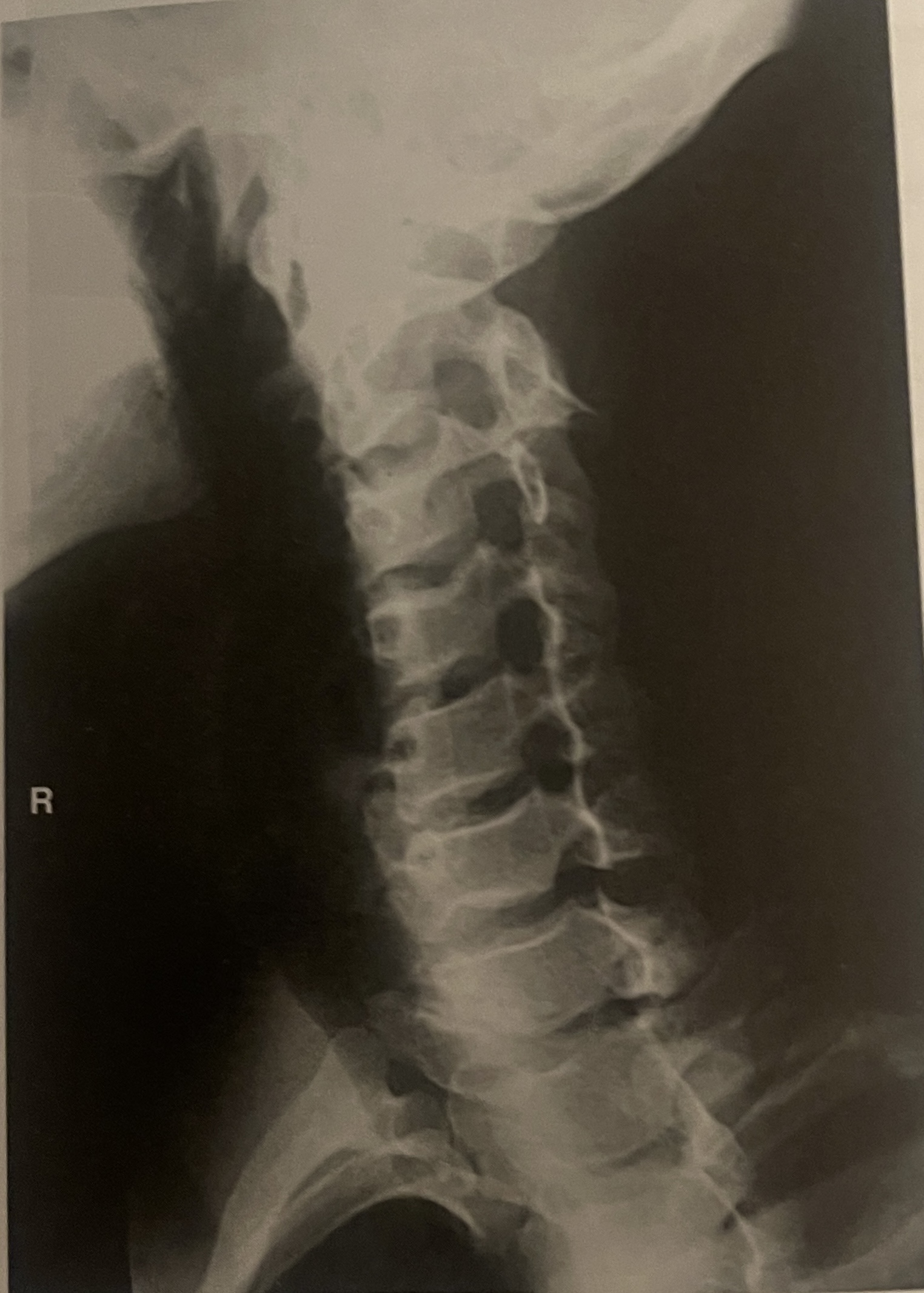
Is the anatomy demonstrated closer or or farther from the IR?
Farther
With the patient positioned in the right anterior oblique position, the intervertebral foramina best demonstrated are those on the patients ______ (right or left) side for the PA axial oblique projection
Right
When the patient is in the standing position, to what level of the patient should the IR be centered for the PA axial oblique projection?
C5
How many degrees should the entire body of the patient be rotated?
45
How many degrees and in what direction should the central ray be angled for the PA axial oblique projection?
15 to 20 degrees caudal
Through which cervical vertebrae should the central ray be directed for the PA axial oblique projection?
C4

What position is shown in the image?
LPO

The intervertebral foramina of which side (left or right) are best demonstrated?
Left
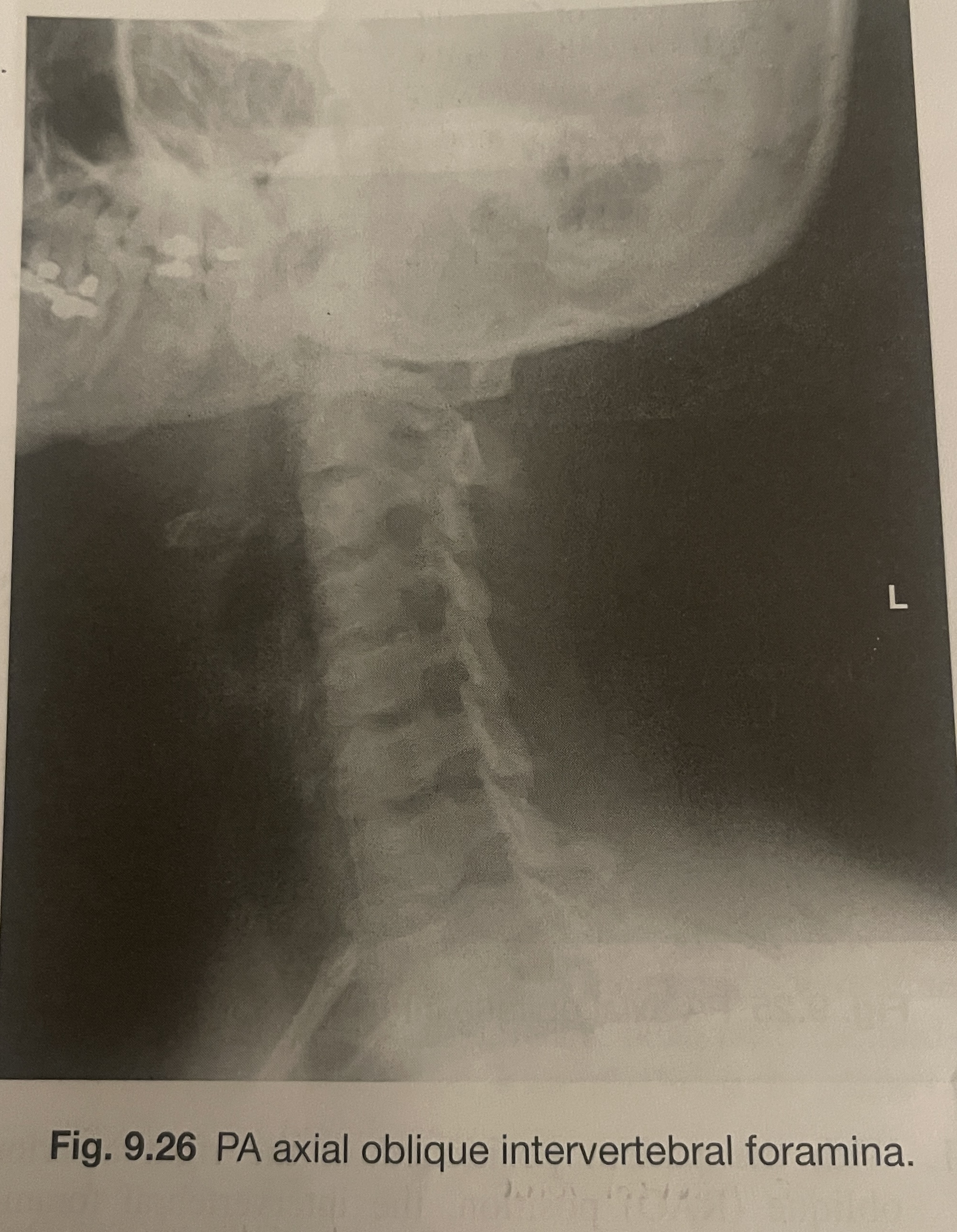
Are the open intervertebral foramina demonstrated in this image closer or farther from the IR?
Closer