BIO 201 final
1/199
There's no tags or description
Looks like no tags are added yet.
Name | Mastery | Learn | Test | Matching | Spaced | Call with Kai | Chat |
|---|
No analytics yet
Send a link to your students to track their progress
200 Terms
Calcium levels in Mr. Gill's blood are dropping to dangerously low levels. The hormone PTH is released and soon blood calcium levels begin to rise. Shortly after, PTH release slows. Is this an example of a positive or negative feedback mechanism? What is the initial stimulus? What is the result?
Negative feedback mechanism. the initial stimulus is the drop in blood calcium levels. PTH is released promoting bones to release calcium. As levels return to normal the parathyroid is signaled to stop the release of PTH
The epidermis is a keratinized stratified squamous epithelium. Explain why that epithelium is much better suited for protecting the body's external surface than a membrane consisting of a simple columnar epithelium would be.
Multiple layers of epithelium compared to just one, increase its ability to protect the external surface of the body. Surface cells have the tough protein keratin to make it waterproof that prevents fluid loss, infection, toxins, and light. Squamous cells are flat, creating a sturdier barrier against physical impact compared to columnar cells which have more absorption abilities.
Osteocytes residing in lacunae of osteons of healthy compact bone are located quite a distance from the blood vessels in the central canals, yet they are well nourished. How can this be explained?
Canaliculi are microscopic channels which allow osteocytes to extend cytoplasmic processes to connect to eachother and the central canal for nutrients, oxygen, and waste disposal.
When a suicide victim was found, the coroner was unable to remove the drug vial clutched in his hand. Explain the reasons for this. If the victim had been discovered three days later, would the coroner have had the same difficulty? Explain.
This is caused by rigor mortis, the natural stiffenning of muscles after death. When ATP is no longer available muscles filaments are locked together. This problem would not be the same 3 days later as muscles would begin to decompose and therefore relax.
Explain the difference between an EPSP and an IPSP.
EPSP (Excitatory Postsynaptic Potential) is the depolarization of the postsynaptic membrane that pushes a neuron to fire and action potential while IPSP (Inhibitory Postsynaptic Potential) is hyper polarization that pushes a neuron away from its threshold and less likely to fire
Indicate the results of sympathetic activation of the following structures: sweat glands, eye pupils, adrenal medulla, heart, bronchioles of the lungs, liver, blood vessels of vigorously working skeletal muscles, blood vessels of digestive viscera, and salivation.
Sweat Glands: produce copious amounts of sweat to cool body
Eye Pupils: Let in more light and improve vision
Adrenal Medulla: release stress hormones (epinephrine and noepinephrine)
Heart: increased heart rate and contraction to pump more oxygen into blood
Bronchioles of lungs: dilate to improve airflow and oxygen intake
Liver: glycogen breakdown to release glucose for energy
Blood Vessels of vigorously working skeletal muscles: dilate to increase blood flow
Blood Vessels of digestive viscera: constrict to shunt blood away from unnecessary areas
Salivation: decreases
relationship between anatomy and physiology
structure determines function
Structural hierarchy of body
chemical, cellular, tissue, organ, organ system, organism
Organ Systems
System | Main Function | Example Organs |
|---|---|---|
Integumentary | Protection, temperature regulation | Skin, hair, nails |
Skeletal | Support, protection, mineral storage | Bones, cartilage |
Muscular | Movement, heat production | Skeletal muscles |
Nervous | Rapid communication | Brain, spinal cord, nerves |
Endocrine | Hormone regulation | Pituitary, thyroid, pancreas |
Cardiovascular | Transport blood | Heart, blood vessels |
Lymphatic/Immune | Defends against disease | Lymph nodes, spleen, thymus |
Respiratory | Gas exchange | Lungs, trachea |
Digestive | Breaks down food | Stomach, intestines, liver |
Urinary | Removes wastes | Kidneys, bladder |
Reproductive | Produces offspring | Ovaries/testes, uterus, penis |
anatomical position
Standing upright
Facing forward
Arms at sides
Palms forward
Feet flat
superior vs inferior
above vs below
anterior (ventral) vs posterior (dorsal)
front vs back
medial vs lateral
closer to midline vs further
proximal vs distal
closer vs farther to attachment
superficial vs deep
closer vs farther from surface
Body planes
Sagittal → left/right
Midsagittal → equal halves
Frontal (Coronal) → front/back
Transverse → top/bottom
Oblique → diagonal
dorsal cavities
cranial and vertebral cavities
ventral cavities
Thoracic: pleural, pericardial, mediastinum
Abdominopelvic: abdominal and pelvic
Components of feedback mechanisms
stimulus → receptor → control center → effector → response
clinical examples of loss of homeostasis
Diabetes mellitus
Hypertension
Heat stroke
Dehydration
types of gradients
concentration gradient: oxygen diffusion
electrical gradient: Na moves towards negative interior of cells
pressure gradient: blood flow
Temperature gradient: warm to cool
electrochemical gradient: neurons
cholesterol in the membrane
stabilizes and maintains fluidity
protein functions in membranes
Functions:
Transport
Receptors
Enzymes
Cell adhesion
Recognition
carbohydrate functions in membranes
Attached to proteins or lipids.
Functions:
Cell recognition
Immune identification
Filtration
Movement driven by pressure.
Example:
Kidney filtration.
primary vs secondary active transport
primary: direct ATP use, Na/K pump (3 Na out and 2 K in)
secondary: stored energy from another ion gradient, glucose absorption in intestine
Bulk Transport
endocytosis (in) or exocytosis (out) of large materials
osmolarity
Total concentration of dissolved particles.
Higher osmolarity = more solutes.
tonicity
Effect of a solution on cell volume.
Hypertonic:higher solute outside cell
Hypotonic: higher solute inside cell
Isotonic: equal
Channal proteins
Allow ions through.
Carrier proteins
Move larger molecules.
Receptor proteins
Receive chemical signals.
Recognition proteins
Identify self from non-self.
Second-messenger system
Hormone binds receptor.
Receptor activates G protein.
G protein activates adenylyl cyclase.
ATP converted into cAMP.
cAMP activates protein kinase.
Cellular response occurs.
Amplifies signals so a small amount of hormone can produce a large response.
Microvilli
Function:
Increase surface area.
Found:
Small intestine
Kidney tubules
Purpose:
Increase absorption.
cilia
Function:
Move substances across cell surface.
Found:
Respiratory tract
Uterine tubes
Examples:
Move mucus out of lungs.
Move egg toward uterus.
epithelial tissue
Tightly packed cells with little extracellular matrix
Forms continuous sheets
Avascular (lacks blood vessels)
Attached to a basement membrane
Exhibits polarity (apical and basal surfaces)
Protection
Absorption
Secretion
cell shapes in epithelial tissue
Squamous: flat cells
Cuboidal: cube-shaped cells
Columnar: tall cells
Transitional: cells change shape when stretched
Simple: one cell layer
Stratified: multiple layers
Pseudostratified: appears multilayered but is one layer
connective tissue
Cells widely separated by extracellular matrix
Matrix contains protein fibers and ground substance
Usually vascular
Support
Protection
Transport
types of connective tissue
Loose
Areolar
Adipose
Reticular
Dense
Dense regular
Dense irregular
Elastic
Supporting Connective Tissue
Cartilage
Hyaline
Elastic
Fibrocartilage
Bone
Fluid Connective Tissue
blood and lymph
Muscle Tissue
skeletal: striated, voluntary
cardiac: striated, involuntary, intercalated discs
smooth: non striated, walls of hollow organs
Nervous tissue
neurons, neuroglia, communication, signaling
Intercellular Junctions
tight junctions
desmosomes
hemidesmosomes
gap junction
tight junctions
seal adjacent cells
prevent leakage
intestinal lining
desmosomes
strong anchoring junctions
resist stretching
skin and cardiac muscle
hemidesmosomes
anchor epithelial cells to basement membrane
gap junctions
Communication channels
Allow ions and small molecules to pass directly between cells
Important in cardiac and smooth muscle
endocrine glands
Definition:
Ductless glands that release hormones directly into the bloodstream.
Examples
Pituitary gland
Thyroid gland
Adrenal glands
Function
Regulate growth, metabolism, reproduction, and homeostasis.
exocrine glands
Definition:
Glands that release secretions through ducts onto epithelial surfaces.
Examples
Sweat glands
Sebaceous glands
Salivary glands
Functions
Lubrication
Cooling
Digestion
Protection
exocrine gland structure
Simple
One unbranched duct
Compound
Branched ducts
functions of integumentary system
Protection
Prevents water loss
Temperature regulation
Sensation
Vitamin D synthesis
Immune defense
Blood reservoir
Excretion of wastes
modes of secretion
Merocrine
Exocytosis
Example: eccrine sweat glands
Apocrine
Part of the cell pinches off
Example: apocrine sweat glands
Holocrine
Entire cell ruptures
Example: sebaceous glands
Layers of the Skin
epidermis
dermis
hypodermis
layers of epidermis
Stratum basale
Stem cells
Melanocytes
Stratum spinosum
Dendritic cells
Stratum granulosum
Keratin production
Waterproofing
Stratum lucidum
Thick skin only
Stratum corneum
Dead keratinized cells
dermis
connective tissue
papillary layer: aerolar connective tissue, capillaries
reticular layer: Dense irregular connective tissue, Hair follicles, Sweat glands
hypodermis
Adipose tissue
Areolar tissue
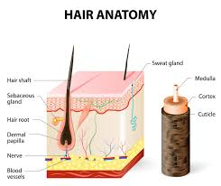
structure of hair
Shaft
Root
Hair follicle
Hair bulb
Dermal papilla
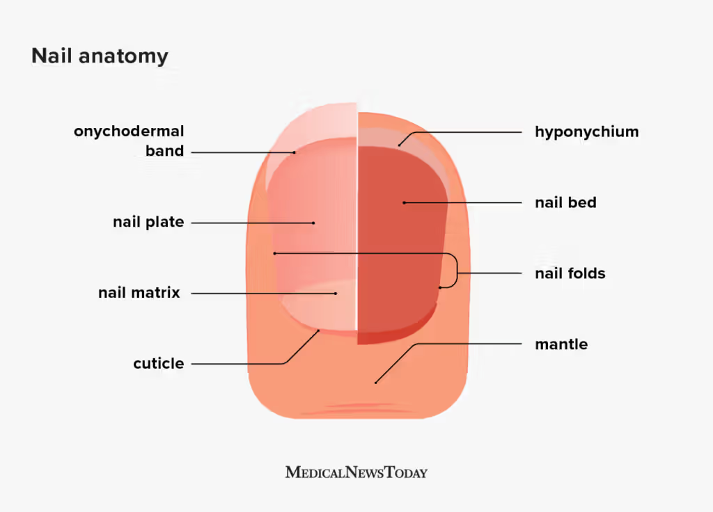
Nail
Nail plate
Nail bed
Nail matrix
Lunula
Cutaneous glands
sebaceous glands
eccrine sweat glands
apocrine sweat glands: groin region
ceruminous: earwax
mammary
Specialized Epidermal Cells
keratinocytes
melanocytes
dendritic cells
merkel cells
keratinocytes
Most abundant epidermal cell.
Function
Produce keratin
Waterproof barrier
Protect against injury
melanocytes
Located in stratum basale.
Function
Produce melanin
Protect DNA from UV radiation
Dendritic Cells (Langerhans Cells)
Immune defense
Detect pathogens
Present antigens to immune cells
merkel cells
Touch receptors
Fine tactile sensation
Basal Cell Carcinoma
Most common
Least dangerous
Originates in stratum basale
Rarely metastasizes
Squamous Cell Carcinoma
Develops from keratinocytes
More aggressive than basal cell carcinoma
May metastasize if untreated
Melanoma
Most dangerous
Originates from melanocytes
Highly metastatic
Early detection is critical
Use the ABCDE rule:
Asymmetry
Border irregularity
Color variation
Diameter greater than 6 mm
Evolving appearance
burns
first degree: epidermis
second degree: epidermis and part dermis
third: epidermis, dermis, and sometimes part of hypodermid
Priorities in Burn Treatment
Stop the burning process.
Maintain airway, breathing, and circulation (ABCs).
Replace fluids to prevent shock.
Prevent infection.
Control pain.
Maintain body temperature.
Promote wound healing and nutrition.
Skin grafting for severe burns.
tissue repair
1) inflammation
2)organization
3) regeneration and fibrosis
functions of skeletal system
protect, support, movement, mineral storage, blood cell formation (hematopoiesis), fat storage, hormone protection
classes of bone
long bones: diaphysis and epiphyses, femur, humerus, tibia
short bones: cube shaped, carpals, tarsals
flat bones: thin and flat, sternum, scapula, ribs
irregular: complex, vertebrae, pelvis
sesamoid bones: develops with tendons, patella
microscopic anatomy of bone
connective tissue of cells in mineralized extracellular matrix
Osteogenic cells
Stem cells
Differentiate into osteoblasts
Found in periosteum and endosteum
osteoblasts
Build new bone
Secrete osteoid (organic bone matrix)
Initiate mineralization
osteocytes
Mature bone cells
Maintain bone tissue
Sense mechanical stress
Communicate through canaliculi
osteoclasts
Break down bone (bone resorption)
Release calcium and phosphorus into the blood
compact vs spongey bone
dense strong outer layer vs porous, contains trabeculae, distributes mechanical stress
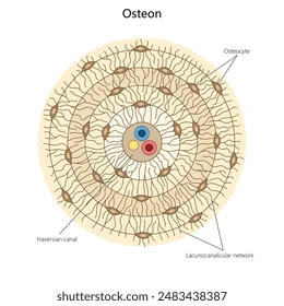
osteon
The osteon is the structural unit of compact bone.
Components:
Central (Haversian) canal: contains blood vessels and nerves
Concentric lamellae: rings of calcified matrix
Lacunae: spaces containing osteocytes
Canaliculi: tiny channels connecting osteocytes
Perforating (Volkmann's) canals: connect central canals
Osteogenesis
intramembranous and endocondrial ossification
intramembranous ossification
flat bones of skull, mandible, clavical
Mesenchymal cells cluster.
Osteoblasts develop.
Osteoid is secreted.
Osteoid calcifies.
Trabeculae form.
Periosteum develops.
Compact bone forms on the surface.
endocondrial ossification
most bones
Hyaline cartilage model forms.
Bone collar develops around diaphysis.
Primary ossification center forms.
Blood vessels invade.
Medullary cavity develops.
Secondary ossification centers form in epiphyses.
Articular cartilage and epiphyseal plates remain.
Postnatal Bone Growth
length growth at epiphyseal plate
Resting cartilage
Proliferating cartilage
Hypertrophic cartilage
Calcified cartilage
Ossification
width growth: Osteoblasts deposit new bone beneath the periosteum while osteoclasts enlarge the medullary cavity.
Bone Deposition
Performed by osteoblasts.
Occurs when:
Calcium is added to bone.
Bone strength increases.
Bone Resorption
Performed by osteoclasts.
Occurs when:
Bone is broken down.
Calcium enters the bloodstream.
Parathyroid Hormone (PTH)
Released when blood calcium is low.
Effects:
Stimulates osteoclast activity indirectly.
Increases calcium reabsorption in the kidneys.
Increases vitamin D activation.
Raises blood calcium levels.
Calcitonin
Released by the thyroid gland when blood calcium is high.
Effects:
Inhibits osteoclast activity.
Promotes calcium deposition in bone.
Lowers blood calcium levels.
Vitamin D (Calcitriol)
Increases calcium absorption from the intestines.
Promotes bone mineralization.
Estrogen and testosterone:
Promote bone growth.
Help close epiphyseal plates.
Maintain bone density in adults.
closed (simple) fracture
Bone does not penetrate the skin.
open (compound) fracture
Bone breaks through the skin.
Higher risk of infection.
complete fracture
Bone is broken all the way through.
incomplete fracture
Bone is partially broken.
greenstick fracture
One side of the bone breaks while the other bends.
Common in children.
communicated fracture
Bone breaks into several pieces.
Fracture Repair
1) hematoma fracture
2) fibrocartilaginous callus formation
3) bony callus formation
4) bone remodeling
osteoporosis
A disease characterized by decreased bone mass and deterioration of bone tissue, resulting in fragile bones and increased fracture risk.
risk factors: aging, females, low calcium intake
functions of muscle tissue
movement, posture, joint stabilization, heat production, control openings