HISTEM W7 Oralfacial Glands - Salivary Glands
1/30
There's no tags or description
Looks like no tags are added yet.
Name | Mastery | Learn | Test | Matching | Spaced |
|---|
No study sessions yet.
31 Terms
What are the 4 characteristics used to classify exocrine glands?
Duct system
Shape of secretory unit
Mode of secretion
Nature of secretion
What are the 2 types of ducts used to classify exocrine glands?
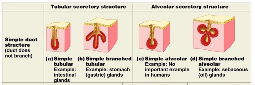
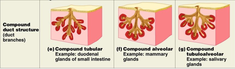
Simple → one duct, no branching
Compound → branching duct, secretory unit empties into smaller ducts which join to form larger duct
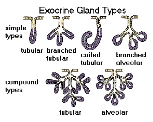
What are the 4 classes of Simple type Exocrine ducts?
Tubular
Branched Tubular
Coiled tubular
Branched alveolar
What are the 2 classes of Compound type Exocrine duct?
Tubular
Alveolar
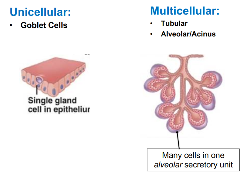
What are the 2 shapes of the secretory unit?
Unicellular → goblet cell
Multicellular → Tubular, Alveolar/Acinus
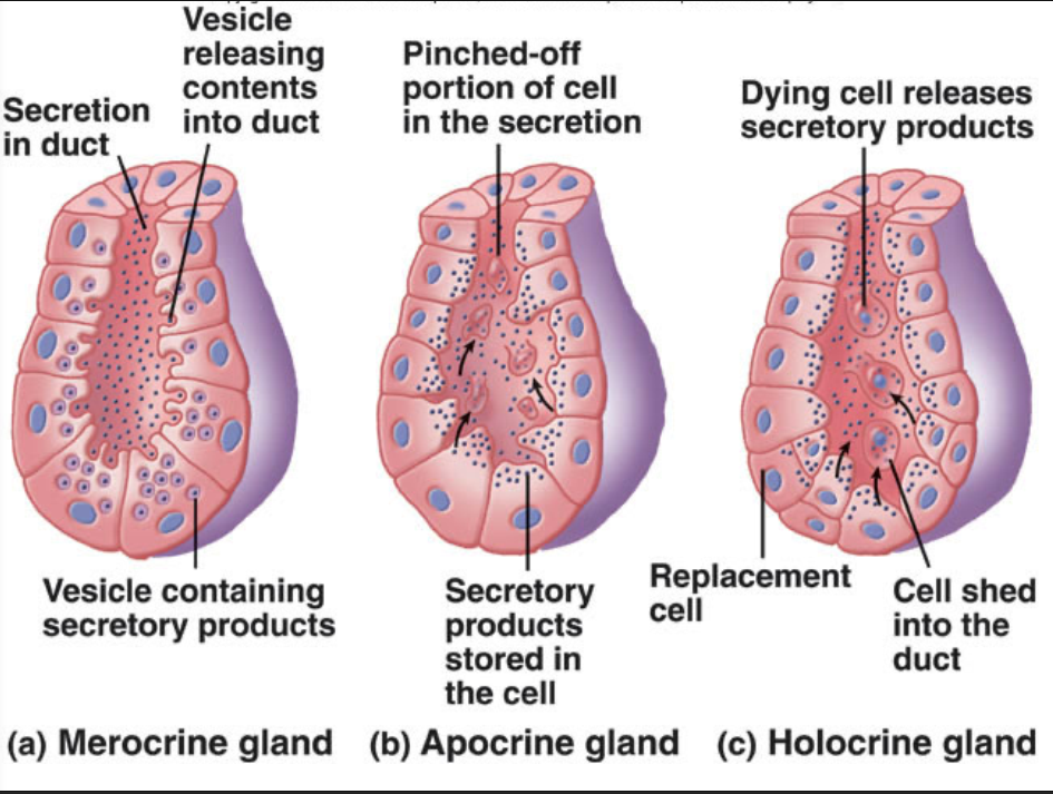
What are the 3 modes of secretion used to classify Exocrine glands? Describe each.
Merocrine
secretes through free surface
no loss of cytoplasm (exocytosis)
ex. Salivary glands, pancreas
Apocrine
secrete small amounts of cytoplasm/cell with secretory product
apical portion of gland pinches off
ex. Mammary glands
Holocrine
entire cells are discharged as secretion
ex. sebaceous gland
What are 3 types of secretions used to classify Exocrine glands?
Serous → contains digestive enzymes (amylase - breaks down carbs)
Mucous → contains mucin (glycoprotein)
Mixed → mix of both
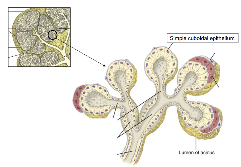
What are Acini/Acinus?
Acini are small, rounded clusters (like grapes) of secretory cells in a gland.
Located at the terminal part of gland connecting to the ductal system
Single layer of CUBOIDAL epithelial cells surrounding a lumen (central opening)
Saliva is deposited into the lumen
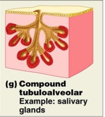
What is the classification for Salivary glands?
Compound Tubular-Alveolar MEROCRINE gland.
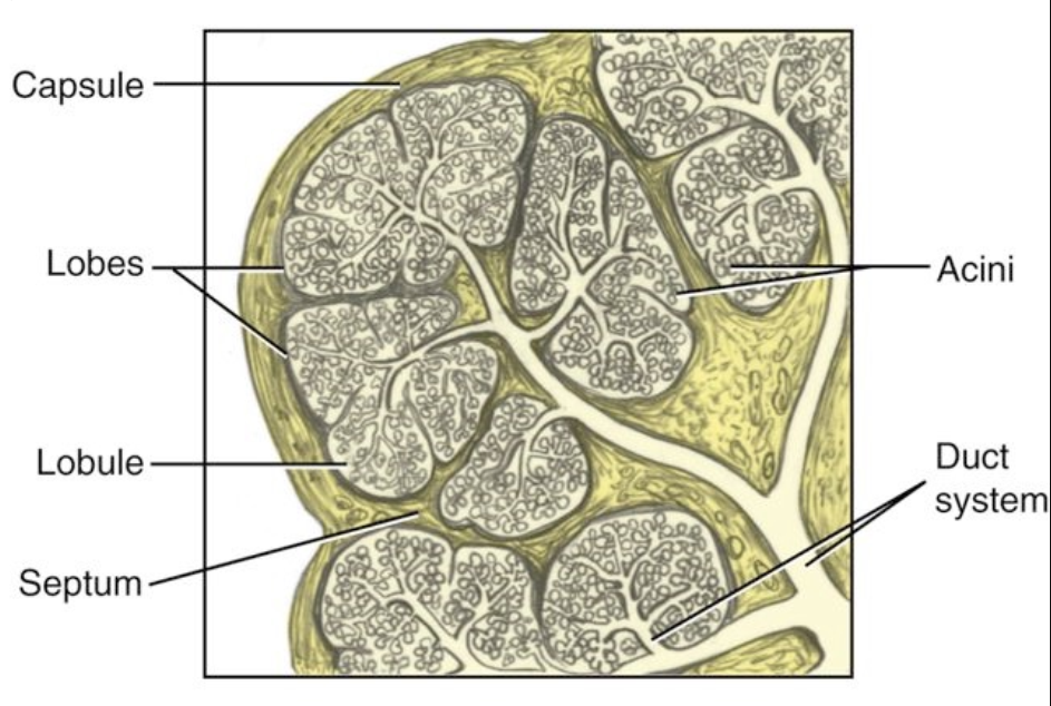
What structures make up the Salivary gland?
Capsule →connective tissue; 20% of gland volume
Lobes
Lobules → contain the glandular units with acini
Septa → interlobar septa and interlobular septa
The duct attached to an acinus is called?
intercalated duct
*acini are terminal ends
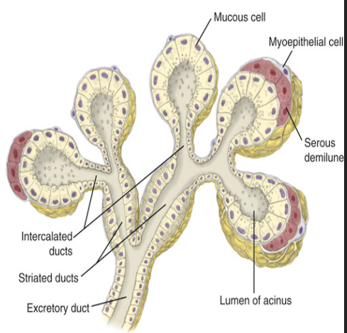
Describe Intercalated ducts of salivary glands
Intercalated ducts
Hollow tube attached to acinus
lined with simple cuboidal epithelium
do not produce secretions
serves as passageway for saliva
Describe Striated ducts of salivary glands. What makes them striated?
connected to intercalated ducts
lined with simple columnar epithelium
cells may have basal striations (basal infoldings of the plasma membrane packed with mitochondria.)
serves as passageway for saliva
cells resorb and excrete electrolytes

Describe the changes in epithelial cell type of Excretory/Secretory ducts in Salivary glands.
Pseudostratified Columnar → Stratified Cuboidal → Stratified squamous (in oral cavity)
What is the difference between major salivary glands and minor salivary glands?
Major:
carries secretions some distance into the oral cavity by a MAIN DUCT
secretes 90% of saliva
Minor:
empties products directly into oral cavity by SHORT DUCT
What are serous demilunes?
Salivary glands that are composed of both serous and mucous cells are called serious demilune.
Serous demilunes are compoased of two different types of cells (serous + mucous) arranged together in the same secretory unit:
Mucous cells form the main body of the acinus
Serous cells cap the end in a crescent shape (the demilune)
What is the functional unit of the salivary gland called?
Alveolus or acinus.
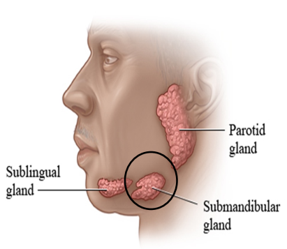
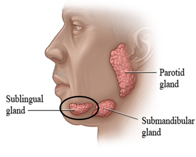
What are the 3 major salivary glands?
parotid, submandibular, sublingual
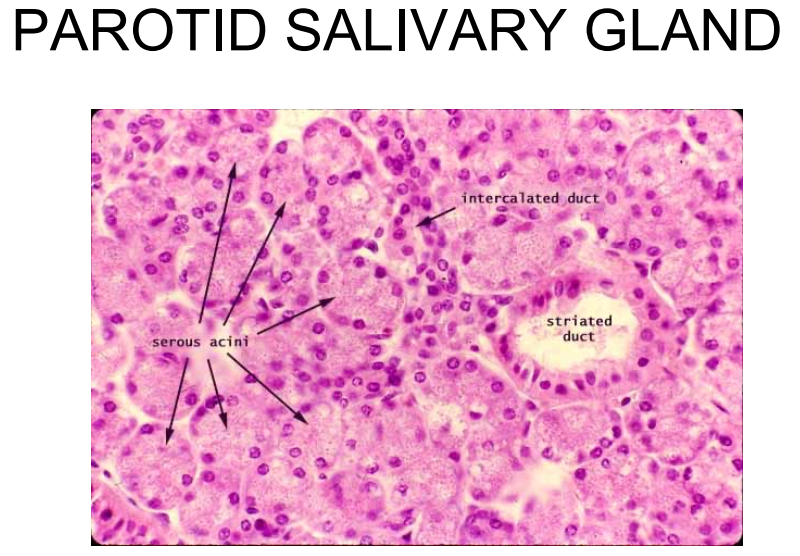
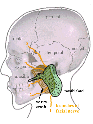
Describe the Parotid gland. Where is it located? What does it secrete?
Largest of the major salivary glands containing only serous acini
Encapsulated
Located behind mandibular ramus, anterior and inferior to the ear, between the skin of cheek and masseter muscle.
Secretions: mainly serious, contributes ~25% of saliva into oral cavity
Describe the duct system of the Parotid gland. Where does the duct open in the oral cavity?
Parotid gland duct system = LONG intercalated ducts with SHORT striated ducts
Stenson’s duct(Parotid duct) opens opposite to 17/27 on buccal mucosa at parotid papilla
Describe the Submandibular salivary gland. Where is it located? What does it secrete?
Second largest of the major salivary glands
Encapsulated
contains mixed glands = serous demilune
Located beneath mandible
Secretions: mainly serous and some mucous secretions; contributing 60-65% of saliva into oral cavity
Describe the duct system of the Submandibular salivary gland. Where does the duct open in the oral cavity?
Submandibular duct system = SHORT intercalated and LONG striated ducts with basal striations
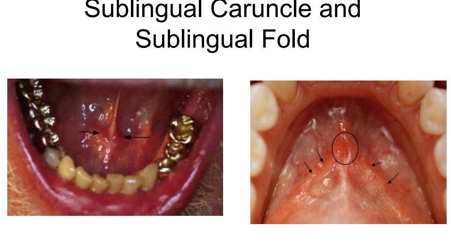
Wharton’s duct (submandibular duct) is located under the tongue near the frenum at the sublingual caruncles.
Describe the Sublingual salivary gland. Where is it located? What does it secrete?
Smallest major salivary gland
NO CAPSULE
mixed glands = mostly mucous
Located on the floor of the mouth in the sublingual fossa, anterior to the submandibular gland
Secretions: mainly mucous secretions and little serous; contributes ~10% of total salivary volume.
Describe the duct system of the Sublingual salivary gland. Where does the duct open in the oral cavity?
Sublingual salivary gland system = 8-30 minor sublingual ducts opening independently into the oral cavity at the sublingual fold.
Bartholin’s duct = formed by several of the small sublingual ducts; opens into oral cavity at the sublingual caruncles
List the minor salivary glands, its location and what it secretes. (5)
Labial → lips → mucous
Buccal → cheek → mucous
Palatine → hard and soft palate → mucous
Lingual → Posterior ?? → mucous
Von ebner’s salivary gland → posterior dorsum of tongue/Zcircumvallate papillae → serous
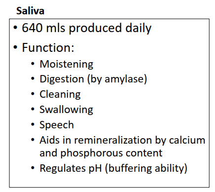
What are the 6 functions of Saliva?
Moisten food and bind food together
Begin digestion (amylase) of carbohydrates
Cleanse mouth and aids in swallowing
Important for speech and articulation
Remineralization of enamel surface (reverse cavities) with the calcium and phosphate in saliva
Regulate pH in the mouth
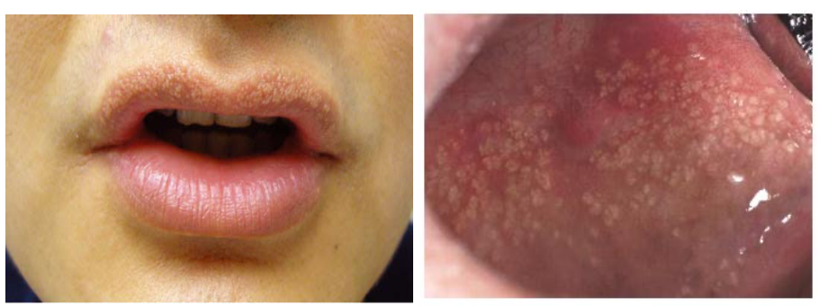
What are misplaced sebaceous glands in the oral cavity called?
Fordyce Granules → mall, yellowish-white, or slightly reddish bumps in oral cavity
60-70% of adults have them
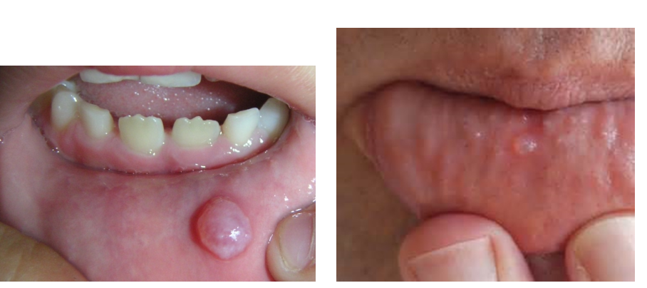
What is a mucocele?
Accumulation of saliva in the mucosa due to trauma to a minor salivary duct
*mucoepidermoid carcinoma kinda looks like a mucocele but its more commonly located by/at/in the parotid salivary gland
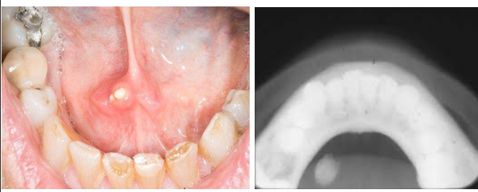
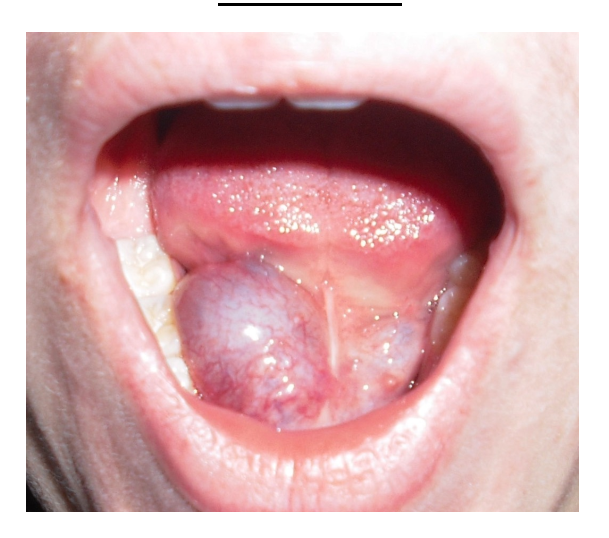
What is a sialolith? What is a ranula?
Salivary stones that block salivary flow
can cause ranula to form.
Ranula - filled with saliva that has leaked from a damaged salivary gland. This leakage leads to a fluid-filled swelling, often appearing bluish or translucent.
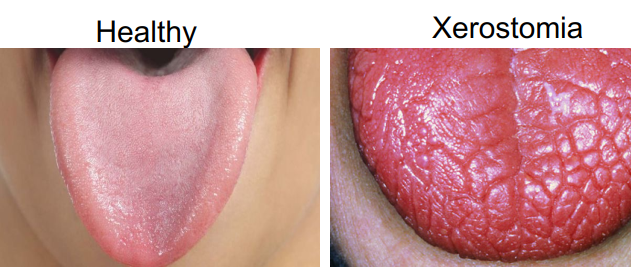
What is Xerostomia?
Dry mouth
caused by decrease in salivary flow or absent.
Decreased salivary flow = increase in bacteria, calculus increase, increased periodontal disease progression
What is Sjogren’s syndrome?
Sjogren’s syndrome is a chronic autoimmune disease where the immune system attacks salivary and lacrimal glands.
Main symptoms: Dry mouth (xerostomia) and dry eyes (xerophthalmia)
Can occur alone (primary) or with other autoimmune disorders (secondary)
Leads to reduced saliva and tear production