Skeleton

0.0(0)
Studied by 0 people
call kaiCall Kai
Locked
learnLearn
examPractice Test
spaced repetitionSpaced Repetition
heart puzzleMatch
flashcardsFlashcards
GameKnowt Play
Card Sorting

1/19

encourage image

There's no tags or description

Looks like no tags are added yet.

Last updated 3:40 AM on 7/23/26
Name
Mastery
Learn
Test
Matching
Spaced
Call with Kai
Chat

No analytics yet

Send a link to your students to track their progress

20 Terms

1
New cards

3 Functions of the skeleton

  1. Protective

  2. Locomotor functions

  3. Manipulating functions

2
New cards

Axial Skeleton

Approx 80 bones

  • Skull

  • Vertebral column

  • Ribs

  • Sternum

Longitudinal Axis of body

Protects:

  • Brain - Skull

  • Spinal cord - Vertebrae

  • Lungs & heart - Ribs & sternum

3
New cards

Apendicular Skeleton

Bones of upper & lower limbs

4
New cards

Compact/Cortical bones

Thick, dense outer layer

  • Protection & strength to bear weight

5
New cards

Spongy/cancellous bone

Inner mesh containing bone marrow

  • highly vascularised

6
New cards

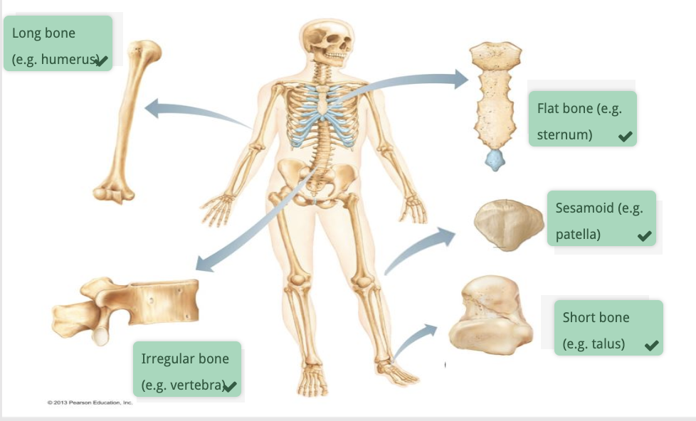
Bone types

<p></p>
7
New cards

Long bones

  • Shaft - Diaphysis

  • Ends - Epiphysis

  • Inner spongy bone - Medullary cavity

  • Growth plates - Made of hyaline cartilage

  • Metaphysis - diaphysis near growth plates

8
New cards

Bone landmarks

  • Articular surfaces - covered with hyaline cartilage

  • Protrusions - attachment sites for tendons and ligaments

    • Tubercles

    • Trochanters

    • Spines

    • Processes

  • Depressions/holes - space for other structures (e.g nerve or blood vessel) to pass by/through

    • Foramen - hole in bone

    • Groove

    • Notches

    • Fossae

9
New cards

Endochondral Ossification

Endo - Inner

Chondral - Cartilage

“Internal cartilage becomes bone”

Hyaline cartilage template → Long, short & most irregular bones

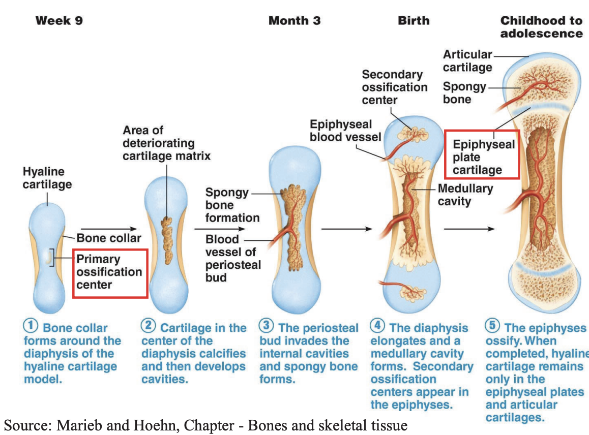
  1. At week 9 of pregnancy, the bone collar of cortical bone forms around the diaphysis of hyaline cartilage models of long, short and most irregular bones before birth.

  1. In the centre of the bone collar is the primary ossification centre, where cartilage calcifies with cavities, hollowing the diaphysis.

  1. At month 3 of pregnancy, a periostal bud implants within the cavities of the diaphysis, growing internal blood vessels that provide the nutrients for the formation of spongy bone.

  1. Once birth occurs, the epiphysis of these bones ossify, becoming the secondary centres of ossification. The diaphysis continues to grow, elongating the bone, thus creating more internal spongy bone that form the medullary cavity

  1. Until adolescence, the diaphysis continues to grow along the cartilagenous growth plate. This growth continues until the diaphysis meets the epiphysis, solidifying into an epiphyseal line in adulthood.
<p>Endo - Inner</p><p>Chondral - Cartilage</p><p>“Internal cartilage becomes bone”</p><p></p><p><span style="background-color: transparent;">Hyaline cartilage template → Long, short &amp; most irregular bones</span><br></p><ol><li><p><span style="background-color: transparent;">At <strong><u>week 9</u> </strong>of pregnancy, the <strong>bone collar of cortical bone</strong> forms around the <strong>diaphysis</strong> of hyaline cartilage models of long, short and most irregular bones before birth.</span></p></li></ol><p></p><ol start="2"><li><p><span style="background-color: transparent;">In the centre of the bone collar is the primary ossification centre, where cartilage calcifies with cavities, hollowing the diaphysis.</span></p></li></ol><p></p><ol start="3"><li><p><span style="background-color: transparent;">At month 3 of pregnancy, a periostal bud implants within the cavities of the diaphysis, growing internal blood vessels that provide the nutrients for the formation of spongy bone.</span><br></p></li></ol><ol start="4"><li><p><span style="background-color: transparent;">Once birth occurs, the epiphysis of these bones ossify, becoming the secondary centres of ossification. The diaphysis continues to grow, elongating the bone, thus creating more internal spongy bone that form the medullary cavity</span></p></li></ol><p></p><ol><li><p><span style="background-color: transparent;">Until adolescence, the diaphysis continues to grow along the cartilagenous growth plate. This growth continues until the diaphysis meets the epiphysis, solidifying into an epiphyseal line in adulthood.</span></p></li></ol><p></p>
10
New cards

Zoom into epiphyseal plate

  1. Resting zone - young chondrocytes

  2. Proliferation zone - young chondrocytes undergo mitosis

  3. Hypertrophic zone - old chondrocytes enlarge

  4. Calcification zone - old chondrocytes calcify, matrix hollows, blood vessels vascularise bone

  5. Ossification zone - New bone forms

Within adults, the growth plate ossifies to become an epiphyseal line.
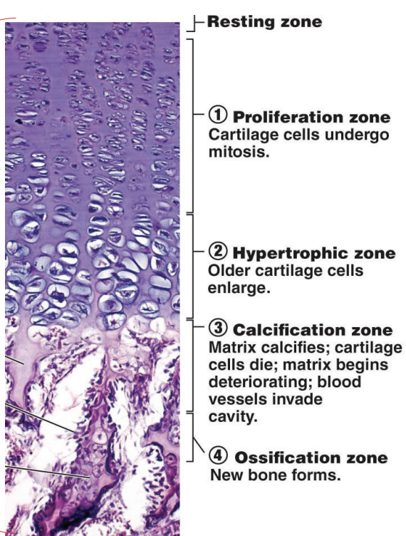
<p></p><ol><li><p><span style="background-color: transparent;">Resting zone - young chondrocytes</span></p></li><li><p><span style="background-color: transparent;">Proliferation zone - young chondrocytes undergo mitosis</span></p></li><li><p><span style="background-color: transparent;">Hypertrophic zone - old chondrocytes enlarge</span></p></li><li><p><span style="background-color: transparent;">Calcification zone - old chondrocytes calcify, matrix hollows, blood vessels vascularise bone</span></p></li><li><p><span style="background-color: transparent;">Ossification zone - New bone forms</span></p></li></ol><p></p><p>Within adults, the growth plate ossifies to become an <strong><u>epiphyseal line.</u></strong></p>
11
New cards

Intramembranous Ossification

Intra - Within

Membranous - Membrane

“Bone forms within a membrane”

Bones develop within a membrane → flat bones & skull bones

  1. Bone cells form within a membrane.

  2. A collagen matrix is laid down which ossifies, where ground substance and minerals like calcium and phosphate are added

  3. Blood vessels vascularise bone to promote growth.

  4. Eventually, the formation of bone is layered as follows: Osteocytes, cortical bone, spongy bone, cortical bone, osteocytes

  5. Bones formed: flat bones of the skull, mandible & clavicles

12
New cards

Appositional growth

  1. Osteons (units of bone) form within cortical bone

  2. More osteons create thicker bone

  3. Layers of lamella cover the circumference of the bone, making it wider
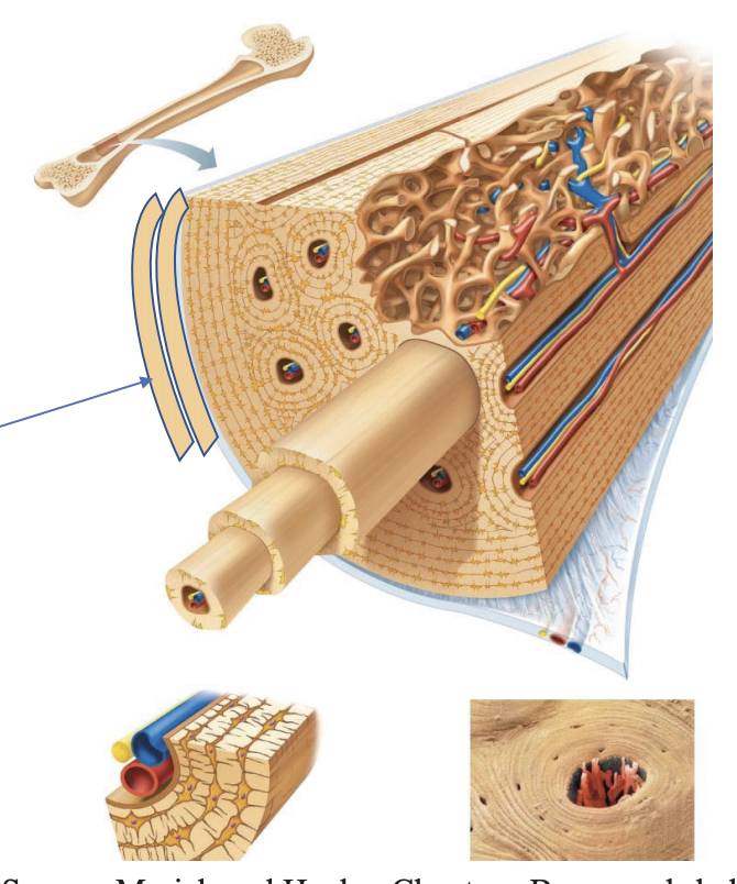
<ol><li><p><strong><em><u>Osteons</u></em></strong> (units of bone) form within cortical bone</p></li><li><p>More osteons create thicker bone</p></li><li><p>Layers of <strong><em><u>lamella</u></em></strong> cover the circumference of the bone, making it wider</p></li></ol><p></p>
13
New cards

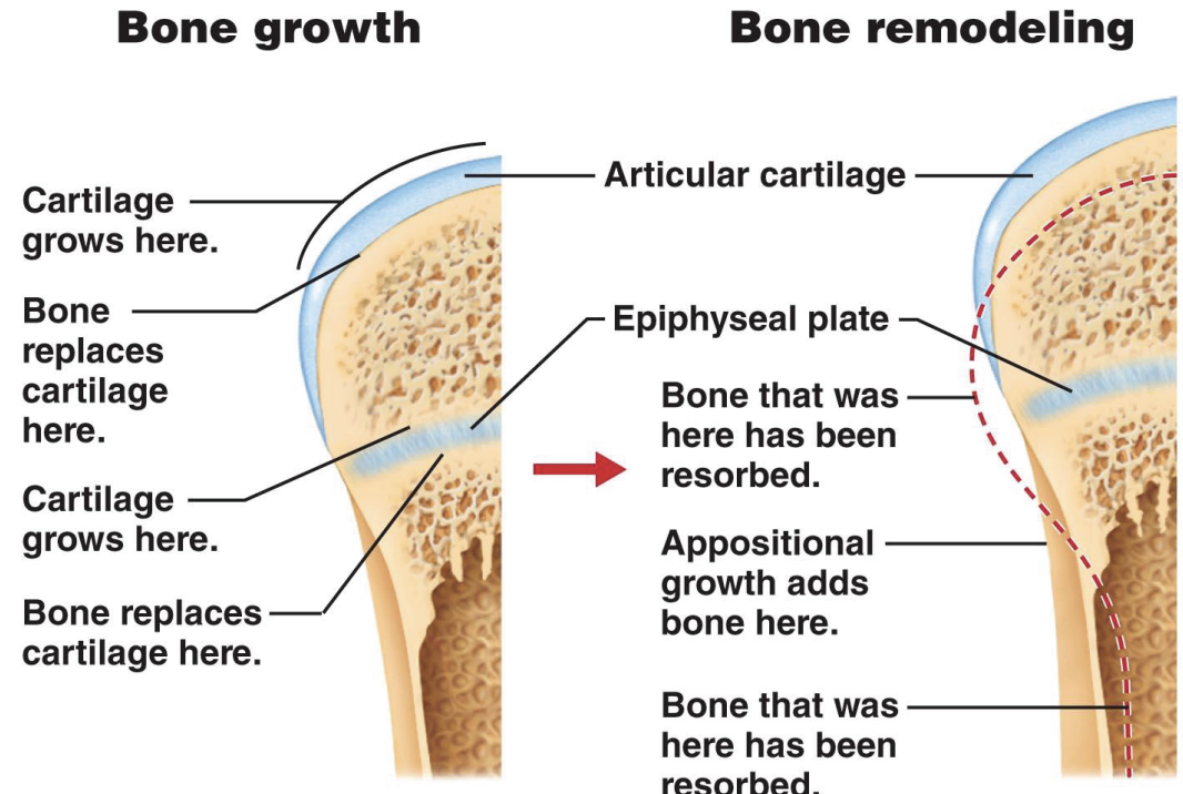
Bone remodelling:

Wolff’s Law

Bone will adapt to the loads under which it is placed

  • Bone is laid down where it’s needed and reabsorbed where it’s not needed

Best to mention Appositional growth:

  • Increased Appositional growth on the arm with greater stress

  • Larger cross-sectional area

  • More cortical bone to bear more strength

  • Thus, greater bone density

<p>Bone will adapt to the loads under which it is placed</p><ul><li><p>Bone is laid down where it’s needed and reabsorbed where it’s not needed</p></li></ul><p></p><p><strong><em><u><mark data-color="yellow" style="background-color: yellow; color: inherit;">Best to mention Appositional growth:</mark></u></em></strong></p><ul><li><p>Increased Appositional growth on the arm with greater stress</p></li><li><p>Larger cross-sectional area</p></li><li><p>More cortical bone to bear more strength</p></li><li><p>Thus, greater bone density</p></li></ul><p></p>
14
New cards

Bone spurs/Osteophytes

Due to Wolff’s law, in the event of increased use of bones (e.g bending back too much):

  • Bones can rub against each other, causing spurred edges called osteophytes

  • e.g osteophytic lipping of the vertebrae
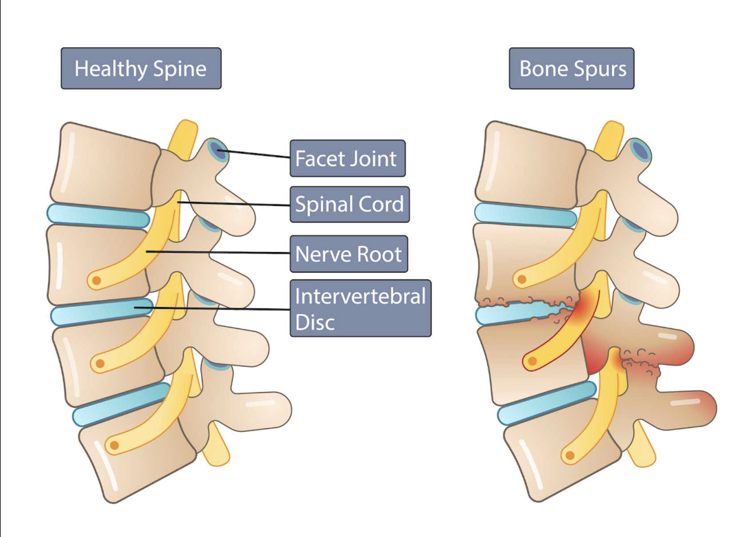
<p>Due to Wolff’s law, in the event of increased use of bones (e.g bending back too much):</p><ul><li><p>Bones can rub against each other, causing spurred edges called <strong><em><u>osteophytes</u></em></strong></p></li><li><p><em>e.g osteophytic lipping of the vertebrae</em></p></li></ul><p></p>
15
New cards

Bone remodelling:

Calcium homeostasis

  • Bones are inherently the calcium reservoirs of the body

  • Bones are laid down - Osteoblasts

  • Bones are reabsorbed - Osteoclasts

    • Osteoclast activity can be up-regulated or down-regulated depending on the scenario

    • Up-regulated - Less stress on bone

    • Down-regulated - More stress on bone

16
New cards

Bones fracture repair

Post-fracture timeline:

6-8hrs: Fracture Hematoma:

  • Proximal blood vessel gathers blood which clots around the fracture

  • This limits the ability for new blood to reach the fracture point, causing the bone lining the fracture point to die.

48hrs: Soft callus:

  • Internal callus: Chrondrocytes within the endosteum (inner lining of the medullary cavity)

    • Secrete fibrocartilaginous matrix between the two ends of bone

  • External callus: More chondrocytes and osteoblasts

    • Create hyaline cartilage (chondrocytes) & bone (osteoblasts)

    • This stabilises the fracture

Few weeks later: Hard callus

  • Osteoclasts absorb dead bone/bony fragments

  • Osteogenetic cells become extremely active

    • Differentiate into osteoblasts

  • Fibrocartilaginous matrix of the internal callus becomes trabecular bone

  • Internal & external callus unite

  • Spongy bone on the outer layers become cortical bone

Final stage: Bone remodelling

  • Any swelling of the outer bone undergoes remodelling to return to the normal shape
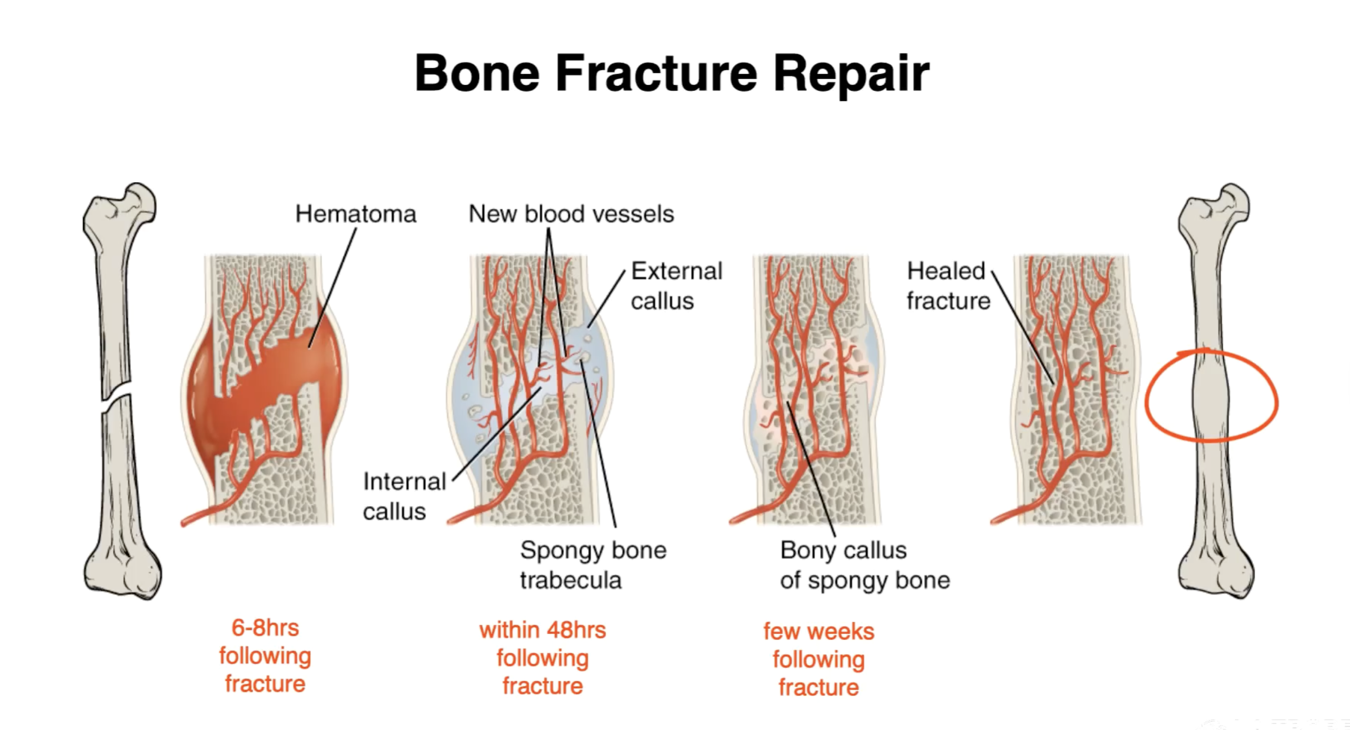
<p>Post-fracture timeline:</p><p>6-8hrs: Fracture Hematoma:</p><ul><li><p>Proximal blood vessel gathers blood which clots around the fracture</p></li><li><p>This limits the ability for new blood to reach the fracture point, causing the bone lining the fracture point to die.</p></li></ul><p></p><p>48hrs: Soft callus:</p><ul><li><p>Internal callus: Chrondrocytes within the endosteum (inner lining of the medullary cavity)</p><ul><li><p>Secrete fibrocartilaginous matrix between the two ends of bone</p></li></ul></li><li><p>External callus: More chondrocytes and osteoblasts</p><ul><li><p>Create hyaline cartilage (chondrocytes) &amp; bone (osteoblasts)</p></li><li><p>This stabilises the fracture</p></li></ul></li></ul><p></p><p>Few weeks later: Hard callus</p><ul><li><p>Osteoclasts absorb dead bone/bony fragments</p></li><li><p>Osteogenetic cells become extremely active</p><ul><li><p>Differentiate into osteoblasts</p></li></ul></li><li><p>Fibrocartilaginous matrix of the internal callus becomes trabecular bone</p></li><li><p>Internal &amp; external callus unite</p></li><li><p>Spongy bone on the outer layers become cortical bone</p></li></ul><p></p><p>Final stage: Bone remodelling</p><ul><li><p>Any swelling of the outer bone undergoes remodelling to return to the normal shape</p></li></ul><p></p>
17
New cards

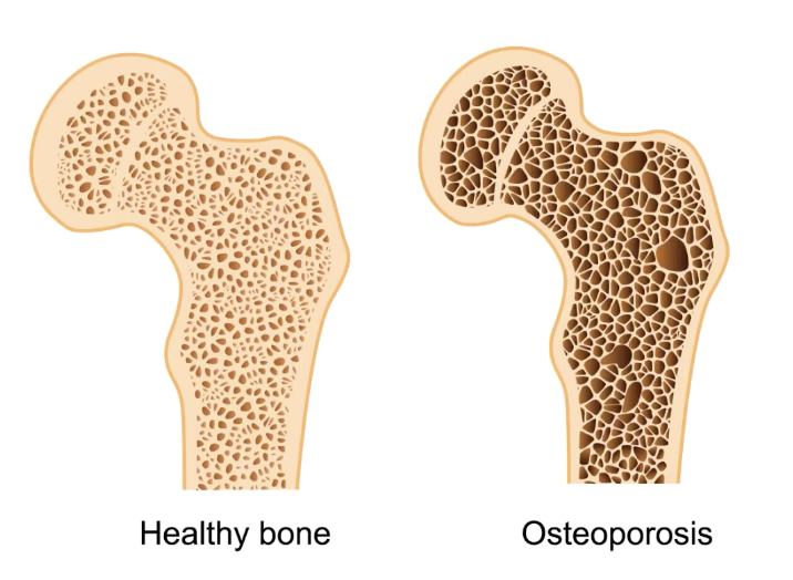
Osteoporosis

Bone resorption overtakes formation. Decreased bone density

Most effected bones:

  • Proximal ends of femur

  • Vertebrae

  • Carpals

<p>Bone resorption overtakes formation. Decreased bone density</p><p>Most effected bones:</p><ul><li><p>Proximal ends of femur</p></li><li><p>Vertebrae</p></li><li><p>Carpals</p></li></ul><p></p>
18
New cards

Who is prone to osteoporosis? Why?

Women over 50.

When menopause occurs, estrogen production stops. Estrogen activates osteoblasts.

19
New cards

How to reduce the risk of osteoporosis?

  • Maintain calcium lvls

  • Maintain Vit D lvls

  • Regular resistance training

Strenghten existing bone

20
New cards