Lecture 5 - Muscle
1/51
There's no tags or description
Looks like no tags are added yet.
Name | Mastery | Learn | Test | Matching | Spaced |
|---|
No study sessions yet.
52 Terms
Characteristics of skeletal muscle.
→ Strong, quick, voluntary contractions
has large, elongated, multinucleated fibers
AKA “striated muscle”

Characteristics of Cardiac muscle.
→ Vigourous, rhythmic, involuntary contractions
has irregular branched cells bound together longitudinally by intercalated discs
intercalated discs connect one fibre with another

Characteristics of smooth muscle.
slow, involuntary contractions
made of groups of smaller, fusiform (spindle/tappered) cells

What fundamental interaction occurs in all types of muscle contraction?
Thick myosin filaments sliding along thin actin filaments
Translate the following into muscle terms.
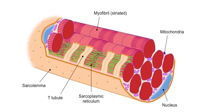
Cytoplasm
Smooth ER
Membrane/Plasmalemma
Cytoplasm = sarcoplasm
Smooth ER = sarcoplasmic reticulum
Membrane/Plasmalemma = sarcolemma
List the 3 CT layers of skeletal muscle.
Epimysium
Perimysium
Endomysium
What is the Epimysium?
Thick layer of dense CT that encloses the entire skeletal muscle
Continuous with fascia and tendon (binding muscle to bone)

What is the Perimysium?
Thin CT
Surrounds and separates fascicles (bundles of muscle fibers)
Nerves, blood vessels, and lymphatics penetrate here to supply fascicles

What is the Endomysium?
Delicate CT
Surrounds individual muscle fibers (elongated multinucleated cells)
may include fibroblast nuclei

What type of collagen do all three CT skeletal muscle layers contain?
Type 1 & Type 3 (reticulin)
How do tendons develop and where do they attach?
develop together with skeletal muscles and attach muscles to the periosteum of bones

What structural feature makes the myotendinous junction strong?
Dense collagen fibers of tendons are continuous with CT around muscle fibers, forming a strong unit that allows contraction to move other structures
How are striations aligned within the skeletal muscle?
Thousands of dark A bands alternate with lighter I bands
Striations may look slightly misaligned between fibers but are straight within each unit

What are myofibrils?
→ parallel, cylindrical bundles inside each muscle fiber, extending the entire length of the fibre
made up of repeating sarcomeres separated by Z discs that contain thick and thin myofilaments
surrounded by parts of the SR
What are myofilaments?
consists of thick & thin filaments
Thick - has 200–500 myosin
Thin - has F-actin, tropomyosin, and troponin
Thick and thin filaments overlap in regions of the sarcomere
organized into contractile protein arrays bundled within myofibrils

What are T-tubules?
deep invaginations in the sarcolemma
Encircle each myofibril near the A-band/I-band boundaries of sarcomeres
each T-tubule is associated with 2 terminal cisternae of the SR → 1 T-tubule + 2 terminal cisternae = a triad
Triads are located along the surface of myofibrils
Seen in TEM images as tubules perpendicular to the fiber surface, extending between myofibrils

How are T-tubules seen in TEM images?
Tubules perpendicular to the fiber surface, extending between myofibrils

What is the function of T-tubules?
Trigger simultaneous Ca²⁺ release from the SR → rapid & uniform contraction of all myofibril
What is a sarcomere?
→ contractile and load-bearing unit of muscle fibers
Extends from Z disc to Z disc

What is the A-band?
→ electron-dense, central region of the sarcomere
Contains thick filaments (myosin) and overlapping thin filaments (actin)
Middle of A band has a lighter H zone

What is the H-zone?
region with only rodlike portions of myosin, no thin filaments

What is the M-line?
Splits the H-zone in half
contains
Myomesin - myosin-binding protein that holds thick filaments in place
Creatine kinase - transfers phosphate from phosphocreatine to ADP → ATP supply

What are the I-bands?
contains only thin filaments
at the ends of the A-band
Each are split in half by a Z disc

What is the Z-disc?
ends of sarcomere
acts as an “anchor” for thin filaments

Label.


What’s the difference b/w thick & thin filaments?
Thin filaments = actin filaments, anchored to α-actinin in the Z disc
Thick filaments = bundles of myosin, spanning the entire A band
What is titin?
→ Giant, springlike protein that connects thick filaments to Z disc across I bands
Provides elasticity and alignment

Outline the features of Extraocular Muscles (EOMs).
Made of 2 main types of muscle fibers
Fast-twitch fibers → fast eye movements
have well-defined myofibrils and well-developed sarcomeres
Slow-twitch fibers → slow/tonic eye movements
have poorly defined myofibrils and poorly developed sarcomeres
Differ histologically from most skeletal muscles
What motor neuron supplies the Fast twitch and slow twitch fibers? How do they differ in innervation and NMJ?
→ Both types supplied by cholinergic motor neurons (release ACh)
Fast-twitch fibers
Innervation: thick, heavily myelinated axons
1 NMJ
Slow-twitch fibers
Innervation: thin axons
Multiple, grapelike clusters of NMJ
What “special feature” of EOMs allows for precise, yoked eye movements?
have the smallest motor units of any skeletal muscle → extremely precise contraction/relaxation and coordinated opposition (e.g., lateral vs. medial rectus)
What are the 2 distinct layers of rectus muscles, and what types of fibers do they contain?
Global layer (inner) - ~62% fast fibers
Orbital layer (outer) - ~81% fast fibers
Both contain mixed fiber types, mostly fast with some slow and a small intermediate category

Where do myelinated motor neurons branch, and what do they form when they reach the muscle fiber?
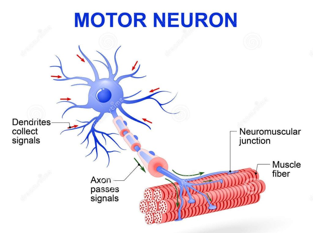
Myelinated motor nerves → branch in perimysium → give rise to unmyelinated terminal twigs → these penetrate the endomysium (the innermost CT layer) → then form synapses (NMJ) with individual muscle fibers
What is a NMJ (aka MEP)?

→ widened end of a nerve branch that sits on the surface of a muscle fiber
contains mit and ACh-filled synaptic vesicles
What role do Schwann cells play at NMJs?
wraps around the unmyelinated axon branches, covering the nerve endings where they meet the muscle fiber.


How is the postsynaptic membrane (sarcolemma) specialized at the NMJ?
has deep junctional folds that ↑SA and contain many ACh receptors
What is the general structure of Cardiac muscle?
Fibers are made of separate cells joined end-to-end at intercalated discs
Cells are often branched, allowing fibers to interweave in a helical arrangement within fascicles → helps efficient contraction of the heart
Central nuclei (euchromatic) present
Myofibrils are scattered and less organized
SR is less organized
Striations are closely spaced but less defined

What is the function of intercalated discs?
Ensure fibers stick together physically and coordinate contraction electrically
Compare the Transverse & Longitudinal regions of the intercalated discs.
Transverse regions:
Highly interdigitated
have desmosomes and fascia adherens → provide strong mechanical adhesion
Longitudinal regions:
have gap junctions → allow electrical communication b/w cells

How do the t-tubules compare in ventricles vs atrial fibers?
Ventricular fibers → well-developed, wide lumens near Z discs → rapid Ca²⁺ delivery for strong contractions
Atrial fibers → smaller or absent
Instead of triads, cardiac muscles have…
Dyads = 1 terminal cisternae + 1 t-tubule
How is contraction in cardiac muscle controlled, and how does innervation affect it?
Cardiac muscle contracts intrinsically and spontaneously
Each fiber contracts in an all-or-none manner
Autonomic innervation modifies rate:
Sympathetic → ↑ impulse frequency
Parasympathetic → ↓ impulse frequency
*since cardiac muscle is under ANS control, it is involuntary*
Outline the descriptive features of smooth muscle.
What control?
Cell shape?
Function?
Where is it found?
Specialized for slow, steady contraction
Involuntary control (b/c it’s under ANS control)
Cells - elongated, tapering, non-striated fibers
covered by an external lamina + network of type I & III collagen (reticulin)
Function
Make ECM components - collagen, elastin, proteoglycans
Contribute to fibroblast-like activity
❌ NMJ, t-tubules, striations
Found in blood vessels, digestive, respiratory, urinary, and reproductive systems
How is smooth muscle in the digestive tract organized, and what does this allow?
2 layers
Inner circular (IC)
Outer longitudinal (OL)
Layers coordinate contraction to allow for peristalsis
What is the role of dense bodies in smooth muscle contraction?
Thin filaments connect to dense bodies located in the membrane and cytoplasm
α-actinin - protein that holds these filaments to the dense bodies
Dense bodies also serve as anchor points for intermediate filaments and cell junctions
This setup enables the tissue to contract smoothly and efficiently as a coordinated unit

List the 2 types of Smooth muscle.
Unitary
Multiunit
What is the Unitary Smooth Muscle?
→ All SM cells acts as a complete unit b/c cells are linked by gap junctions
Eg: GI tract, bladder, uterus, ureter, and iris sphincter

What is the Multiunit Smooth Muscle?
→ each SM cell acts as an individual unit (like skeletal fibres) b/c they’re innervated by post-ganglionic fibers of PNS & SNS
Eg: ciliary muscle and vas deferens

How does smooth muscle compare to skeletal muscle in strength and endurance?
→ Less strong than skeletal muscle but can sustain longer contractions.
Ex: levator palpebrae superioris lifts the eyelid; Mueller’s muscle keeps it open between blinks
What are myoepithelial cells and where are they found?
→ True epithelial cells with smooth muscle-like processes
express keratins and smooth muscle actin
function under SNS control
Ex: found in exocrine gland ducts and iris dilator muscle
How does diabetes affect myoepithelial cells vs. true smooth muscle?
Myoepithelial cells (like the dilator) accumulate excess glycogen, impairing their function
True smooth muscle (like the sphincter) is not affected
explains why diabetics respond bad to dilating drops but constrict normally

What are myofibroblasts and where are they found?
→ Cells found sub-epithelially in mucosal surfaces (liver, lung, kidney)
involved in fibrosis (b/c they’re fibroblasts) and wound contraction

What markers are expressed by myofibroblasts?
Vimentin (mesenchymal marker)
α-smooth muscle actin (ACTA2)
Palladin (actin cytoskeleton scaffold protein)