L1 - Skeletal Musc Anatomy & L2 - Neuromuscular Anatomy and Signaling
 Call Kai
 Learn
Learn Practice Test
 Spaced Repetition
 Match
Match Flashcards
 Knowt Play
1/31

There's no tags or description
Looks like no tags are added yet.
Name | Mastery | Learn | Test | Matching | Spaced | Call with Kai | Chat |
|---|
No analytics yet
Send a link to your students to track their progress
32 Terms
What are the primary functions of a skeletal muscle and a motor neuron?
Muscle: Generate force and allow for movement.
Motor Neuron: Signal the muscle to contract and allow for voluntary control.
What is the general chemical composition of skeletal muscle tissue by percentage?
Approximately 75% water, 20% protein, and 5% salt and other substances (phosphates, ions, etc.).
List the structural hierarchy of skeletal muscle from largest/macro structure down to the smallest/protein level (6 levels).
Muscle —> Fascicle —> Fiber (Cell) —> Myofibril —> Sarcomere —> Filaments/Proteins.

What are the three layers of connective tissue in skeletal muscle?
Epimysium (surrounds the whole muscle)
Perimysium (surrounds fascicles)
Endomysium (surrounds individual muscle fibers)

What are the 4 main functions of connective tissue in skeletal muscle?
Scaffolding for fiber formation during growth; determines muscle structure.
Conduit for blood vessels and nerves supplying fibers.
Aids in resistance to passive stretch, helps reform shape, and distributes force to minimize damage.
Conveys contractile force to tendons.
What is the sarcolemma and what are its key properties?
It is the plasma membrane (lipid bilayer) surrounding each muscle fiber beneath the endomysium. It conducts action potentials (excitable) and contains receptors for hormones and other molecules.
What are the functions of the basement membrane in muscle fibers?
Fuses with the epimysium to attach muscle fibers to the neuromuscular junction (NMJ) and tendons.
Termination of synaptic transmission.
Acts as scaffolding for fiber regeneration.
Regulates the NMJ and guides the regeneration/insertion of acetylcholine (ACh) receptors.
What makes up a "Triad" in skeletal muscle, and what is its purpose?
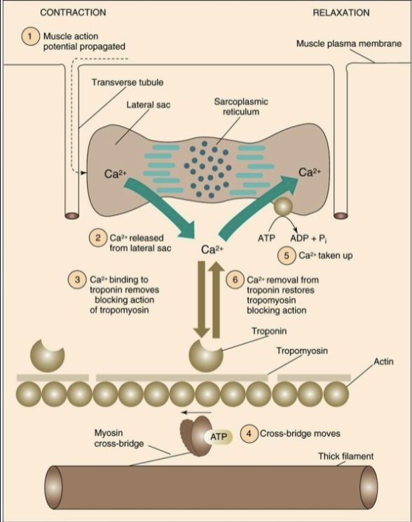
Composition: 2 terminal cisternae (ends of the sarcoplasmic reticulum) and 1 Transverse (T)-tubule.
Purpose: Allows depolarization (action potential) to spread internally from the sarcolemma to the inside of the fiber.

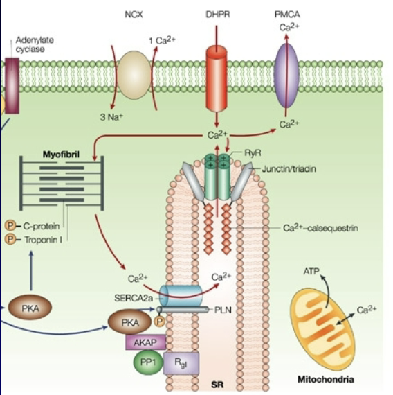
What are the roles of the DHP receptor, RyR receptor, and SERCA pump?
DHP (Dihydropyridine) Receptor: Voltage-sensitive receptor located in the T-tubule that signals the RyR receptor.
Ryanodine (RyR) Receptor: Signals for calcium (Ca2+) to be released from the sarcoplasmic reticulum (SR).
SERCA (Calcium ATPase): Enzyme/pump that pulls calcium back into the SR.
What are satellite cells and what is their primary function?
They are muscle "stem" cells found near the membrane. They are important for regeneration, repair, and muscle growth, and they help control protein synthesis (DNA —> RNA —> Proteins).
What are the two main parts of a myosin (thick) filament and their characteristics?
Light Meromyosin: Forms the tail; made of 2 intertwined heavy chains.
Heavy Meromyosin: Forms the cross-bridge, ends in 2 globular heads (containing 2 heavy chains + 2 sets of light chains). It has ATPase activity and moves/tilts to create movement.

What are the three protein components of the thin filament and their roles?
Actin: Double-stranded helix (G-actin is globular, F-actin is filamentous).
Tropomyosin (TM): Long rope-like protein (1 TM per 7 G-actins) that covers myosin-binding sites at rest.
Troponin (Tn): Has 3 subunits — TnC binds Ca2+, TnT binds tropomyosin, and TnI is inhibitory.

Cross Bridge Formation

What are the 4 steps of the cross-bridge cycle?
Myosin attaches to Actin: Calcium binds troponin, moving tropomyosin, allowing myosin to attach.
Power Stroke: Release of Pi, myosin head pivots (~5nm movement), then ADP is released.
Cross-Bridge Detachment: ATP binds myosin, causing it to detach from actin.
ATP Hydrolysis: ATP is broken down into ADP and Pi (via ATPase action), pivoting the myosin head back into its "cocked" position.

What is the difference between Histochemistry and Immunohistochemistry in muscle biopsies?
Histochemistry: Uses incubations with substrates or stains to look at Myosin ATPase, metabolic enzymes (SDH, LDH, Hexokinase), or energy substrates (glycogen, lipids).
Immunohistochemistry: Uses reactions between specific protein isoforms and antibodies to look for Myosin Heavy Chain (MHC) isoforms.
What are the four primary fiber types classified by Myosin Heavy Chain (MHC) and enzymatic activity?
Type I (Slow)
Type IIa (Fast oxidative)
Type IIx (Fast glycolytic)
Type IIb (and Hybrids like IIax, IIxa)
Compare the oxidative capacity, anaerobic capacity, and fatigue resistance of Type I, Type IIa, and Type IIx fibers
Type I: High oxidative capacity, low anaerobic capacity, very fatigue-resistant.
Type IIa: Moderate oxidative capacity, high anaerobic capacity, somewhat fatigue-resistant.
Type IIx: Low oxidative capacity, high anaerobic capacity, very easy to fatigue (fastest/least resistant).
What factors determine muscle fiber type, and can they change?
Primary Determinant: Genetics determines which motor neurons innervate fibers (all fibers in a motor unit are the same type).
Plasticity/Training: Endurance training or inactivity can cause minor shifts (e.g., Training: Type IIx —> Type IIa; Inactivity: Type IIa —> Type IIx).
Aging: Loss of fast fibers/denervation can lead to re-innervation by slow motor neurons, increasing ST percentage.
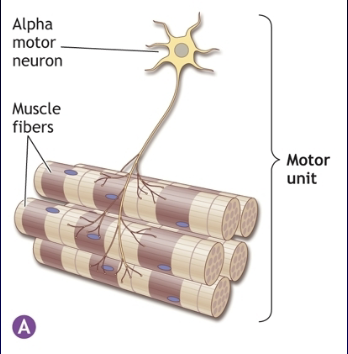
What is the general ratio between muscle fibers and motor neurons in the human body?
Approximately 250 million muscle fibers and 450,000 motor neurons. Every muscle fiber is connected to a motor neuron, and each motor neuron connects to multiple fibers.
What are the five primary structural parts of a motor neuron?
Cell Body (Soma)
Dendrites
Axon
Myelin
Nodes of Ranvier

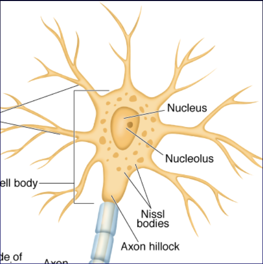
What are the three key components of the neuron's cell body and their functions?
Nucleus: Contains nucleolus (rRNA), cannot divide after birth, directs protein synthesis.
Nissl Bodies: ER and ribosomes near the axon hillock; aid transport of materials down the axon.
Cytoskeletal Proteins: Microtubules, actin, and neurofilaments; provide support, regeneration, and axoplasmic transport.
Where are motor neuron cell bodies located in the spinal cord?
They are grouped together in specific regions of the spinal cord, specifically lying in the ventral horn of the gray matter.

What is a "parent" axon, collateral branches, and the connection point called?
Parent Axon/Collateral Branches: A single axon that branches out to innervate hundreds of muscle fibers within the same muscle.
Synapse / Motor End Plate / Neuromuscular Junction: The connection point between an axon and a muscle fiber.
What produces myelin, and how does it speed up action potential conduction?
Myelin is a lipid layer produced by Schwann cells.
Nodes of Ranvier have a very high concentration of voltage-gated Na+ channels, allowing the action potential to "jump" from node to node via saltatory conduction, increasing conduction velocity.
What are the five structural features of the neuromuscular junction (NMJ)?
Primary Cleft: Area between nerve and muscle (~70nm, no direct contact).
Secondary Cleft: Indentation of the sarcolemma to increase surface area.
ACh Receptors: Ion channels in 1º and 2º clefts that bind 2 ACh molecules.
Basement Membrane: Contains acetylcholinesterase to end the nerve signal.
Vesicles: Located in the axon terminal, containing acetylcholine.

What defines a motor unit, and how does its size affect movement?
Definition: A motor neuron and all of the muscle fibers it innervates.
Effect on Movement: The ratio of muscle fibers to motor neurons affects movement precision (small ratios = fine control; large ratios = gross/powerful movements).
What is the resting membrane potential of a neuron, and what two conditions are required to maintain it?
Resting Potential: -70 mV on the inside of the cell.
2 Conditions:
Impermeable membrane (requires channels to move ions).
Ion pumps (actively move ions against their concentration gradient).
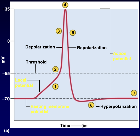
What are the 7 chronological steps of an action potential in a neuron?
Graded Potential
Threshold stimulus (exceeds critical value, "all-or-none")
Depolarization (Na+ and K+ channels open; Na+ moves faster)
Spike/Peak (~ +35 mV)
Repolarization (Na+ channels close, K+ permeability increases and moves out)
Hyperpolarization ("Overshoot" of K+ moving out)
Restore ion gradients via the Na+/K+ pump
What is the difference between the absolute and relative refractory periods during an action potential?
Absolute Refractory Period: No stimulus of any strength can trigger an AP as long as Na+ gates are open.
Relative Refractory Period: Only an especially strong stimulus can trigger a new AP because K+ gates are still open (hyperpolarization phase).

Why does an action potential move in only one direction away from the cell body?
Because of the refractory periods trailing behind the action potential, preventing it from moving backward.
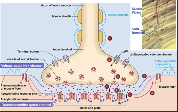
What happens when an action potential reaches the axon terminal (bouton) at the NMJ? (List the main steps)
AP moves into the bouton and opens voltage-gated Ca2+ channels.
Influx of Ca2+ triggers ACh vesicle release into the NMJ.
ACh binds to ACh receptors (2 molecules required), opening channels for Na+ and K+.
This creates an end-plate potential, depolarizing the sarcolemma and sending an AP in both directions.
Signal is terminated via acetylcholinesterase.
What are the primary steps of Excitation-Contraction Coupling and the immediate subsequent events leading to a contraction?
E-C Coupling Steps:
Depolarization of T-tubules (via DHP receptors signaling RyR receptors).
Opening of SR channels and release of Ca2+.
Subsequent Events:
Ca2+ binds to troponin (TnC).
Tropomyosin pulls away from actin binding sites.
Cross-bridge attachment occurs.
Power stroke generates force.