17.7 The hypothalamus and the Pituitary gland
 Call Kai
 Learn
Learn Practice Test
 Spaced Repetition
 Match
Match Flashcards
 Knowt Play
1/64

There's no tags or description
Looks like no tags are added yet.
Name | Mastery | Learn | Test | Matching | Spaced | Call with Kai | Chat |
|---|
No analytics yet
Send a link to your students to track their progress
65 Terms
Hypothalamus
An almond-sized component of the brain that is sandwitched between the inferior regions of the cerebral hemispheres and positioned beneath the thalamus.
Pituitary gland/hypophysis
Lies inferior to the hypothalamus
is connected to the hypothalamus by a very thin stalk called infundibulum/infundibular stalk.
This small slightly oval gland, which is approximately the size of a large pea, is housed within the hypophyseal fossa formed by the sella turcica of the sphenoid bone.
The pituitary gland is partitioned both structurally and functionally into 2 parts.
The posterior pituitary and an anterior pituitary.
The posterior pituitary is composed of ___ and the anterior pituitary is composed of ___.
neurons; hormone-producing endocrine cells.
The posterior pituitary makes up approximately ____ of the mass of the pituitary gland.
one-quarter.
How many neurons extend from the hypothalamus to the posterior pituitary?
Approximately 10,000 neurons, with the dendrites and cell bodies of these neurons located within the hypothalamus. These cell bodies compose 2 specific hypothalamic nuclei.
Supraoptic nucleus
Hypothalamic nuclei, located superior to the optic chiasm
Paraventricular Nucelus
Located adjacent to the 3rd ventricle.
Hypothalamo-hypophyseal tract
Where the unmyelinated axons from these neurons extend through the infundibulum. (A tract is a bundle of axons within the CNS) The ends of the axons, including synaptic knobs, are within the posterior pituitary.
The anterior pituitary is about ___ of the mass of the pituitary gland.
three-quarters. Endocrine portion of the pituitary gland which is why it is also called the adenohypophysis.
The connection between the hypothalamus and the anterior pituitary involved 2 capillary plexuses interconnected by portal veins.
The primary plexus and secondary plexus.
Plexuses
Branching networks of microscopic blood vessels.
Portal veins
(Any vessel located between 2 capillary beds)
Primary plexus
The capillary network associated with the hypothalamus.
Secondary plexus
The capillary network associated with the anterior pituitary.
Hypophyseal portal veins
Blood is drained by them from the primary plexus of the hypothalamus and transported to the secondary plexus of the anterior pituitary. This blood vessel network is collectively called the hypothalamo-hypophyseal portal system.
Hypothalamo-hypophyseal portal system
Provides a direct blood pathway between the hypothalamus and the anterior pituitary.
What 2 hormones does the posterior pituitary store?
Antidiuretic hormone and oxytocin.
Antidiuretic hormone and oxytocin are synthesized where?
The hypothalamus.
The supraoptic nucleus primary forms antidiuretic hormone
The paraventricular nucleus primarily produces oxytocin.
Neurosecretory cells
Neurons in the hypothalamus.
Following their synthesis in the hypothalamus…
The hormones are packaged within secretory vesicles and transported by fast axonal transport through the axons of the hypothalami-hypophyseal tract to their synaptic knobs within the posterior pituitary.
Hormone is released from the posterior pituitary when…
Nerve signals are sent from the hypothalamus along axons of the hypothalamo-hypophyseal tract. Specifically, nerve signals from the supraoptic nucleus cause release of antidiuretic hormone, and those from the paraventricular nucleus stimulate release of oxytocin. They then enter the blood when released.
Antidiuretic Hormone (ADH)
Functions to help maintain fluid balance, blood volume, and blood pressure.
It is released when there is increased blood concentration, which indicates a state of dehydration.
The hypothalamus detects this change in blood concentration as blood moves through this region of the brain and, in response imitates nerve signals to the posterior pituitary to release ADH into the general circulation.
Target cells of ADH include:
The kidneys, which are stimulated to decrease urine output
The thirst center, which then relays signals to the cerebral cortex for us to become conscious of being thirsty and our need to increase our fluid intake.
ADH in high doses…
Causes vasoconstriction.
ADH is regulated by?
Negative feedback.
Following fluid intake
We become rehydrated and our blood concentration returns to preexisting well-hydrated levels, the hypothalamus relays fewer nerve signals to the posterior pituitary, resulting in a lower amount of ADH released.
Oxytocin (OT)
Functions in both the delivery of a baby and the ejection of milk from mammary glands of the breast. During labor, OT is related into the blood in progressively higher amounts. Sensory input is relayed from the uterus to the hypothalamus; in response, the hypothalamus iniititates nerve signals to the posterior pituitary to release OT into the general circulation. Oxytocin stimulates the smooth muscle of the uterus to contract with increasing force until the baby is delivered.
In addition, after a baby is born when a baby suckles at the breast, sensory input is initiated from the breast to the hypothalamus, and in response the hypothalamus imitates nerve signals to the posterior pituitary to release OT into the general circulation. Here, OT stimulates smooth muscle contractions within the breast to cause ejection of breast milk. This release requires prolactin, which stimulates milk production.
What does oxytocin do in males?
Oxytocin facilitates the movement of sperm from the testes through the reproductive ducts.
What does research has shown about oxytocin?
That physical contact between individuals(hugging, holding hands) causes the release of OT, which improves our mood and alters our physiology. *lowers our levels of stress hormones, reduces our blood pressure, increases our tolerance for pain). Oxytocin is even released when a persons pets a dog, in both the individual and the dog.
What triggers the release of hormones from the anterior pituitary?
Hormonal stimulation which occurs when regulatory hormones produced within the hypothalamus are released into the primary plexus and then transported via the hypophyseal portal veins to reach the secondary plexus, within the anterior pituitary, which recall is collectively the hypothalamo-hypophyhseal portal system. The anterior pituitary then releases its hormones into the blood of the general circulation, through which they reach target cells.
Regulatory hormones produced and released from the hypothalamus fall into one of 2 groups:
Releasing hormones(RHs) and inhibiting hormones (IHs)
Releasing hormones (RHs)
Hormones that stimulate the production and secretion of specific anterior pituitary hormones
Inhibiting hormones (IHs)
Hormones that decrease the production and secretion of specific anterior pituitary hormones.
How many Rhs and IHs are there?
5 RHs and 2 IHs.
The anterior pituitary synthesizes and releases its own what?
Hormones.
What are the hormones of the Anterior pituitary?
Growth hormone (GH)
Thyroid-stimulating hormone(TSH)
Adrenocorticotropic hormone(ACTH)
Follicle-stimulating hormone(FSH)
Luteinizing hormone(LH)
Prolactin(PRL)
Growth Hormone (Somatotropin)
is secreted by cells called somatotropes. GH stimulates the liver to release both insulin-like growth factor 1 and 2 (IGF-1 and IGF-2, also called somatomedins) (so'mă-tõ-me'din). Both GH and IGFs function synergistically to stimulate cell growth and cell division, particularly within the skeletal and muscular systems
Thyroid-stimulating hormone(TSH) (Thyrotropin)
TSH is synthesized by specific cells called thyrotropes. TSH stimulates both growth of the thyroid gland and the thyroid gland's release of thyroid hormone (TH), which is the hormone that functions primarily to establish the body's metabolic rate
Adrenocorticotropic hormone(ACTH)
(also called corticotropin) is secreted by cells called corticotropes. ACTH stimulates the adrenal cortex to produce and secrete glucocorticoids (e.g., cortisol), which increase blood levels of nutrient molecules, including glucose, glycerol, fatty acids, and amino acids
Follicle-stimulating hormone(FSH) & Luteinizing hormone (LH)
are collectively called gonadotropins. Both are secreted by cells called gonadotropes. These hormones act on the gonads. In the ovaries, FSH and LH control (a) development of the oocyte and the follicle (the spherical structure that encloses the oocyte), (b) ovulation, which is the release of oocyte from the follicle, and (c) the release of estrogen and progesterone (see L section 28.3b). In the testes, FSH and LH regulate the development of sperm and the release of testosterone
Prolactin (PRL)
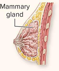
primarily regulates mammary gland growth and breast milk production (see l section 29.8c). During pregnancy, the specific cells that produce prolactin (lactotropes) increase in size with greater amounts of prolactin produced. Milk production (lactation), however, does not occur until after the baby is born.
Melanocyte-stimulating hormone (MSH)
This hormone typically has little effect in healthy adult humans because secretion ceases prior to adulthood. Melanocyte-stimulating hormone is of significance in humans only in selected health conditions, such as the hypersecretion of MSH, which causes a darkening or bronzing of the skin in Addison disease
The secretion of each of the hormones from the anterior pituitary is controlled by what?
Specific releasing hormones of the hypothalamus.
Tropic hormones
Hormones that target another endocrine gland to secrete its hormone(s). Tropic means having an attraction to or affinity for, and these hormones have an affinity for the receptors of other endocrine glands. GH- for the liver, TSH for the thyroid gland, ACTH for the adrenal cortex, and FSH and LH for the gonads.
Prolactin directly stimulates the mammary glands of the breast (and not another endocrine gland)for milk production.
All hormones released from the anterior pituitary except prolactin.
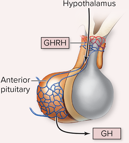
Homeostatic system: GHRH-GH-IGFs
The hypothalamus releases growth hormone-releasing hormone (GHRH) into the hypothalamo-hypophyseal portal system and when GHRH reaches the anterior pituitary it stimulates specific cells of the anterior pituitary (somatotropes) to release growth hormone (GH) (or somatotropin) into the general blood circulation.
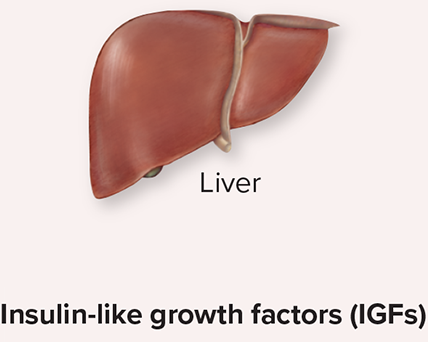
Growth hormone(GH) stimulates the liver to release insulin-like growth factors(IGFs) (or somatomedins). Growth hormone and IGFs functions synergistically to stimulate cell growth and cell division, particularly within the skeletal and muscular systems.

Homeostatic system: TRH-TSH-TH
The hypothalamus releases thyrotropin-releasing hormone(TRH) into the hypothalamo-hypophyseal portal system, and TRH stimulates the anterior pituitary(specifically thyrotropes) to release thyroid-stimulating hormone(TSH) or (thyrotropin) into the general blood circulation.
Thyroid-stimulating hormone(TSH) stimulates the thyroid gland to release thyroid hormone (TH). TH functions to establish the body’s metabolic rate.


Homeostatic system: CRH-ACTH-Cortisol
The hypothalamus releases corticotropin-releasing hormone (CRH) into the hypothalamo-hypophyseal portal system, and CRH stimulates the anterior pituitary (specifically corticotropes) to release adrenocorticotropic (ă-drẽ nõ-kô r'i-ko-tro pik) hormone (ACTH)
(or corticotropin) into the general blood circulation. Adrenocorticotropic hormone (ACTH) stimulates the adrenal gland (specifically, the adrenal cortex) to release glucocorticoids (e.g., cortisol). Cortisol increases blood levels of nutrient molecules, including glucose, glycerol, fatty acids, and amino acids

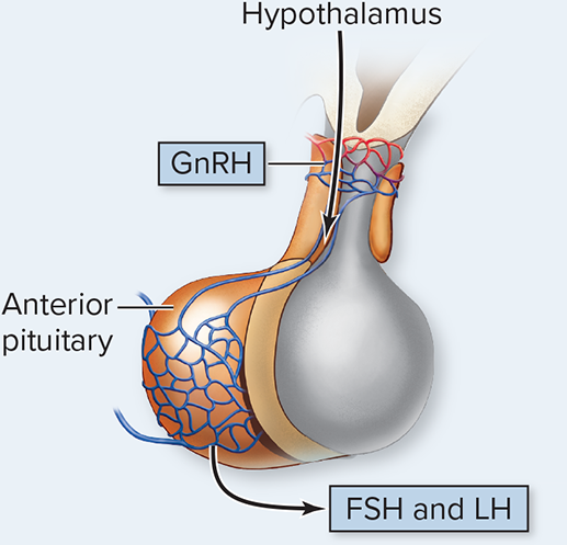
Homeostatic system: GnRH-FSH and LH-gonads
The hypothalamus releases gonadotropin-releasing hormone (GnRH) into the hypothalamo-hypophyseal portal system, and GnRH stimulates the anterior pituitary (specifically gonadotropes) to release two hormones: follicle-stimulating hormone (FSH) and luteinizing (lữ'tē-i-ni-zing) hormone (LH), which are collectively called gonadotropins. The gonadotropins are released into the general blood circulation and act on the gonads. FSH and LH act on the ovaries to control
(a) development of the oocyte and the follicle (the spherical structure that encloses the oocyte), (b) ovulation, which is the release of oocyte from the follicle, and (c) the release of estrogen and progesterone (see l section 28.3b). FSH and LH act on the testes to regulate the development of sperm and the synthesis and release of testosterone


Homeostatic System: PRH-PRL
The hypothalamus releases prolactin-releasing hormone (PRH) into the hypothalamo-hypophyseal portal system, and PRH stimulates the anterior pituitary (specifically
lactotropes) to release prolactin (pro -lak'tin; lac = milk) (PRL) into the general blood
circulation. Prolactin primarily regulates mammary gland growth and breast milk production, which is called lactation
Step #1 in the regulation of growth hormone release
The stimuli that are monitored by the hypothalamus for controlling growth are varied and are described shortly.
Step #2 in the regulation of growth hormone release
The stimuli are detected by the hypothalamus, which serves as the receptor.
Step #3 in the regulation of growth hormone release
In response to the various stimuli, the hypothalamus, serving also as the control center, releases growth hormone-releasing hormone (GHRH) into the hypothalamo-hypophyseal portal system.
Step #4 in the regulation of growth hormone release
The anterior pituitary, in response to GHRH, releases growth hormone (GH) into the blood of the general circulation.
Step #5 in the regulation of growth hormone release
GH stimulates the liver to release insulin-like growth factors (IGFs).
Step #6 in the regulation of growth hormone release
Both GH and IGFs are water-soluble proteins and readily dissolve in the aqueous environment of the blood to be transported throughout the body (thus, a transport protein is not required).
GH and IGFs stimulate targets cells (effectors) as described shortly.
Step #7 in the regulation of growth hormone release
The net result is stimulation of growth and release of nutrients into the blood to supply the energy required for the growth.
Step #8 in the regulation of growth hormone release
Increased blood levels of GH and IGFs inhibit the release of additional GHRH from the hypothalamus. In addition, Gh inhibits release of additional GH from the anterior pituitary. Thus, both GHRH and GH release are regulated by negative feedback.
What are the other stimuli that regulate the release of GHRH
A person's age. The levels of GHRH released by the hypothalamus change over our lifetime, as measured by growth hormone levels in the blood.
Observe in L figure 17.14a, which is a graph of changing growth hormone with age in years, that children and adolescents experience the highest amounts of GH, with progressive decreases in growth hormone as we age. Young adolescents have almost double the GH levels of young adults (700 ug/mL per day in young adolescents versus 400 ug/mL per day in young adults).
Time of day. GHRH released by the hypothalamus also changes throughout the day, which in turn alters GH levels. At any age, there are daily fluctuations in the release of GH.
Observe in E figure 17.14b, which is a graph of the blood level of growth hormone as it changes throughout a day, that in a typical sleep-wake cycle, peak GH levels correspond to the early stages of the sleep cycle. This peak allows for the most growth to occur while we are sleeping.
Nocturnal (nightly) peaks account for the majority of the GH released daily. Perhaps, after considering this, it does seem advantageous that most of our growth occurs at night when we sleep and are less active.
Nutrient levels. Blood levels of nutrients, which are monitored by the hypothalamus (see section 13.4c), influence GHRH release.
Additional GHRH is released in response to either an increase in amino acid levels (e.g., following a high-protein meal) or a decrease in glucose levels (e.g., when experiencing hypoglycemia). Thus, both of these changes in blood nutrient levels increase growth. (If we consider that amino acids are required in protein synthesis for growth and growth hormone results in increasing blood glucose, as described shortly, these two stimuli are better understood.)
Stress and exercise. Emotional, physical, and chemical stress (including surgery, trauma, or exercise) are monitored by the hypothalamus, as the master control of the autonomic nervous system and the center of the limbic system (the emotional brain; see l section 13.4c). Stress and exercise increase the release of GHRH. Thus, when we are stressed, growth is stimulated.
Although it should be noted that severe emotional stress can cause a decrease in GHRH release in children, and chronic stress can stunt a child's growth.
Growth hormone (GH) and Insulin-like factors (IGFs)
causes growth and initiate the release of nutrients to supply the energy required for this growth.
Both are water-soluble protein molecules and bind to cell surface receptors of their target cells to activate signal transduction.
The primary target cells of GH and IGFs
All cells
cells of cartilage, bone, and skeletal muscle tissue
hepatocytes of the liver
adipocytes of adipose connective tissue.
All cells (GH and IGFs)
All cells of the body have receptors for GH, IGFs, or both. The physiologic processes that cause growth in all of our cells include: increasing cell size through protein synthesis (see L section 4.8b) and increasing cell number through cellular division (see L section 4.9b). These hormones also stimulate cell differentiation, which initiates cells to change to their more mature form
Cartilage, bone, and skeletal muscle tissue (GH and IGFs)
Cartilage, bone, and skeletal muscle tissue are especially affected by growth hormone and IGFs.
Cartilage growth results from hyperplasia (increase in number) of chondrocytes and formation of extracellular matrix within cartilage (see
L section 7.3). Linear growth of bone occurs due to cartilage growth within the epiphyseal plate of bones with the cartilage then replaced with bone tissue. Appositional growth (growth in diameter) of bone is also stimulated (see L section 7.5a and ( section 7.5c). In skeletal muscle fibers, uptake of amino acids increases, which stimulates the synthesis of contractile proteins composing myofibrils (L section 10.8a), resulting in skeletal muscle hypertrophy. Some athletes have been known to illegally use growth hormone-in an attempt to increase skeletal muscle size and strength. Sometimes GH is used in conjunction with anabolic steroids (see
Liver(GH and IGFs)
GH and IGFs stimulate hepatocytes within the liver to release nutrients from storage into the blood.
Glycogen (a polymer of glucose molecules stored in the liver) is chemically digested by enzymes within the liver to glucose molecules. Recall from section
2.7c that this process is called glycogenolysis.
Glucose is also formed within the liver from noncarbohydrate sources (e.g., amino acids), through the process of gluconeogenesis. Glucose molecules formed either by the breakdown of glycogen or from noncarbohydrate sources is released into the blood. As a result, blood glucose levels rise. (Note that glycogenesis, which is formation of glycogen from glucose molecules, is inhibited within the liver by GH and IGFs.) The resulting increase in blood glucose levels is referred to as a diabetogenic (di'ă-bet'õ-jen-ik) effect due to the similarity to elevated blood glucose levels in individuals with diabetes mellitus (see
Adipose Connective Tissue (GH and IGFs)
Adipocytes (fat cells) composing adipose connective tissue are stimulated by GH and IGFs to increase lipolysis, which is chemical digestion of triglyceride molecules to glycerol and fatty acids (see L section 2.7b). Glycerol and fatty acids are released from adipocytes, causing an increase in the level of glycerol and fatty acids in the blood.
(Lipogenesis, which is formation of triglycerides from glycerol and fatty acids with the subsequent storage of triglycerides, is inhibited within the adipocytes by GH and IGFs.)
The function of releasing nutrient molecules (i.e., glucose from hepatocytes of the liver, and glycerol and fatty acids from adipose connective tissue) is to provide the fuel molecules required in cellular respiration to synthesize ATP molecules (see L section 3.4). Cellular growth is a high-energy process, and cells need significant amounts of ATP to support these processes.
Note that we have been describing the effects of both GH and IGFs on target cells, and that these two hormones have overlapping synergistic interactions (see section 17.6b). However, although we might predict that growth hormone is the more significant of the two hormones (given its name), IGFs are responsible for the greater response from the target cells. This difference in response results in part from the difference in their individual biological half-lives: GH (a protein hormone) has a biological half-life of 6 to 20 minutes, and IGFs (also protein hormones) have a biological half-life of approximately 20 hours. This longer biological half-life of IGFs is due to IGFs being transported in the blood by transport proteins that, although not required, help protect these hormone molecules from destruction (see L section 17.4a). Additional responses to the release of GH and IGFs are listed in the summary table in the reference section, which directly follows this chapter (see