MYCO | 1.3 Ultra- & Internal structures, 1.4 Fungal Growth
1/76
There's no tags or description
Looks like no tags are added yet.
Name | Mastery | Learn | Test | Matching | Spaced |
|---|
No study sessions yet.
77 Terms
T/F: Mycelium to yeast conversions are just as common as yeast dimorphisms
FALSE
Mycelium-to-yeast conversions are not as common as yeast dimorphisms
_ is the most well-studied model for mycelium-to-yeast conversion triggered by heat shock/shift
Paracoccidioides brasiliensis
Mycelium to yeast conversion is usually a response to strenuous conditions, including _
Heat stress (26-30 C to 37-40 C)
Oxidative stress (e.g., putting dilute solution of hydrogen peroxide)
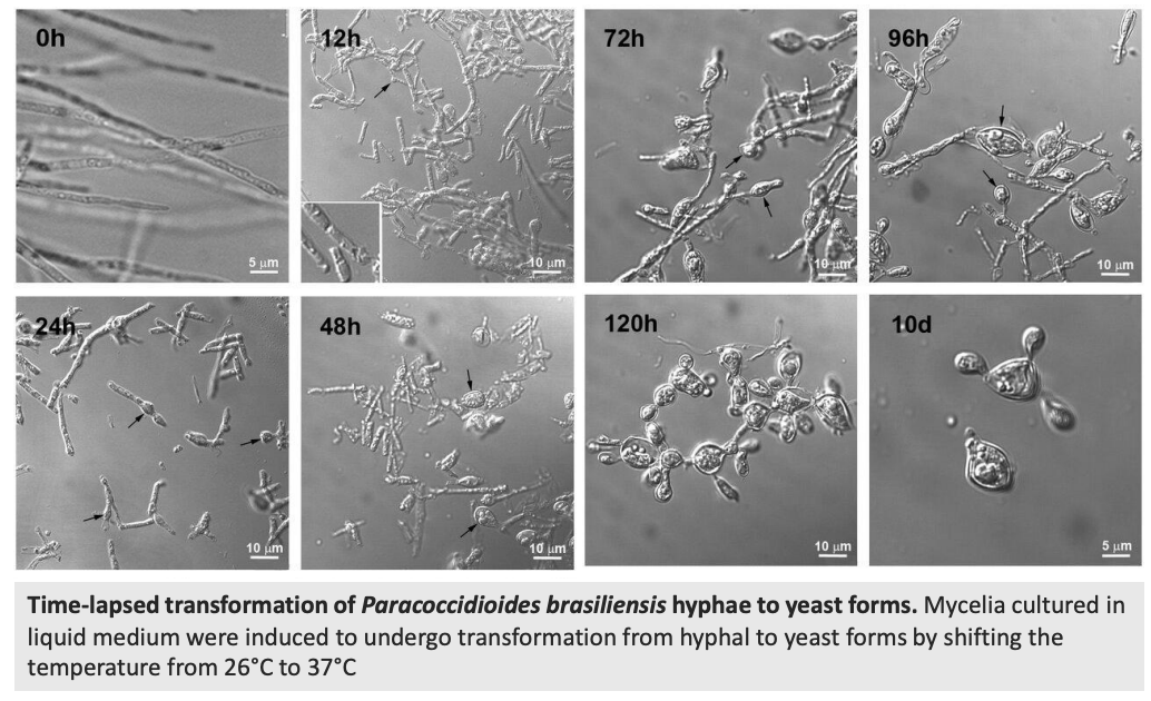
Apart from Paracoccidioides brasiliensis, what other species can exhibit mycelium to yeast conversion? Also, describe figure shown
Time-lapsed transformation of Paracoccidioides brasiliensis hyphae to yeast form
Mycelium cultured in liquid medium was induced to undergo transformation from hyphal to yeast form by increasing temp from 26 C → 37 C
Other species bhsp
Blastomyces dermatitidis
Histoplasma capsulatum
Sporothrix schenckii
Penicillium marneffei
T/F: The hyphal wall is composed of chitins, glucans, and different sugars and proteins
TRUE
Fungal walls are composed of _
Matrix components (sugars, i.e., mannan; B-1,3- and B-1,6 glucan that are embedded and linked to scaffolds of
Fibrous load-bearing polysaccharides (protein-bound chitin)
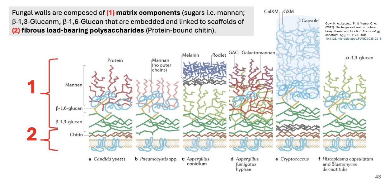
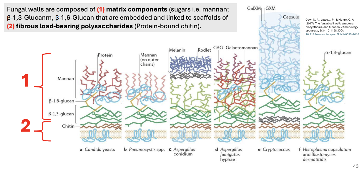
Explain figure shown
Variations in hyphal & yeast cell wall compositions
General
Matrix components (mannan, B-1,3-, B-1,6)
Fibrous load-bearing polysaccharides (protein-bound chitin)
C. albicans yeast = chitin, B-1,3- & B-1,6-glucans, mannan proteins
Pneumocystis sp. = no chitin, B-1,3- & B-1,6, mannan (no outer chains)
Aspergillus conidium = Melanin (UV protection), rodlets
Aspergillus fumigatus hyphae = GAG, Galactomannan (with antigenic properties)
Cryptococcus = Capsule (immune defense evasion), GalXM/GXM polysaccharides
Histoplasma capsulatum, Blastomyces dermatitidis = a-1,3-glucan
_ catalyze chitin formation and transformation across the membrane
Chitin synthases
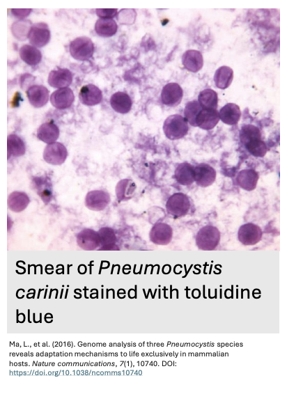
_ is the first identified member of the fungal kingdom that does not have chitin in its cell wall
Describe the figure shown
Pneumocystis
Smear of Pneumocystis carinii stained with toluidine blue
Why does Pneumocystis sp. have no chitin?
pin
It’s because their parasitic lifestyle has adapted them to the nutrient-rich environment of their host tissues, where chitin’s rigidity would be unnecessary and could hinder flexibility.
Additionally, the absence of chitin reduces immune recognition by host defenses and allows for a more efficient nutrient uptake, which is key to their survival within host’s lungs
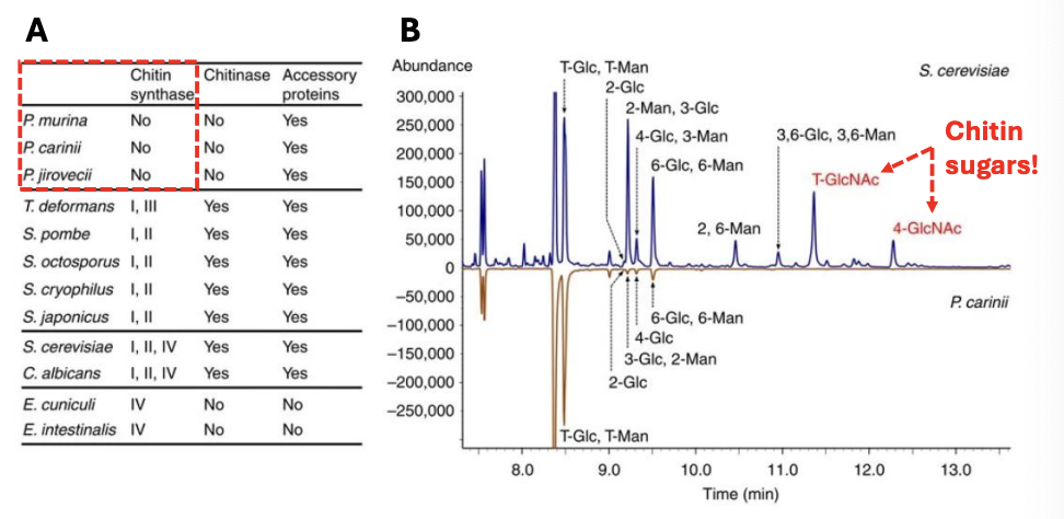
Explain the figure shown
Special wall of Pneumocystis sp.
(A) Enzymes and accessory proteins (i.e., chitin synthase, chitinase) involved in chitin metabolism in fungi not found in Pneumocystis sp.
(B) Gas chromatograms of partially methylated alditol acetates of P. carinii and S. cerevisae
Terminal and 4-linked N-acetylglucosamine signals (T-GlcNAc, 4-GlcNAc) were detected in S. cerevisiae but not in P. carinii
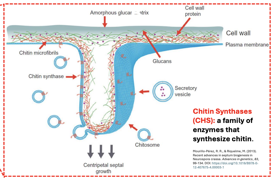
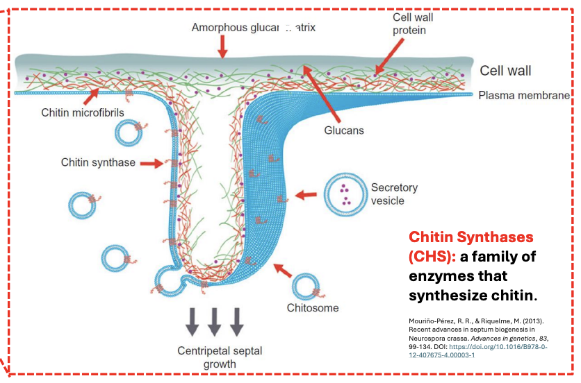
Explain chitin formation to create septum
Blue = plasma membrane
Chitosomes deliver chitin synthases within secretory vesicles, fusing to plasma membrane
Chitin synthases, once proteolytically activated, will start synthesizing chitin
Chitin then lines the septum, allowing centripetal septal growth (from edge to center)
T/F: Chitin microfibrils provide flexibility to the fungal cell wall.
FALSE
Chitin microfibrils provide rigidity and structural strength to the cell wall, not flexibility. Flexibility is contributed by other components, such as glucans.
T/F: The centripetal growth of the septum in fungi refers to its expansion outward from the center to the edges.
FALSE
Centripetal growth refers to growth starting from the edges and moving inward toward the center E→C

T/F: Chitosomes are responsible for transporting glucans to the plasma membrane during septum formation.
FALSE
Chitosomes are responsible for transporting chitin synthases to the plasma membrane during septum formation.

T/F: The plasma membrane plays no role in septum formation.
FALSE
The plasma membrane is critical for septum formation as it houses enzymes like chitin synthase and coordinates the assembly of wall components.
T/F: The matrix component of yeast cell walls are generally similar across different taxonomic groups
FALSE
The matrix component of yeast cell walls varies per taxonomic group
Matrix components of yeast cell walls are critical for various life processes, its main roles include _
isrp
Interaction with other organisms
Survival and resistance to immune defense
Prevention of parasitism (by other fungi)
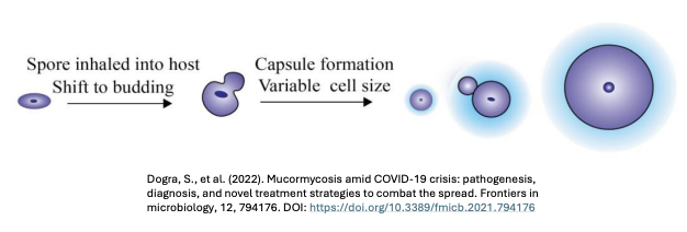
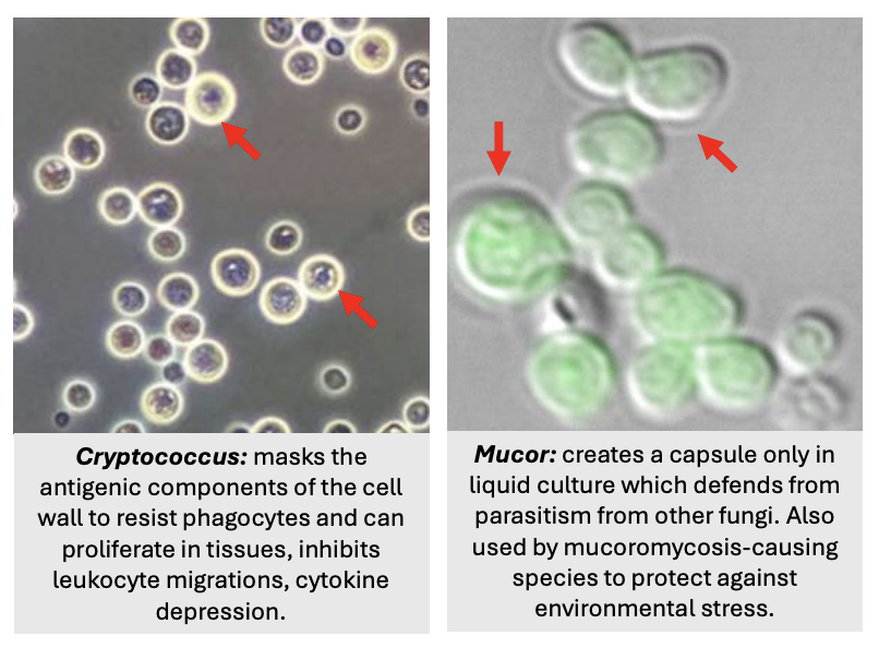
Some infectious yeasts couple _ to resist phagocytosis; explain figure
Cell size change with capsule formation
Spore inhaled into a host shifts to budding,
Producing yeast cells of varying sizes and
Forming capsule to evade immune defense
2 types of extrahyphal matrix (capsule in cell wall) of yeasts
Discrete polysaccharide capsule
Diffuse polysaccharide / glycoprotein
_’s extrahyphal matrix (contains sugary capsule) masks the antigenic components of its cell wall (e.g., glucans) and can proliferate in tissues, inhibiting leukocyte migration and suppressing cytokine production
Cryptococcus
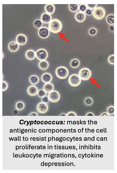
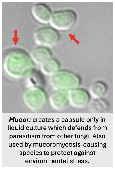
T/F: Cryptococcus is a vertebrate pathogen, while Mucor is a plant pathogen
TRUE
Usually spore → filamentous once inside host, but to evade immune recognition, Cryptococcus & Mucor couples cell size change with capsule formation to resist phagocytosis
T/F: Generally, fungal capsule does not trigger immune defense but fungal cell wall does
TRUE
_ creates a capsule only in liquid culture to defend itself against parasitism from other fungi; other _-causing species use this capsule to protect themselves against environmental stress
Mucor
Mucoromycosis-
How is mycelium-to-yeast conversion triggered in Paracoccidioides brasiliensis?
Heat shock/shift, specifically by increasing temperature from 26 C to 37 C
Fungi possess vacuoles of _ for multiple functions, existing in both yeast and hyphal forms
varied shapes and arrangements

4 main function of fungal vacuoles
spia
Solute storage
Protein turnover
Ion homeostasis
Apoptosis

T/F: Tubular vacuolar systems are unique to fungi
TRUE
Fungal vacuoles are similar to _
mammalian lysosomes & plant vacuoles

Fungal vacuoles have a wide variety of _ in different species, reflecting their ecological specialization
architecture & roles
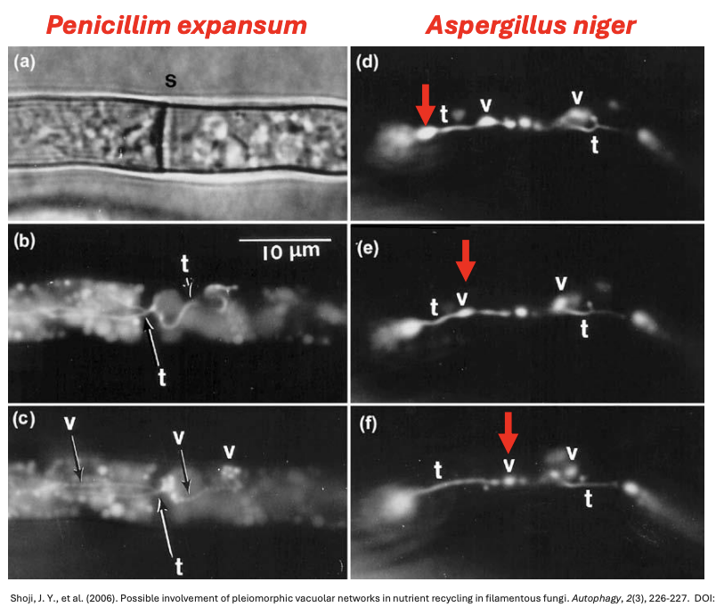
Explain the figure shown
Pattern of vacuole distribution in yeast and filamentous fungi
(A) Network of spherical and tubular vacuoles in many filamentous fungi, e.g., Aspergillus oryzae
(B) Migratory cytoplasm in hyphae of C. albicans
(C) Empty-looking vacuolated sub-apical compartments of Ustilago sp.
(D) Oval/spherical vacuoles of S. cerevisiae
(E) C. albicans vacuole segregation structure extending from mother to daughter cell during vacuole inheritance
(D) Numerous small vacuoles of fission yeast Schizosaccharomyces pombe
_ is a family of enzymes that synthesize chitin
Chitin synthases (CHS)
Fungi have dynamic _ used for exocytosis
tubular vacuolar system

Explain the figure shown
Dynamic tubular vacuolar system of living fungal hyphae
a) Hypha showing the presence of septum
b) Same hypha showing narrow tubule passing through the septum and then branching
c) Same hypha showing dilated vacuoles after tubules pass through the septum
def) Hypha photographed at 8-sec intervals showing successive peristalsis-like movements causing vacuolar dilations to travel along hypha from left to right
Without _, growth in the hyphal tip is not possible
tubular vacuolar system that allows the peristaltic motion for the delivery of vesicles
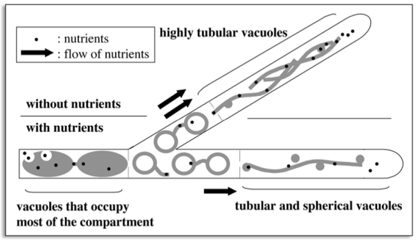
Explain figure shown
Vacuoles can either fuse together or elongate to create long vacuolar structures extending to hyphal tip
Tubular vacuolar system, through the peristaltic movement it creates, essentially squeezes nutrients, allowing these to flow towards the apical tip and constantly supply materials SPK needs to extend
Same is true for branching, semi-circular tubular vacuoles can participate in branching and septation
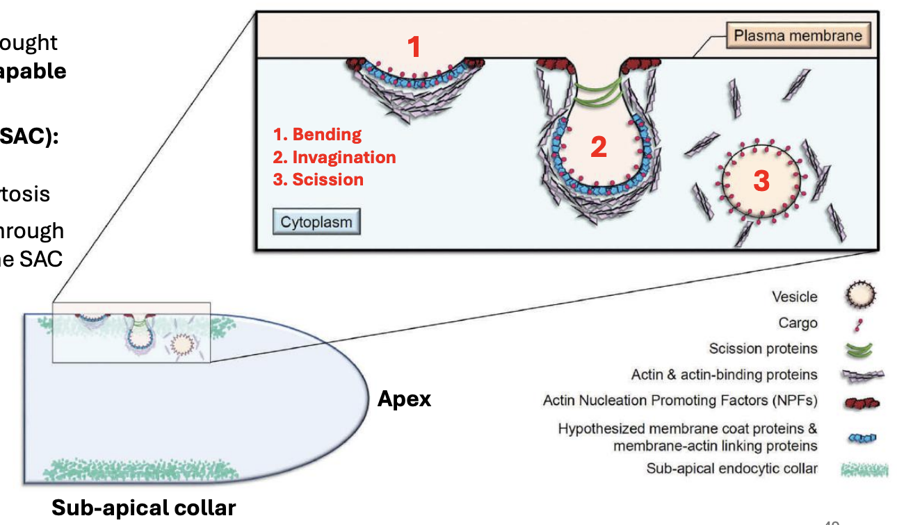
_ is an important protein-rich region for endocytosis
Sub-apical collar (SAC) of the hypha

T/F: It was previously thought that fungi were incapable of endocytosis
TRUE
But then they discovered SAC of the hypha

_ are enriched with proteins for endocytosis
Sub-apical collar (SAC) of the hypha
T/F: Polarity is entire controlled by SPK
FALSE
SPK and SAC directs polarity of the tip
_ points to evidence for endocytosis using steryl dyes
Tubular vacuolar system
T/F: Fungi have dynamic tubular vacuolar system used for endocytosis
FALSE
Fungi have dynamic tubular vacuolar system used for exocytosis

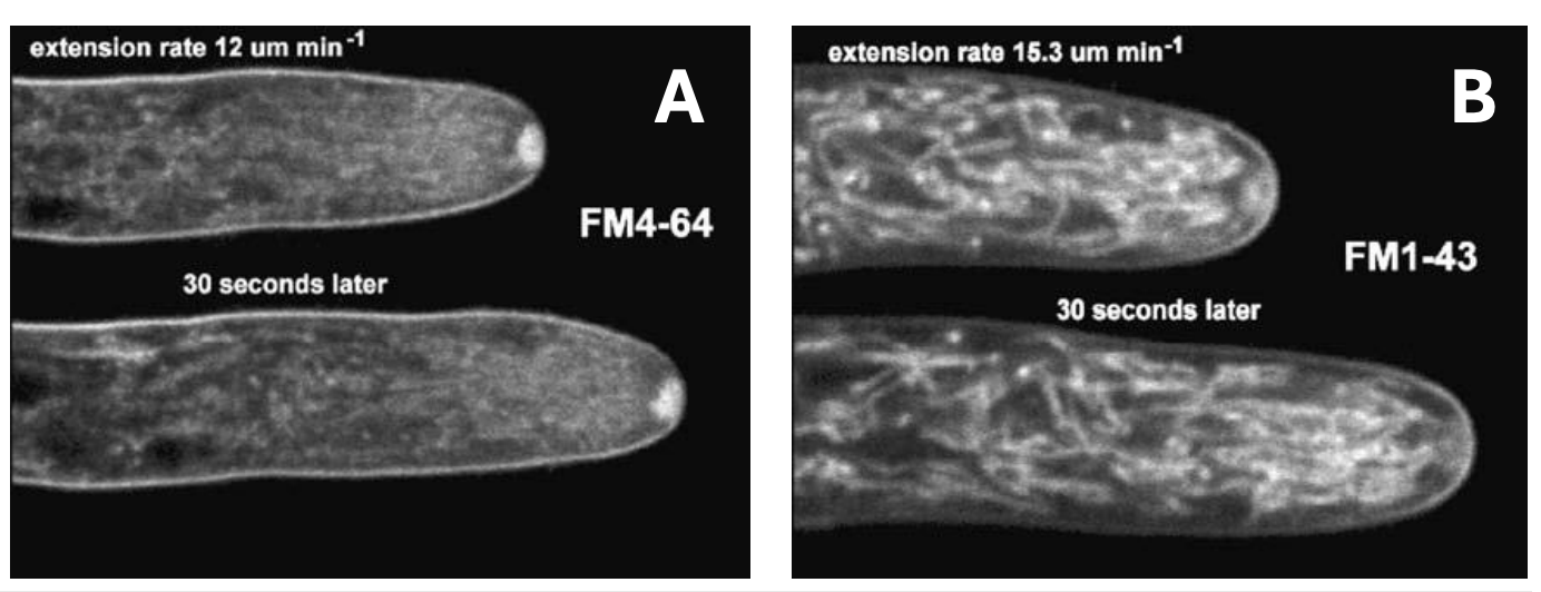
Explain figure shown
Hyphal tips of Neurospora crassa treated with 2 steryl dyes to detect putative endocytosis
Both steryl dyes (A) FM4-64 and (B) FM1-43 became internalized, and both hyphae treated with steryl dyes continued to grow during confocal laser imaging, as shown in photos taken at 30-sec intervals

How are (1) internalization of steryl dyes and (2) subsequent continued growth of hyphal tips evidences for endocytosis actually occurring?
This is because steryl dyes specifically bind to the plasma membrane and are taken into the cell via endocytic vesicles, thus observing dye internalization indicates active endocytosis occurring at the site.
Meanwhile, continued growth at hyphal tips also is evidence for endocytosis because polarized growth at the hyphal tip requires a balance between exocytosis (adding of membrane and materials for expansion) and endocytosis (recycling of excess membrane and retrieving of materials), thus observing continued growth at hyphal tips while dyes are internalized indicates that endocytosis is actively supporting this process of polarized growth along with exocytosis occurring at the tip
The fungal cell membrane has _ that are antifungal targets
sterols (ergosterol) and transporters (cytosine permease)
Fungal cell membrane is similar to a typical Eukaryotic bilayer: _
lipids + proteins
_ is the distinguishing component of fungal cell membrane that functions to module permeability and fluidity and is thus the target of several drugs
Ergosterol (steroid bc it has 4 rings)
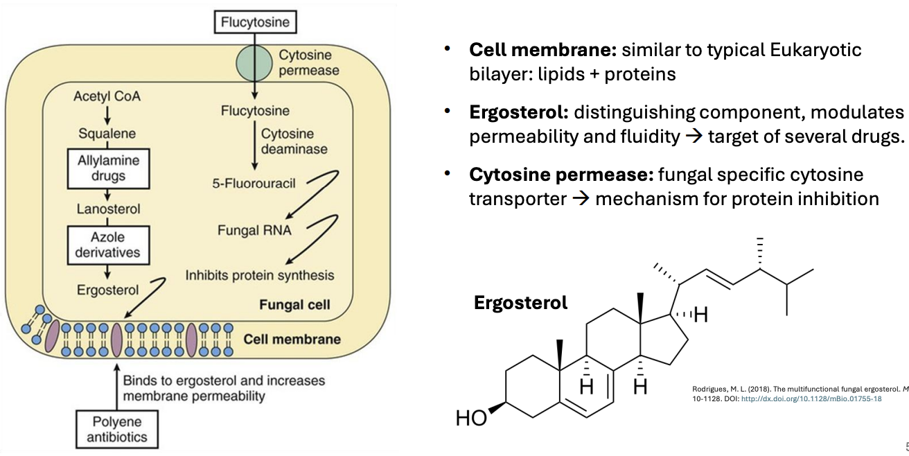
T/F: Humans have ergosterol and cytosine
FALSE
Human PM don’t have ergosterol and cytosine deaminase
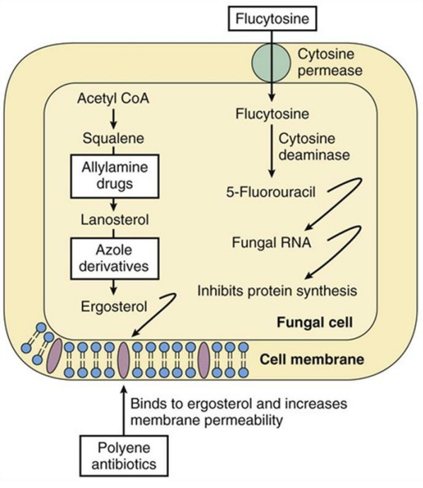
Enumerate 4 common antifungal substances
faap
Flucytosine (work from in → out bc u need all fungi inside u to have their protein synthesis interrupted)
Flucytosine (cytosine analog) enters fungal cell via cytosine permease
Once inside, flucytosine is converted into 5-fluorouracil by cytosine deaminase
Once 5-FU is incorporated into fungal RNA, this results in inhibition of RNA synthesis and, subsequently, protein synthesis, impairing fungal cell functions
Selective drug: Mammalian cells don’t have cytosine deaminase, making flucytosine specific to fungi
Allylamine drugs
Inhibit squalene epoxidase that converts squalene → lanosterol, thus *ultimately inhibiting ergosterol synthesis, leading to decreased membrane permeability and thus leakage of ions & essential nutrients, eventually leading to cell death*
Azole derivatives (e.g., ketoconazole) topical
Inhibit enzyme converting lanosterol → ergosterol, thus *
(works from out → in, affecting membrane’s resistance to turgor pressure)
Polyene antibiotics (e.g., Amphotericin B)
Directly binds to and break down ergosterol, increasing membrane permeability
T/F: Azoles work slower than polyene antibiotics
TRUE
Bc it will take more time to completely inhibit synthesis of all ergosterols in PM than directly breaking it down
T/F: Sub-apical collar (SAC) of hypha is used for exocytosis, while tubular vacuolar system (TVS) is used for endocytosis
FALSE
Sub-apical collar (SAC) of hypha is used for endocytosis, while tubular vacuolar system (TVS) is used for exocytosis
_ is a fungal specific cytosine transporter that serves as mechanism for protein inhibition (via action of competitive inhibitor flucytosine)
Cytosine permease

Fungi utilize _ for their unique nutrient acquisition strategy
cell membrane enzymes, i.e., H+ ATPases and H+ symporters and antiporters
The most common proteins in the fungal plasma membrane function for:
ssca
Solute transport (bc of constant growth)
Signal transduction (bc anastomoses)
Cell wall synthesis (hyphal tip extension)
Anchorage for cytoskeleton (support for hyphal growth)
_ is the fungal cell membrane enzyme that allows fungi to secrete H+ ions, increasing external H+ concentrations, decreasing pH (more acidic, pH 4 - 5.5)
H+ ATPases
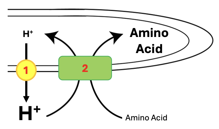
T/F: H+ ATPases do primary active transport
TRUE
H⁺ ATPases perform primary active transport because they directly use the energy from ATP hydrolysis to pump protons (H⁺ ions) across a membrane

_ is the fungal cell membrane enzyme that co-transports important materials, such as amino acids, ions, etc.
H+ symporters (but antiporters are also present)

T/F: H+ symporter is a primary active transport
FALSE
H+ symporter is a form of secondary active transport because it just relies on energy stored in the proton gradient generated by the action of H+ ATPases

Ergosterol is the distinguishing component of fungal cell membrane that is the target of several drugs and functions to _
modulate permeability and fluidity
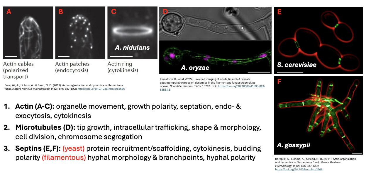
Fungal cytoskeleton has 3 major polymers, namely _
ams
Actin ogsec
Organelle movement, growth polarity = actin cables (polarized transport)
Septation
Endo- & exocytosis = actin patches
Cytokinesis = actin rings
Microtubules tism cc
Tip growth, intracellular trafficking, shape & morphology, cell division, chromosome segregation
Very close to nucleus bc during cell division, microtubules make up spindle fibers
Septin y pcb f hmbp
Yeast = protein recruitment/scaffolding, cytokinesis, budding polarity
Filamentous/hyphal = hyphal morphology, branching, & polarity
3 steps involved in fungal endocytosis
bis
Bending
PM begins to bend inward, forming small depression
Initiated by recruitment of actin and actin-binding proteins to PM, providing structural support to membrane curvature
Cargo molecules bind to PM
Invagination
Membrane deepens into vesicle-like structure
Facilitated by membrane coat proteins & actin nucleation-promoting factors to stabilize curvature and drive invagination
Cargo molecules are selectively captured during this stage
Scission
Scission proteins, e.g. dynamin-related proteins, cut neck of invaginated vesicle, releasing into cytoplasm as fully formed endocytic vesicle
Vesicle transports cargo for processing / recycling
Shed out actin & actin-binding proteins
When making small batches of fungi culture, it’s important to do it less than 2 weeks because _
As nutrients in the medium get depleted and as the fungus is continuously secreting H+ ions, the medium will get hyperacidified and fungi dies due to lack of nutrient supply and hyperacidity (unless u add buffer to medium to deal with excess pH)

Apical hyphal growth is unique in its _
structural plasticity, complex organization
No other living group of organisms exhibits _ through a localized synthesis of cell wall components
continuous tubular growth

No other living group has extreme structural plasticity, displayed through _
Ballooning of structures to form spores/yeasts,
Tapering hyphae to penetrate plants

_ can give rise to complex tissues and infectious structures, growing fast by being supplied with protoplasm continuously
Hyphae
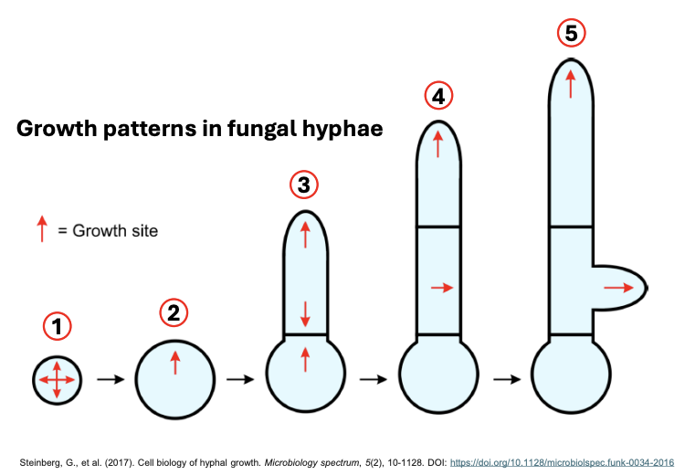
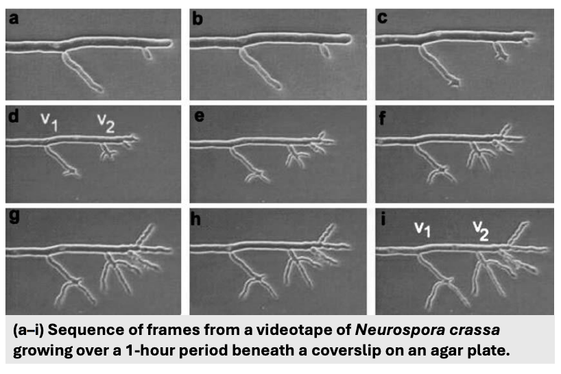
Explain the growth patterns in fungal hyphae
Growth occurs in an isotropic (equal) fashion during spore germination (1)
Specification of polarity axis ultimately results in formation of hypha that continues to grow at the tip (2-3)
While tip growth is maintained, specification of additional polarity axes and compartmentalization enables formation of septa and lateral branches
While septum formation is transient, branching results in the formation of secondary hypha that also continues to grow at the tip
Structural variation in Mycota are made from _
hyphal growth + fusion
Elongation, compartmentalization, fusion ecf
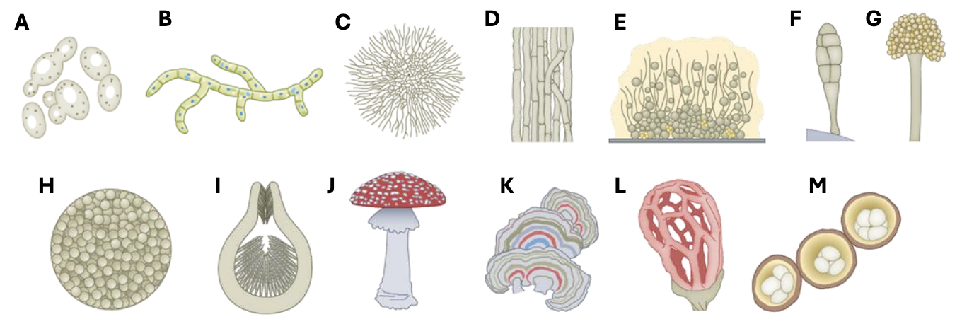
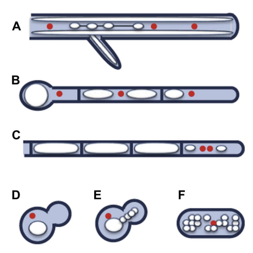
Describe figure shown
Structural variations resulting from hyphal growth + fusion (micro- & macroscopic fungal forms)
a) Unicellular yeasts
b) Pseudohyphae
c) Filamentous mycelia
d) Fungal parenchyma (hyphae anastomoses in parallel direction)
e) Biofilm aggregates (not all fungi can form)
reproductive structures f-g
f) Compartmentalized macroconidia
g) Conidiophores
h) Spherules
fruiting bodies i-j
i) Perithicium
j) Fruiting bodies
k) Brackets
L) Cages
M) Nests
Apical tip growth and its plasticity involves _
cell wall expansion from secreted vesicles
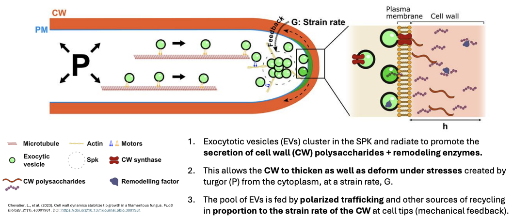
Explain Tip Growth Model of the Hyphal Apex
tsvc
For the hyphal tip to continuously grow, 3 important components are necessary:
Turgor pressure (fluid in protoplasm pushing against CW)
SPK (determines direction) & constant delivery of vesicles (release chitin synthases and other materials) to hyphal tip
Cytoskeleton & TVS to direct all vesicles to proper area
So what happens is:
Actin, microtubules, motor proteins deliver diff vesicles from internal protoplasm to the tip where there’s SPK, hence SPK accumulate lots of vesicles at tip
Once these vesicles cluster at tip, containing materials for PM + cell wall synthesis, vesicles specifically containing transporters (CW synthase) fuse with PM, movement of materials commence, including chitosomes embedded into CW
Hyphal tip usually becomes thinnest point of CW bc its components move to sides, causing strain on hyphal tip, which forces elongation to move forward, coupled with turgor pressure bc protoplasmic movement also exerts pressure against CW
Strain is important to determine whether hypha continues to grow because once it becomes very low, in the case that fungus hits a wall, then no more vesicles are brought to the tip.
But with the strain constant, vesicles also continue to move forward.
Exocytotic vesicles (EV) cluster in the SPK and radiate to promote secretion of cell wall polysaccharides + remodeling enzymes
This allows CW to thicken and deform under stresses by turgor pressure from cytoplasm, at a strain rate
The pool of EVs is fed by polarized trafficking and other sources of recycling in proportion to the strain rate of the CW at the cell tips (mechanical feedback)
T/F: It’s not only turgor pressure and CW formation driving hyphal motion forward
TRUE
There’s also actin
T/F: Polarized vesicle trafficking ensures that the majority of materials are delivered to the hyphal sides for wall expansion.
FALSE
Materials are delivered to the tip, not directly to the sides, but are redistributed from the tip.
Area where germ tube comes out and hyphae develops
Polarity axis

(3) When 2 polarity axes go against one another, this can trigger _
septum formation
T/F: Septum formation is more transient than secondary hypha / branching formation
TRUE
Bc secondary hypha will also continue to grow at the tip