Anatomy Lab - Unit 3
1/115
Name | Mastery | Learn | Test | Matching | Spaced |
|---|
No study sessions yet.
116 Terms
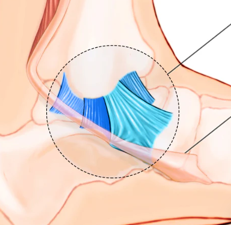
dorsal interossei
abduction of metatarsophalangeal joints of toes 3 & 4
flexion of metatarsophlangeal joints of toes 2-4
extension at the interphalangeal joints of 2-4
subtalar ligaments
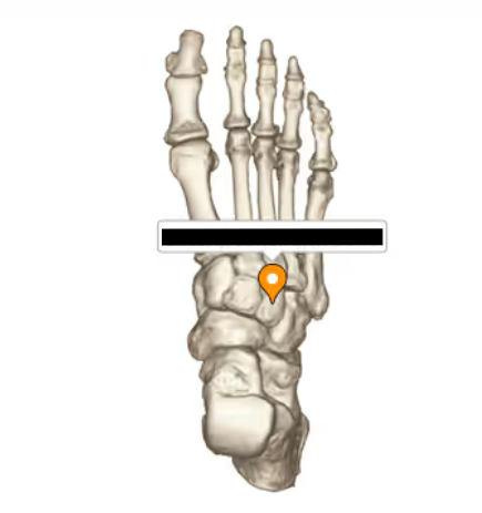
navicular bone
tuberosity of fifth metatarsal
tuber calcanei
most posterior part of calcaneus; “the heel”
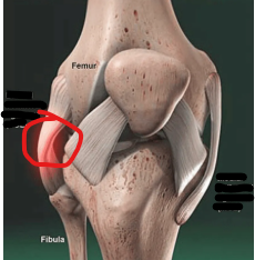
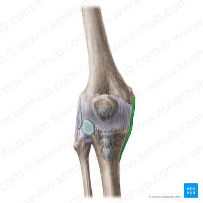
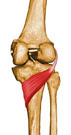
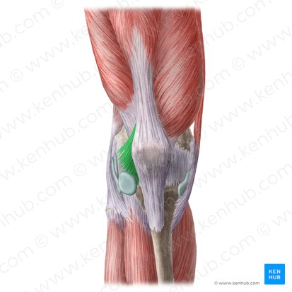
lateral collateral ligament of knee
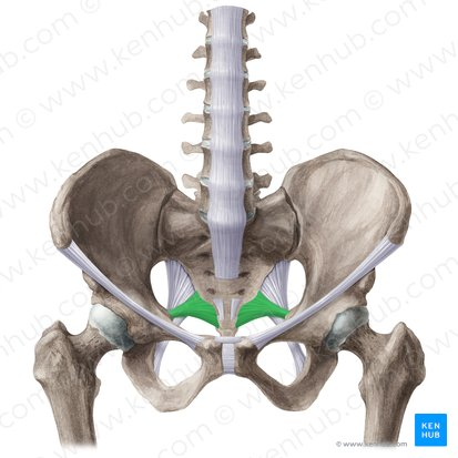
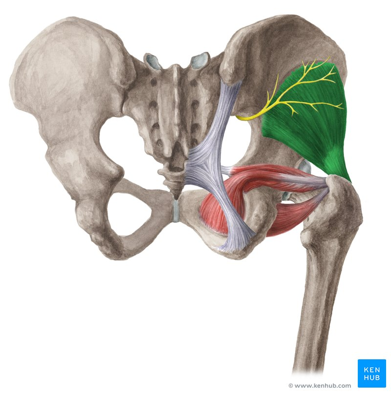
sacrotuberous ligament - goes from PSIS to lower sacrum to ishial tubersosity of pelvis
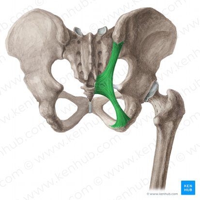
sacrospinous ligament
medial meniscus

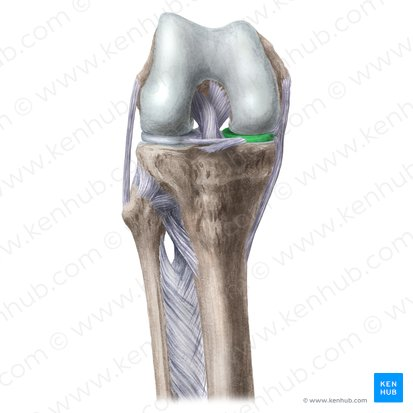
posterior cruciate ligament (PCL)
has 2 heads
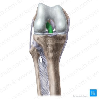
anterior cruciate ligament (ACL)
tibia to femur
stabilizes joint
tibial collateral ligament (medial collateral ligament or MCC)
patellar ligament
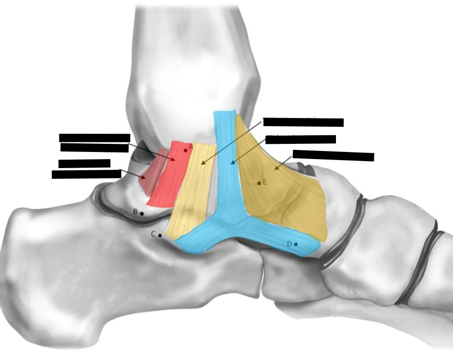
deltoid ligament
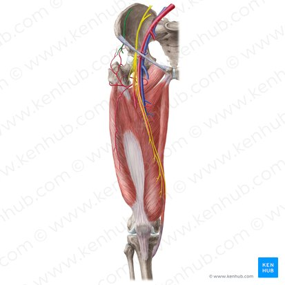
sciatic nerve - largest and longest nerve in body
emerges through greater sciatic notch from under piriformis muscle
in 15% of ppl it goes through the piriformis
curves over 3 muscles
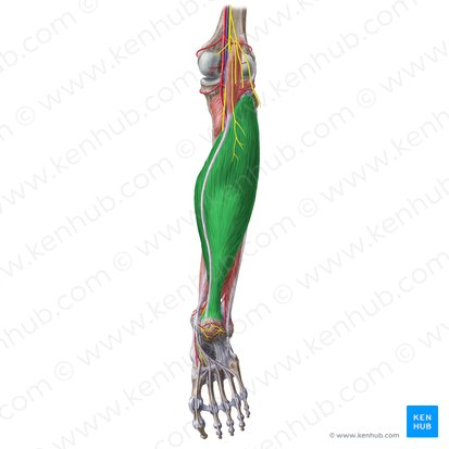
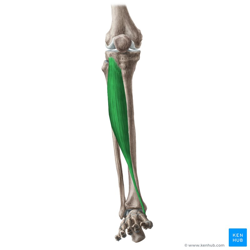
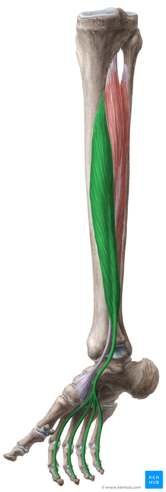
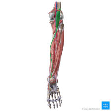
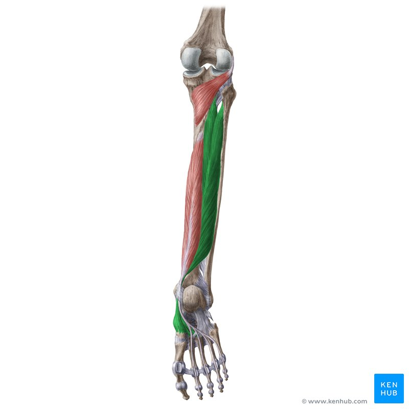
soleus
plantar flexion at ankle
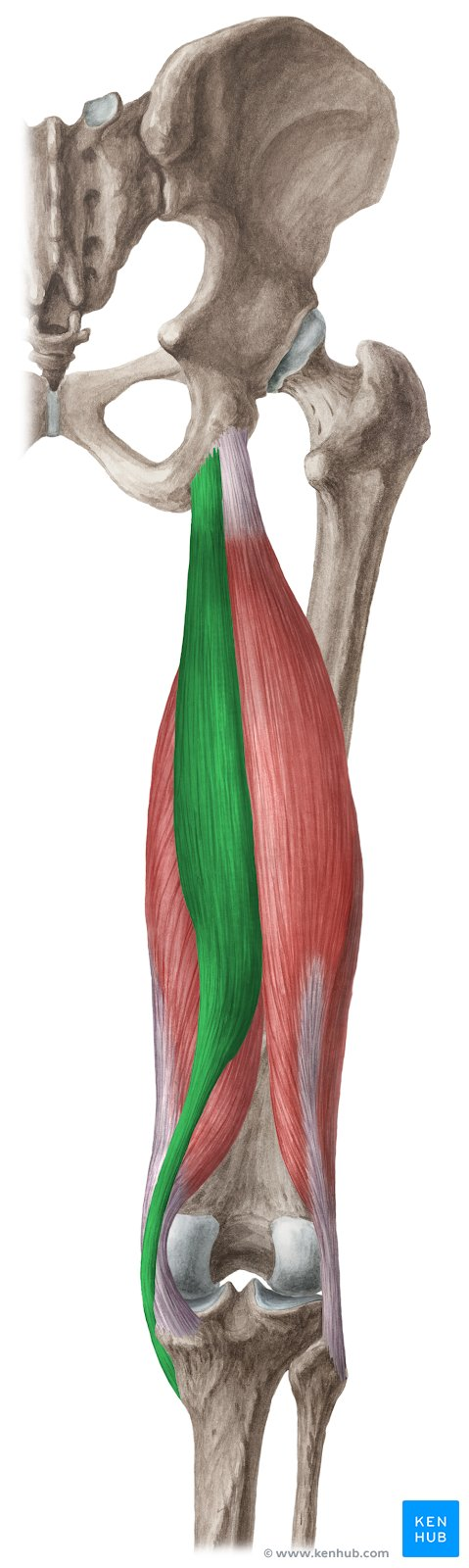
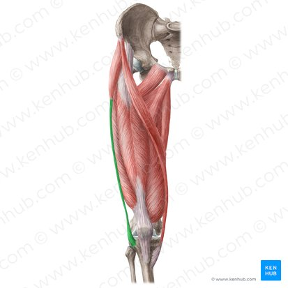
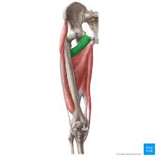
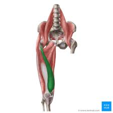
sartorius
seatbelt muscle
flexion at knee
abduction, flexion, lateral rotation at hip
gluteus minimis
abduction and medial rotation at hip
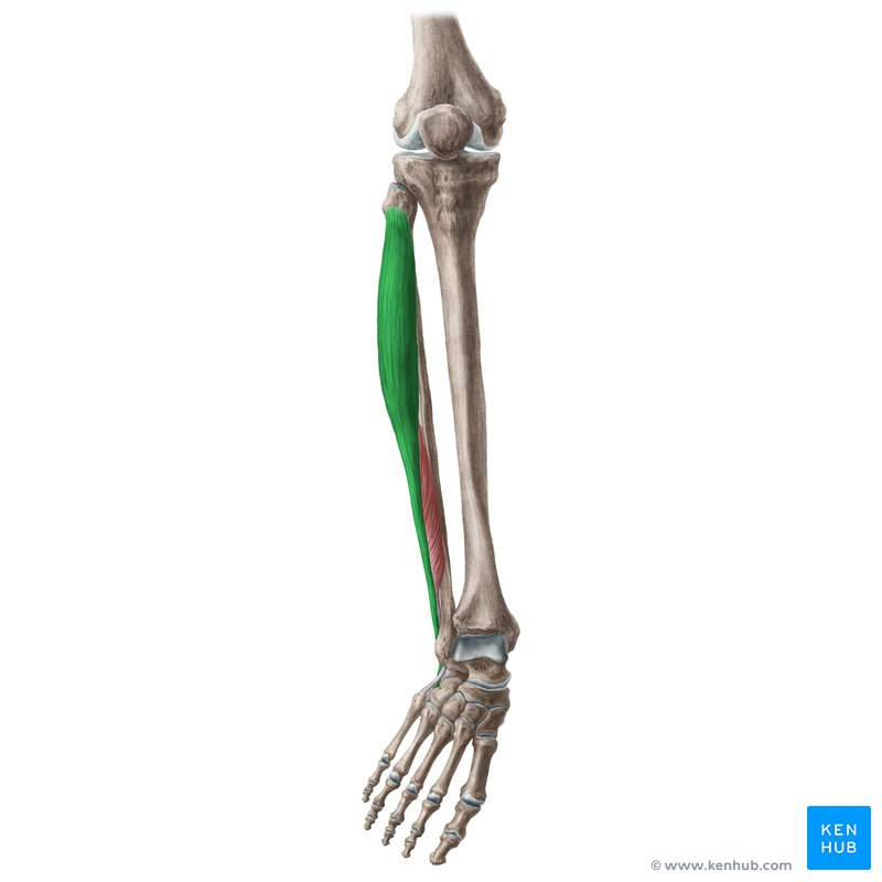
fibularis brevis
eversion of foot and plantar flexion at ankle
tibialis anterior
dorsiflexion of ankle
inversion of foot
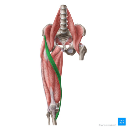
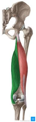
biceps femoris (long head in blue, short head in yellow)
flexion at knee
extension and lateral rotation at hip
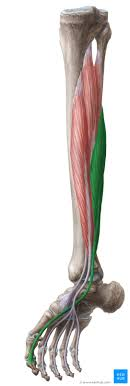
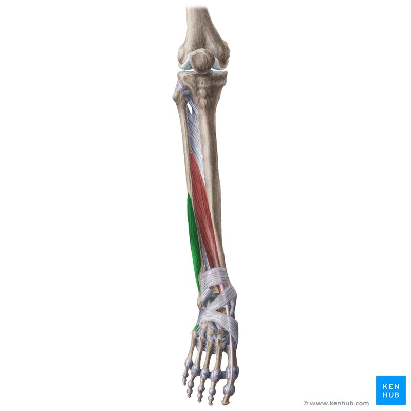
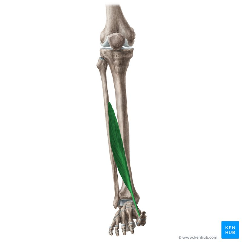
flexor hallucis longus
flexion at joints of great toe
plantar flexes ankle
popliteus
medial rotation of tibia (or lateral rotation of femur) at knee
flexion at knee
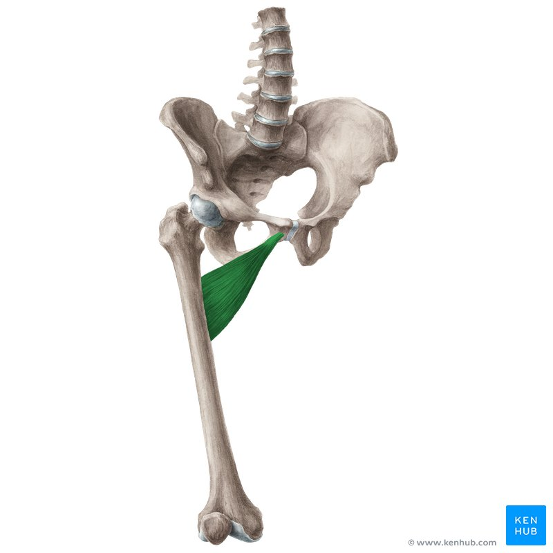
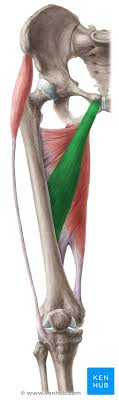
adductor brevis
adduction and flexion at hip
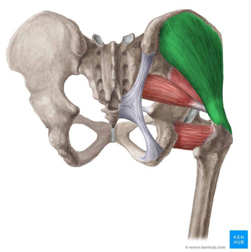
ischiofemoral ligament
on posterior side
connects trochanter femor to body
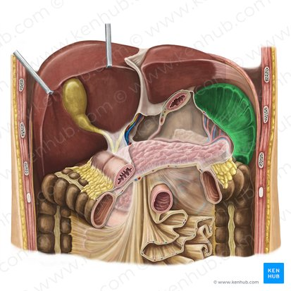
The small intestine absorbs _% of _?
90%, nutrients we eat
What are the small intestine parts, their length, and descriptions?
Duodenum, Jejunum, Ileum
1ft long, shortest and widest part
3ft long, thick wall of DNA rich blood supplu
6ft long, thin walls
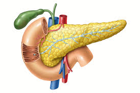
pancreas
behind stomach
secretes enzyme rich pancreatic juice which gets delivered into the duodenum
Abdominal Aortic Aneurysms (AAA) are the __ most common
second
spleen
most lateral and in contact with the tail of the pancreas
important for RBC (red blood cell) turnover
you can live without it, but have to be careful
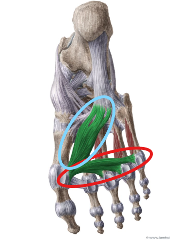
quadratus plantae
flexion at joints of toes 2-5
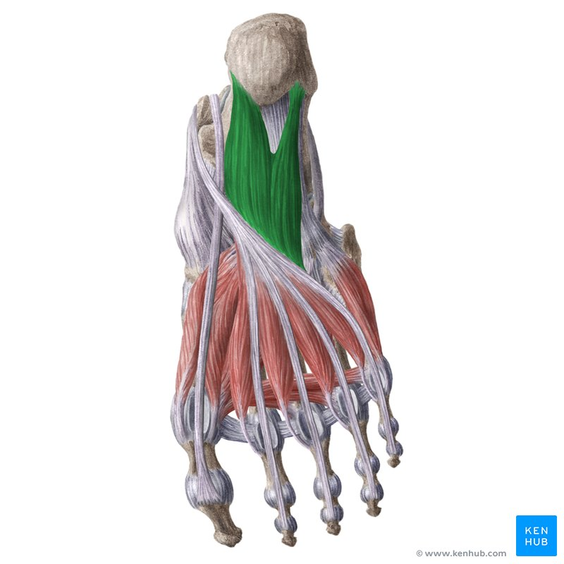
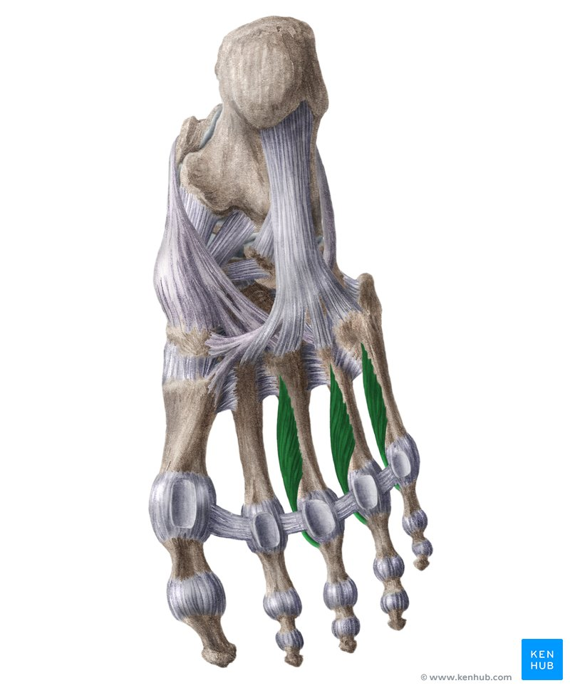
lumbricals
flexion at metatarsophalangeal joints; extension at interphalangeal joints of toes 2-5
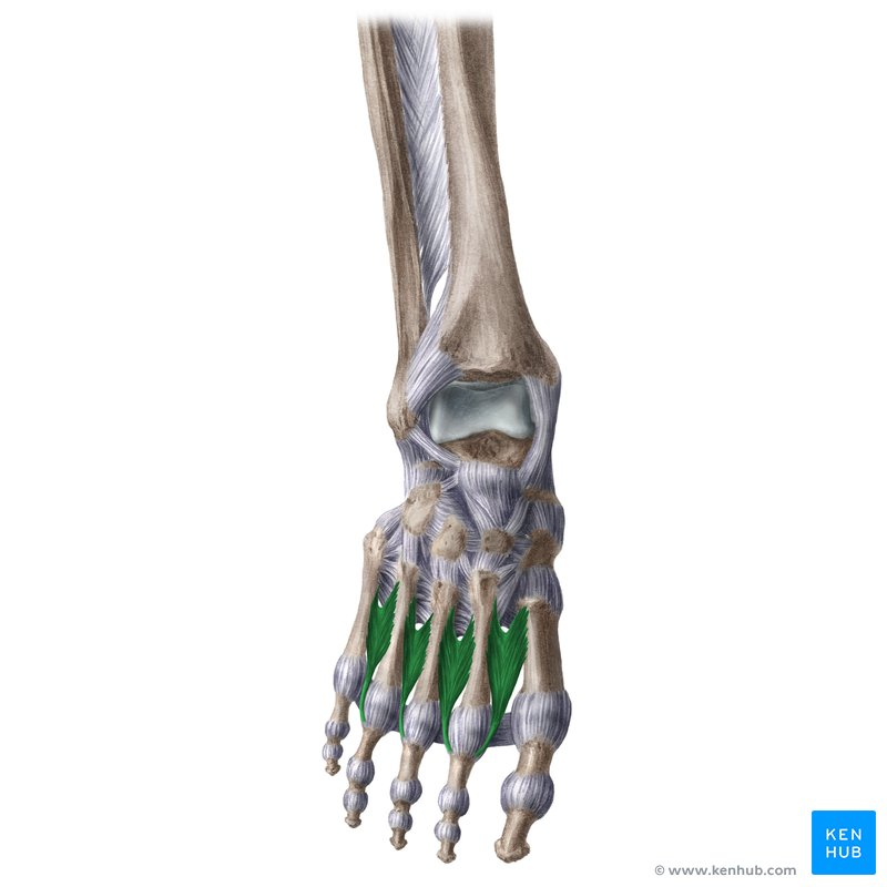
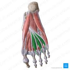
plantar interossei (3 of these)
adduction at metatarsophalangeal joints of toes 3-5
flexion of metatarsophalangeal joints
extension at interphalangeal joints
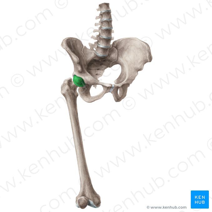
head of femur
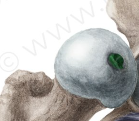
fovea capitis of femur
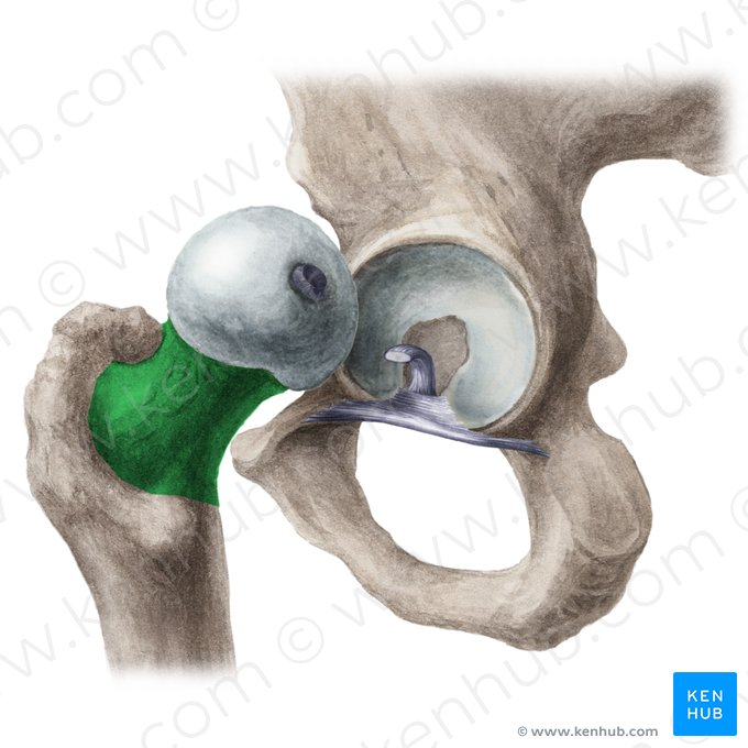
neck of femur
greater trochanter
lesser trochanter
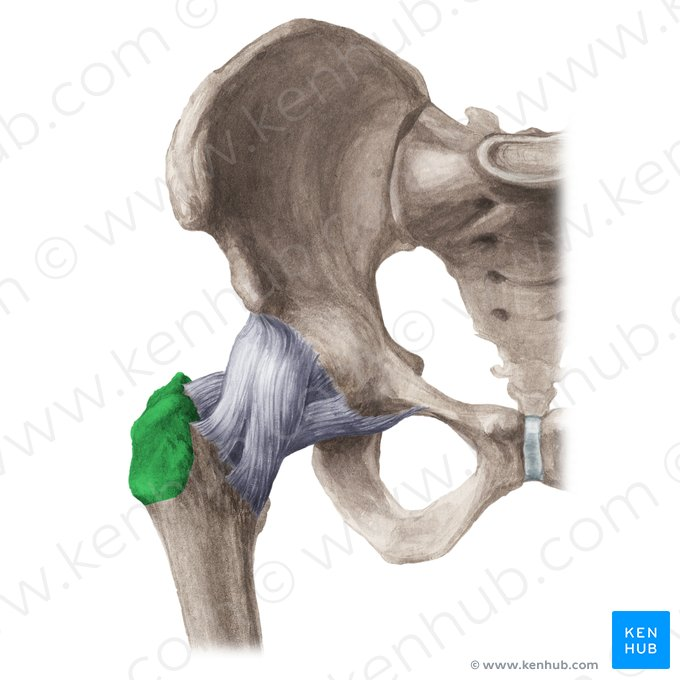
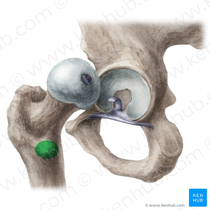
gluteal tuberosity of humerus
pectineal line of femur
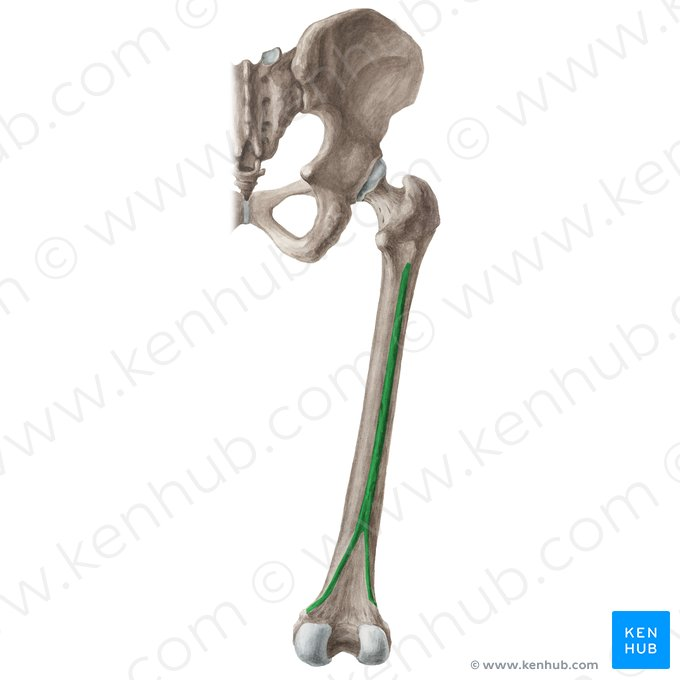
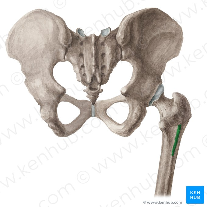
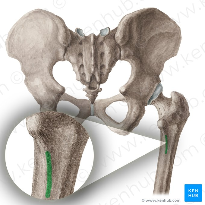
linea aspera of femur
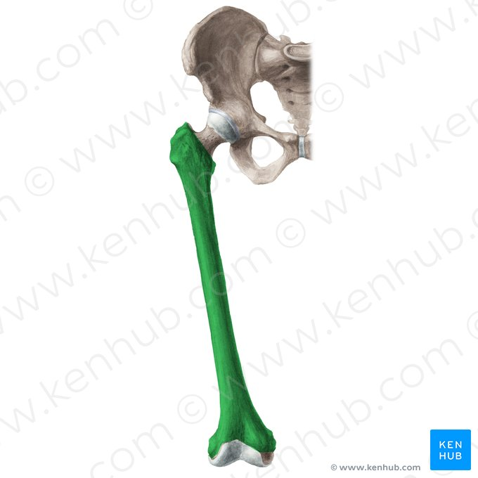
body of femur
intertrochanteric crest of femur
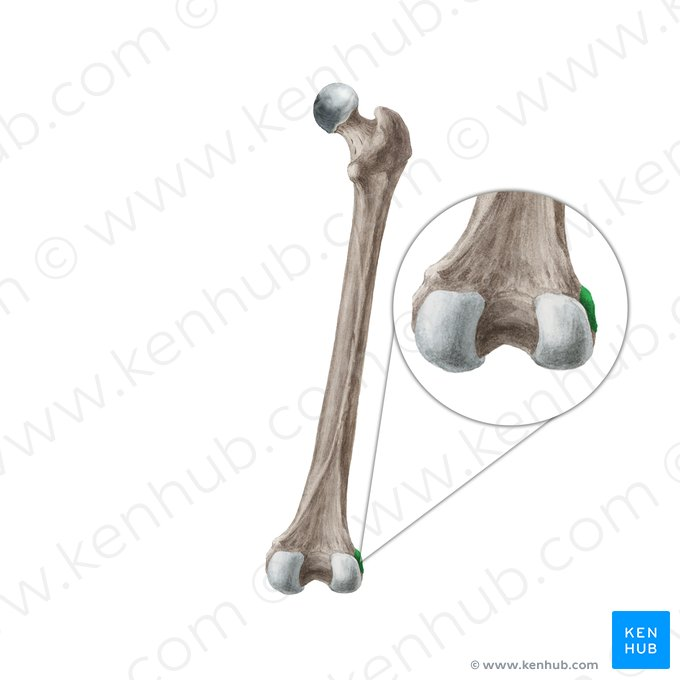
lateral epicondyle of femur
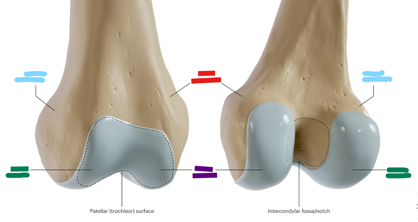
intercondylar fossa of femur
medial epicondyle of femur
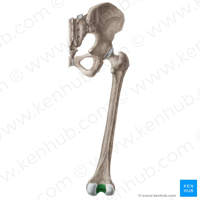
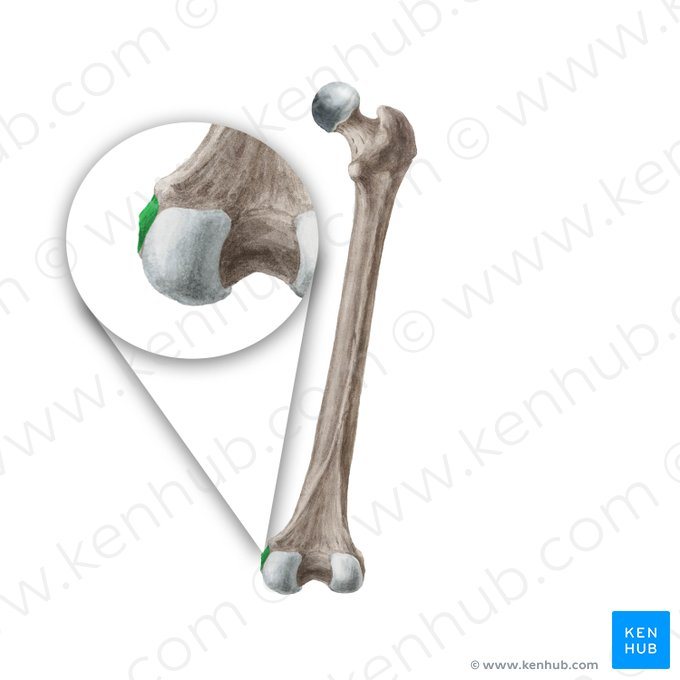
red: medial condyles
purple: medial epicondyles
blue: lateral epicondyles
green: lateral condyles
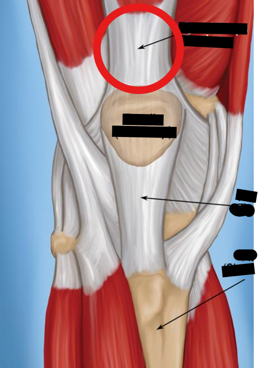
quadriceps tendon
semitendinosus
flexion at knee
extension and medial rotation at hip
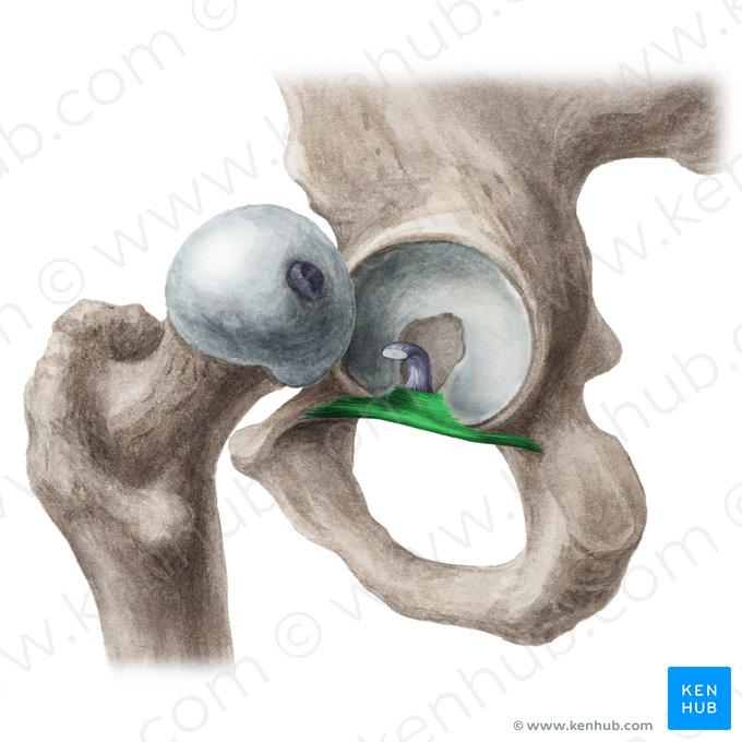
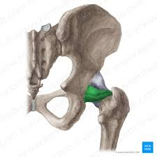
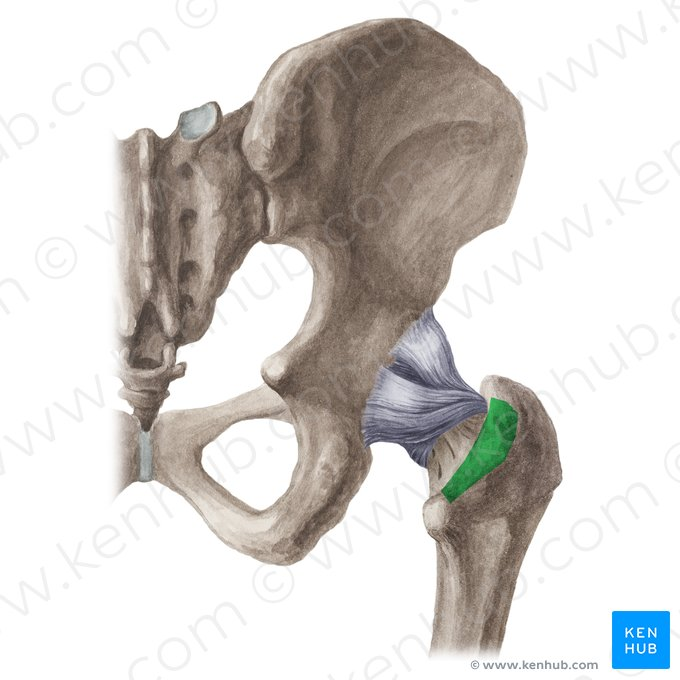
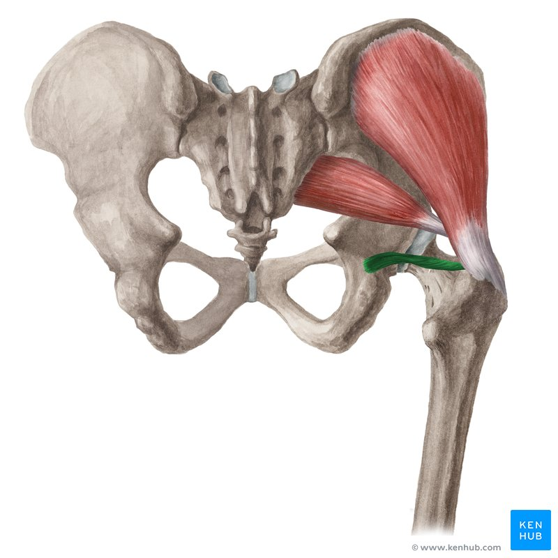
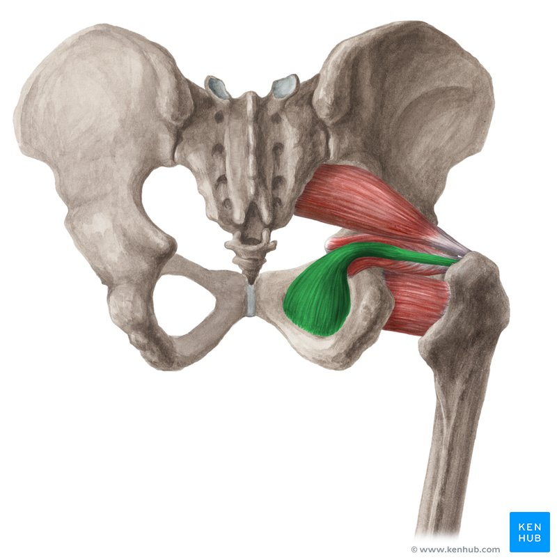
transverse acetabular ligament
on inferior portion of the acetabulum
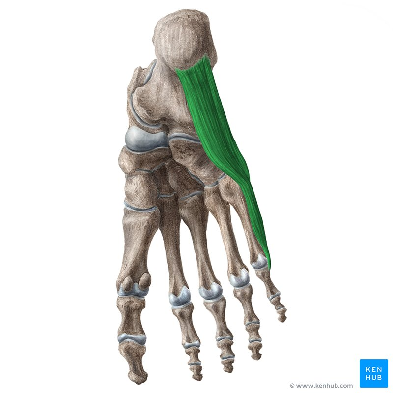
extensor hallicus longus
extension at joints of great toe
dorsiflexes ankle
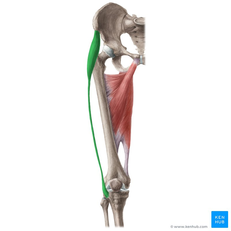
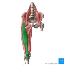
gracilis
flexion and medial rotation at knee
adduction and medial rotation at hip
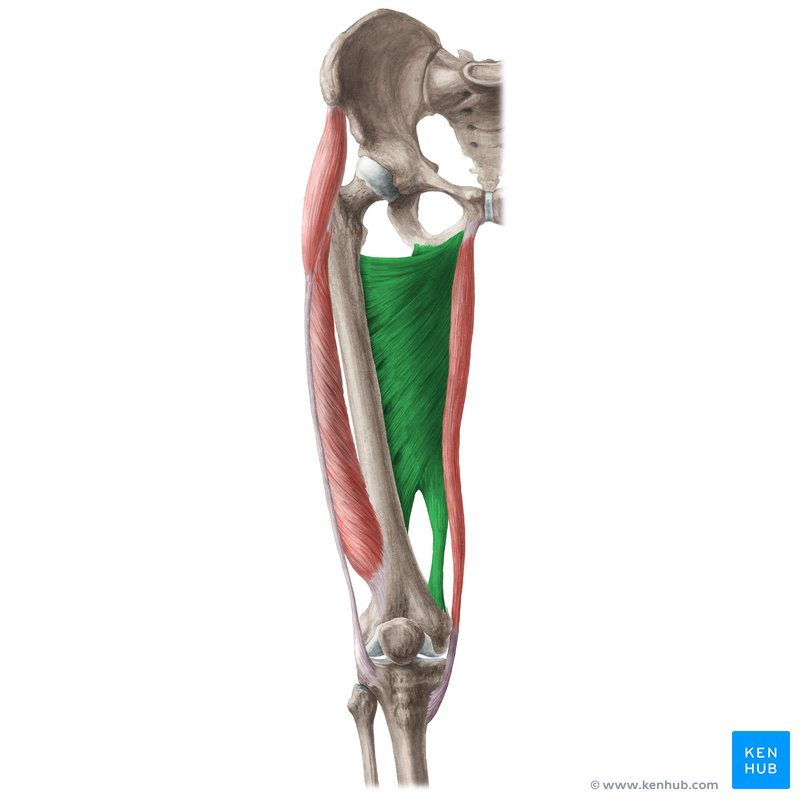
adductor longus
adduction, flexion, and medial rotation at hip
psoas major
flexion at hip and/or lumbar intervertebral joints
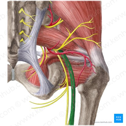
obturator nerve
destination: obturator foramen
medial to psoas groupm, slightly lateral & posterior to the common iliac artery
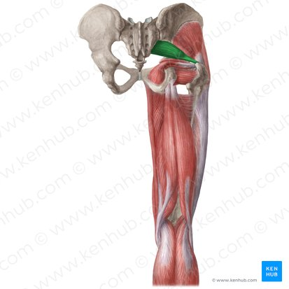
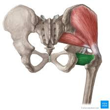
piriformis
lateral rotation and abduction of hip
helps maintain stability of hip
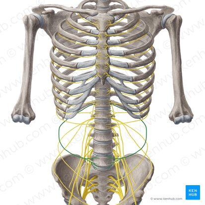
subcostal nerve
inferior to rib 12
gluteus medius
abduction and medial rotation at hip
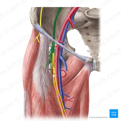
lateral femoral cutaneous nerve (of the thigh)
originates from deep to the psoas group
crosses the iliacus & continues under the inguineal ligament to the lateral thigh
“cutaneous” indicates that it is sensory to the skin of that region
tensor fascia latae
extension at knee and lateral rotation of leg (through IT band)
abduction and medial rotation of thigh
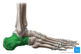
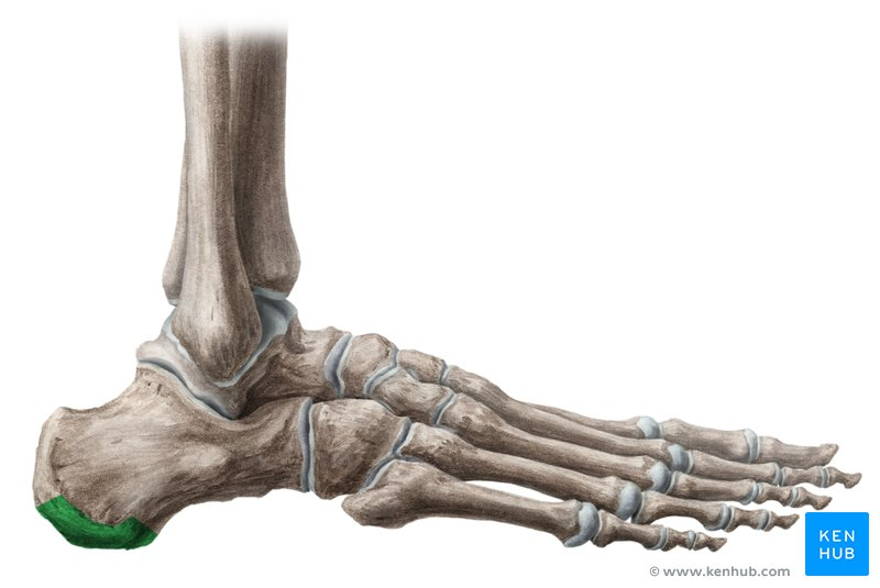
calcaneus
heel bone
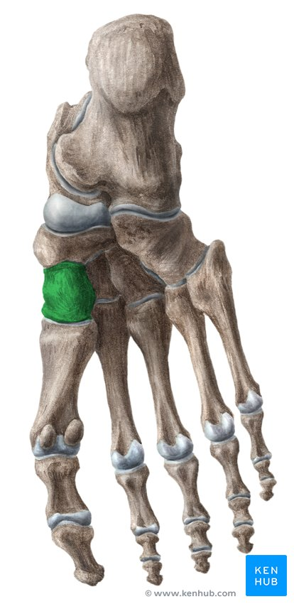
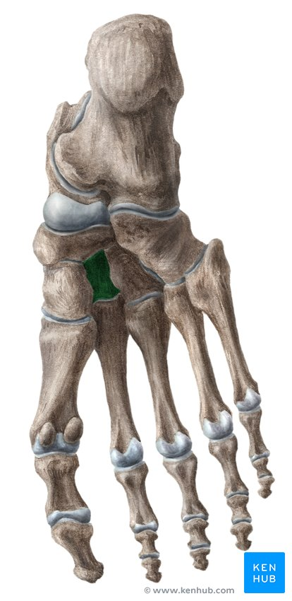
lateral cuneiform bone
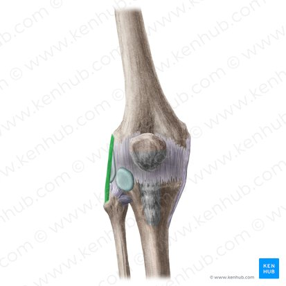
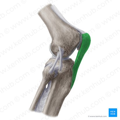
fibular collateral ligament (LCL)
fibula to femur
patellar retinaculae (lateral)
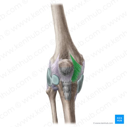
patellar retinaculae (medial)
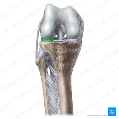
lateral meniscus
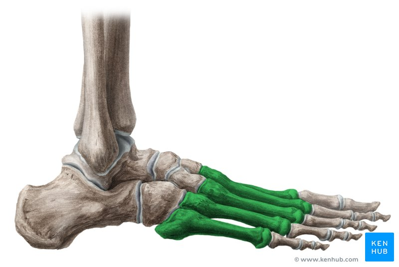
metatarsals
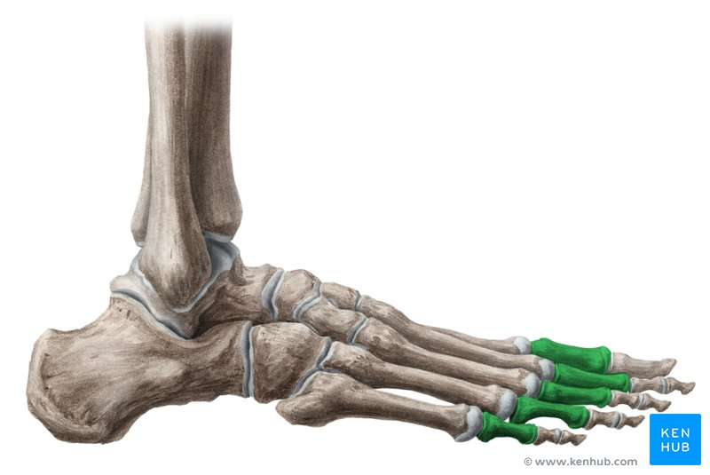
proximal phalanges
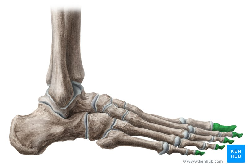
distal phalanges
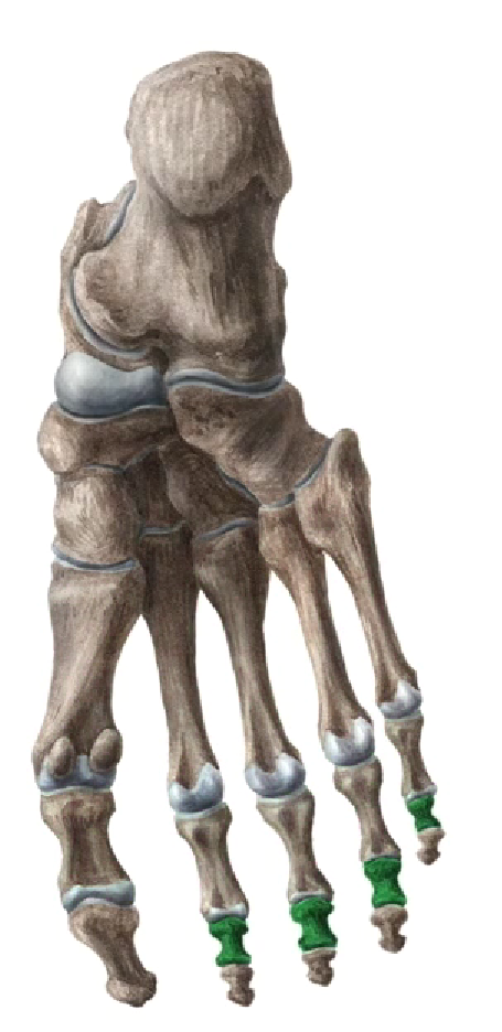
middle phalanges
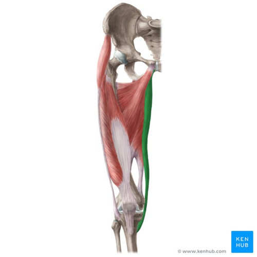
iliotibial tract/band
a band of collagen fibers that extends along the lateral surface of the thigh and inserts on the tibia
stabilizes knee (when balancing on one foot)
femoral nerve
huge nerve headed to quadriceps
partially hidden by psoas major, crosses under the inguineal ligament into the femoral triangle of the anterior thigh
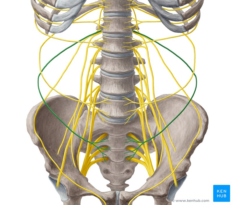
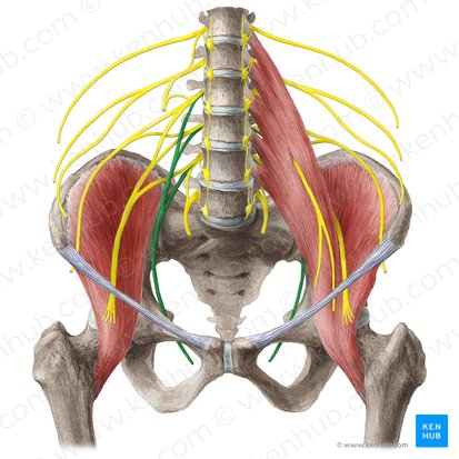
iliohypogastric nerves
begins under stomach, ‘hypogastric’ & typically dives into the fascia between QL and transverse abdominis
subtalor ligaments
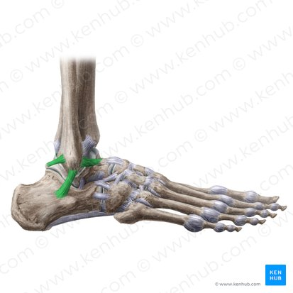
lateral collateral ligament of ankle joint
medial cuneiform (note: inferior view)
intermediate cuneiform (note: inferior view)
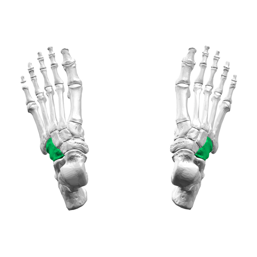
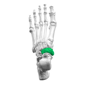
cuboid
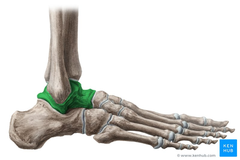
talus
on top of calcaneus
articulates w/ tibia & fibula
pectineus
flexion and adduction at hip
adductor hallucis (red - transverse head, blue - oblique head)
adduction and flexion of metatarsophalangeal joint of great toe
fibularis longus
eversion of foot and plantar flexion at ankle
iliosacaral ligament
forms SI joint
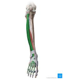
flexor digitorum longus
flexion of joints of toes 2-5
plantar flexes ankle
adductor magnus
adduction at hip
one part does flexion and medial rotation
one part does extension
inferior gemellus
lateral rotation and abduction of hip
vastus medialis
extension at knee
rectus femoris
extension at knee; flexion at hip
quadratus femoris
lateral rotation of hip
semimembranosus
flexion at knee
extension and medial rotation at hip
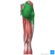
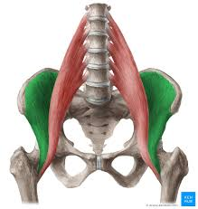
gluteus maximus
extension and lateral rotation at hip, helps stabilize extended knee
abduction at the hip
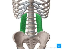
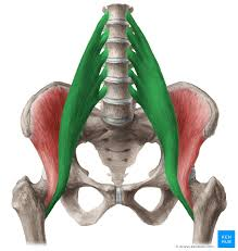
quadratus lumborum
flex the spine and depress the ribs
obturator internus
lateral rotation and abduction of hip
plantaris
plantar flexion at ankle
flexion at knee
tibilalis posterior
inversion of foot
plantar flexion at ankle
extensor digitorum longus
extension of toes 2-5
dorsiflexes ankle
iliacus
flexion at hip and when working w/ psoas major, flexes the intervertebral joints
abductor digiti minimi of foot
abduction and flexion at metatarsophalangeal joint of toe 5