Ch. 1 Microscopy
 Call Kai
 Learn
Learn Practice Test
 Spaced Repetition
 Match
Match Flashcards
 Knowt Play
1/12

There's no tags or description
Looks like no tags are added yet.
Name | Mastery | Learn | Test | Matching | Spaced | Call with Kai | Chat |
|---|
No analytics yet
Send a link to your students to track their progress
13 Terms
histology
study of microscopic structure and function of tissues
Formation of Organ Systems
Cells → Tissues → Organs → Organ Systems
4 types of tissues
epithelial
connective
muscle
nervous
Fixation
cross-linking fixatives perserve tissue before embedding
Common fixatives
glutaraldehyde and osmium tetroxide (EM)
paraformaldehyde (LM)
Prep order of a biopsy
Fixation → dehydration → cleaning → infiltration → embedding
sectioning tool
microtome or cryostate
Types of Staining
histological: genetic tissue characteristic
electron microscopy “stains”: heavy metal (osmium tetroxide embedded means electrons cannot pass)
immunohistochemical: specific molecule
Hematoxylin
basic dye, stains basophilic components blue because of GAGs and nucleic acids
Eosin
acid dye, stains acidophilic tissue components red/pink
mitochondria, secretory granules, and collagen
Transmission Electron Microscopy (TEM)
powerful imaging technique that shoots a beam of electrons through an ultra-thin sample to view its internal structures at the atomic level
lipid bilayer appears black due to osmium tetroxide (EM Stain)
Scanning Electron Microscope (SEM)
beam scans tissue surface 3D using electrons
EM stain = gold (coats specimen)
Sectioning Terms
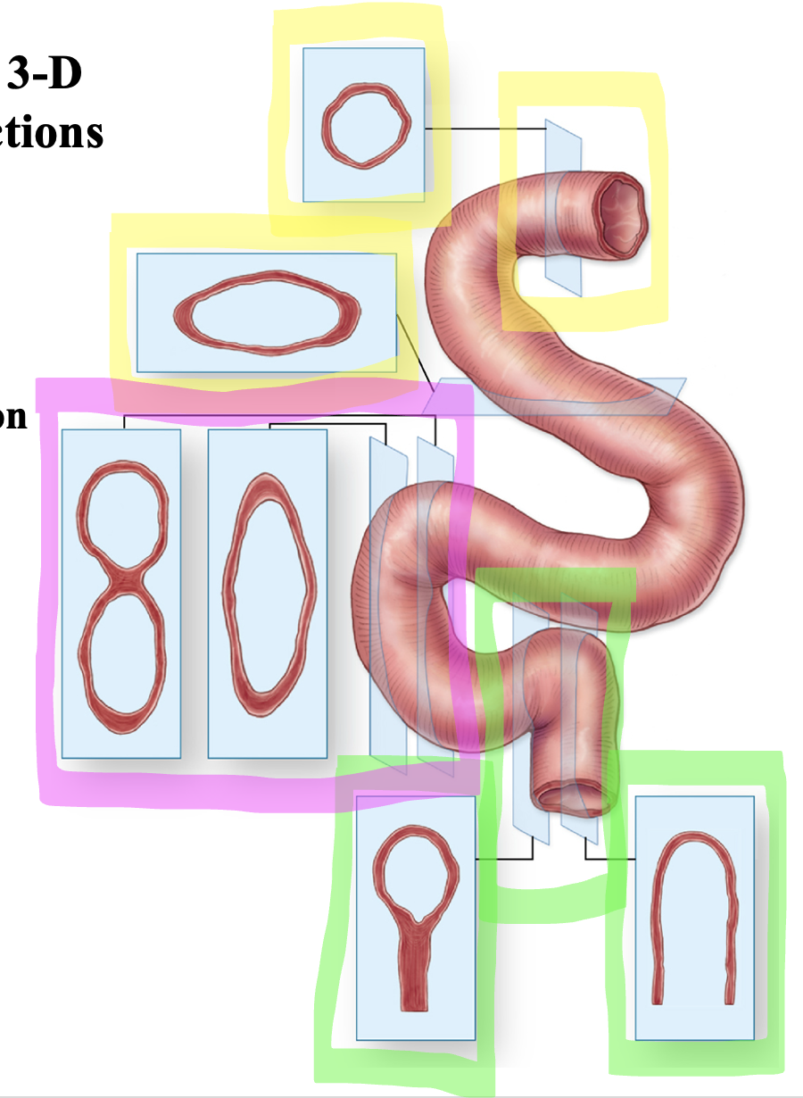
Cross (transverse) section” 90 degree angle
longitudinal (horizontal) section - follows long axis
oblique section: angle of arm