3.4.1 Mass transport in animals
1/47
There's no tags or description
Looks like no tags are added yet.
Name | Mastery | Learn | Test | Matching | Spaced |
|---|
No study sessions yet.
48 Terms
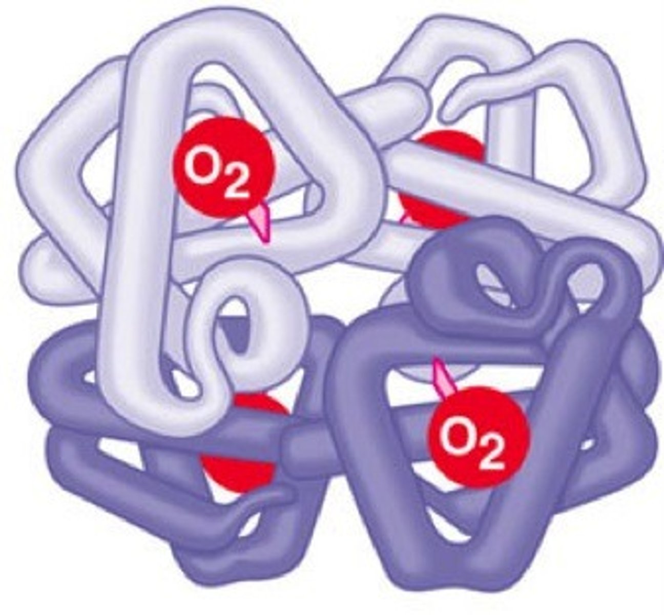
describe the structure of haemoglobin
- Protein with quaternary structure
- made of 4 polypeptide chains (2 alpha and 2 beta chains)
- Each chain contains a Haem prosthetic group containing an iron ion

what are the haemoglobins?
A group of chemically similar molecules found in many different organisms
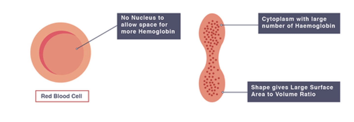
How are red blood cells adapted to transporting oxygen?
- biconcave disc shape: increases surface area to volume ratio and reduces length of diffusion pathway
- contain lots of haemoglobin
- have no nucleus so more space for haemoglobin
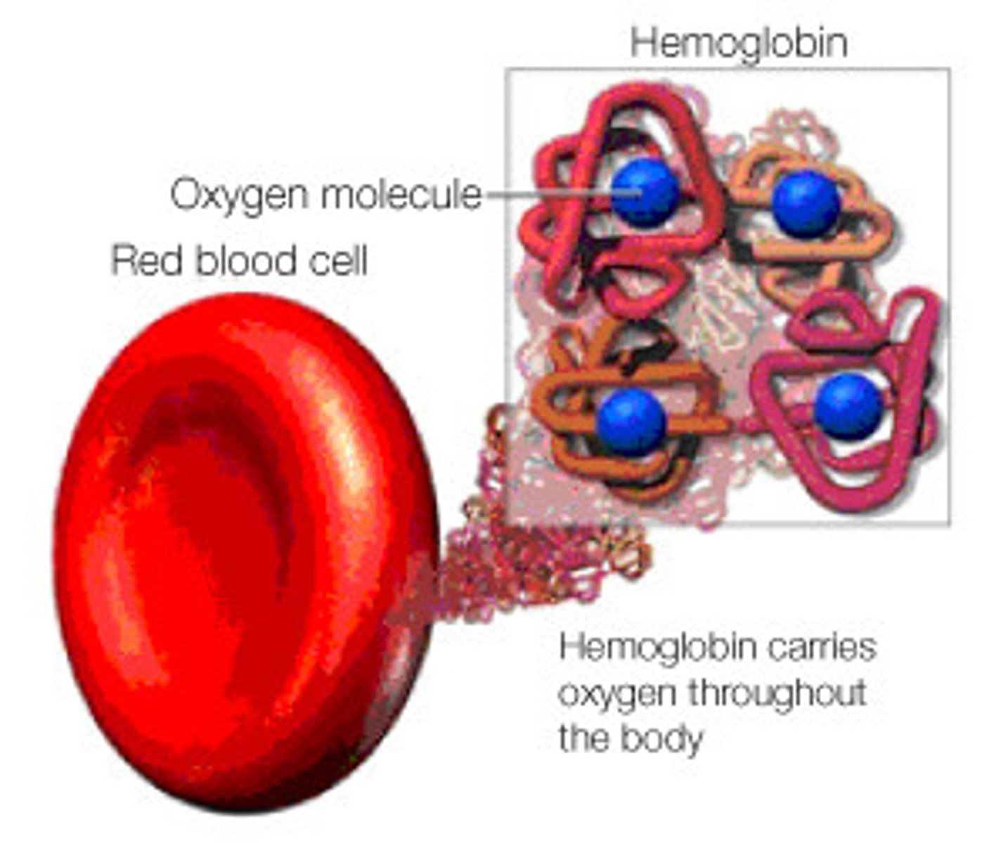
Describe the role of haemoglobin and red blood cells in transport of oxygen
- RBCs contain lots of haemoglobin.
- Hb associates/ loads oxygen at gas exchange surfaces where partial pressure of oxygen is high
- This forms oxyhemoglobin, which transports oxygen
- Hb disassociates/ unloads oxygen near cells or tissues where partial pressure of oxygen is low
what is affinity for oxygen?
How readily haemoglobin loads and unloads oxygen
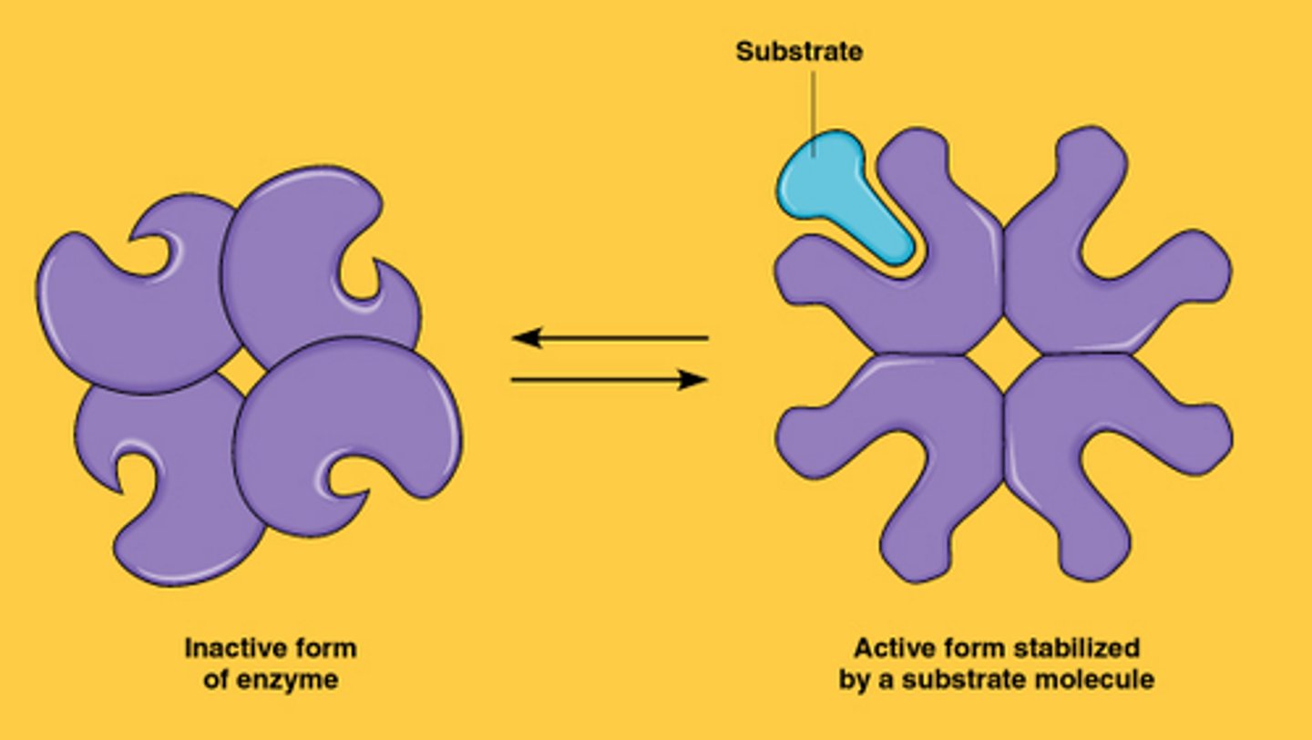
how and when does haemoglobin change its affinity for oxygen?
haemoglobin can change its affinity for oxygen under different conditions by changing its tertiary structure
how many oxygen molecules can haemoglobin carry?
4 (one on each haem group)
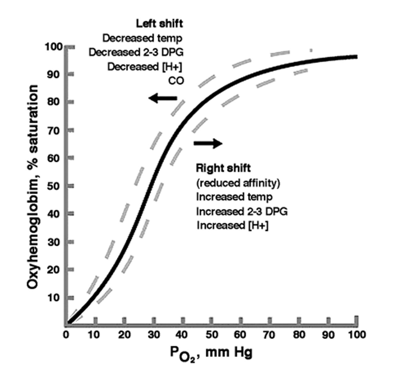
describe the loading, transport and unloading of oxygen in relation to the oxyhemoglobin dissociation curve
In areas with low pO2, eg. respiring tissues, Hb has a lower affinity for oxygen so it readily unloads and percentage saturation is low
In areas with high pO2, mainly gas exchange surfaces, Hb has a higher affinity for oxygen so it readily loads and percentage saturation is high

explain the s-shape (sigmoidal) of the oxyhaemoglobin dissociation curve
1. at low pO2, there is little increase in percentage saturation of Hb with oxygen as oxygen increases. The gradient of the curve is therefore shallow.
2. however, after the first oxygen binds, it takes a smaller increase in pO2 to bind the second oxygen. The gradient of the curve steepens.
3. this is because binding of first oxygen changes tertiary structure of haemoglobin
4. this exposes the haem group binding sites, making further binding of oxygens easier
5. the gradient of curve levels off because it is harder for the last oxygen to bind, since it is less likely to find an unoccupied binding site
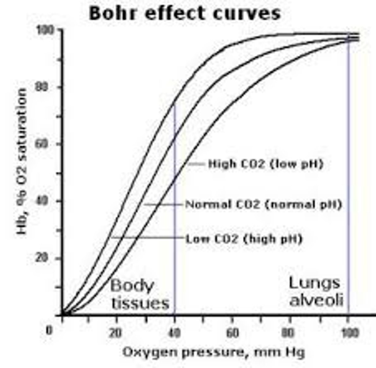
what is the Bohr effect
the effect of carbon dioxide concentration on dissociation of haemglobin - if concentration increases the curve shifts to the right as haemoglobin's affinity for oxygen decreases
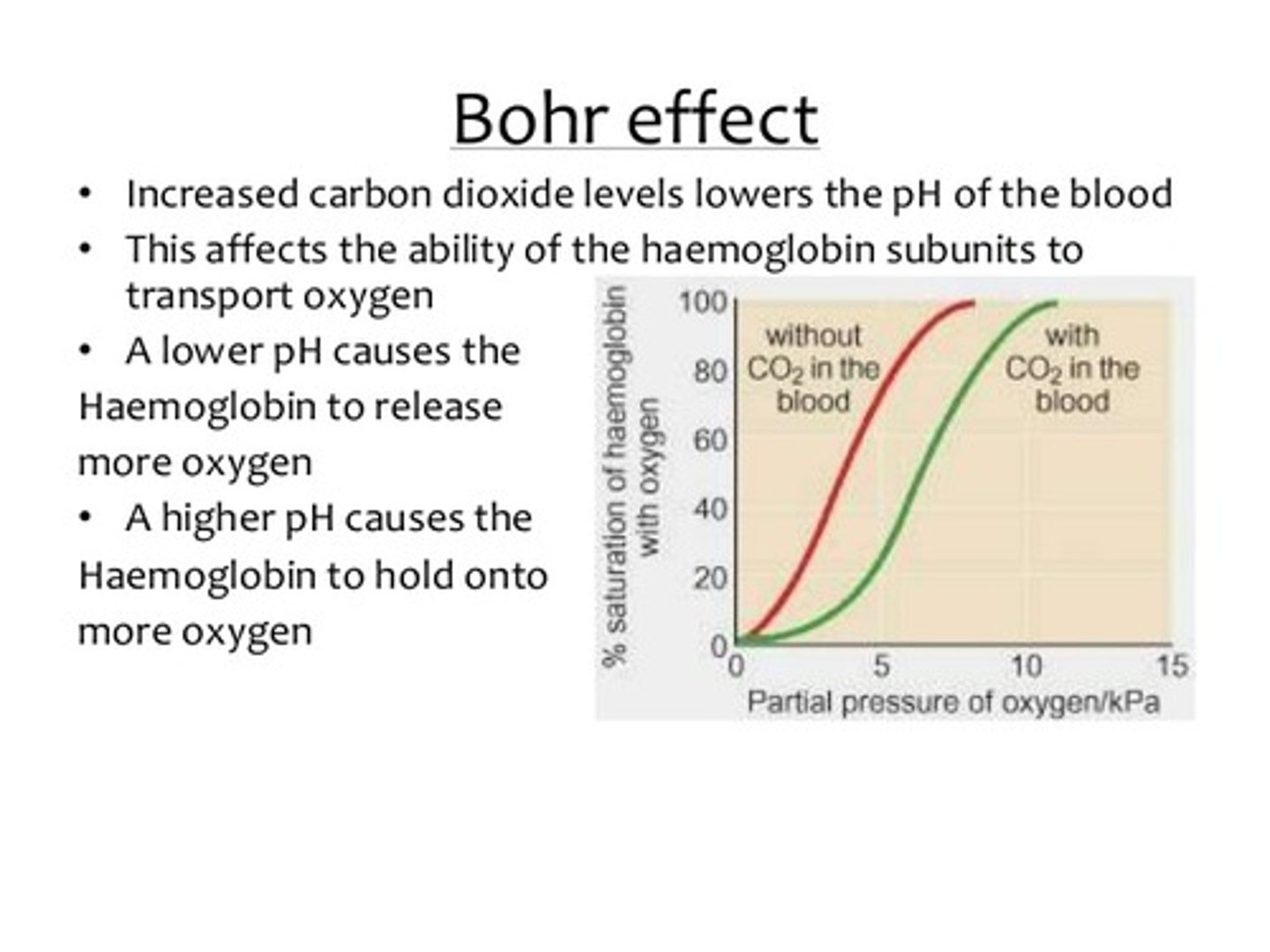
explain effect of carbon dioxide concentration on the dissociation of haemoglobin (Bohr's effect)
1. blood carbon dioxide concentration may increase eg. due to increased rates of respiration
2. this lowers the blood pH as CO2 is acidic in solution
3. Hb's affinity for oxygen decreases as its tertiary structure changes
4. this means oxygen is more readily unloaded at respiring tissue at any given pO2
what is the advantage of the Bohr effect?
during exercise, more oxygen will be unloaded, so higher rate of aerobic respiration, so more ATP is produced
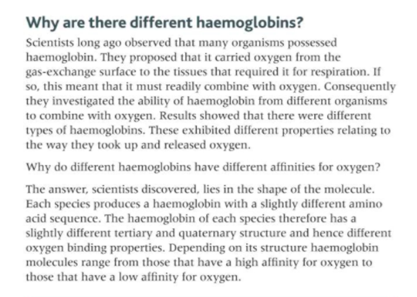
why do different types of haemoglobins have different oxygen transport properties?
- different types of Hb are made of polypeptide chains with slightly different amino acid sequences (different primary structures)
- which results in different tertiary and quarternary structures
- so different affinities for oxygen
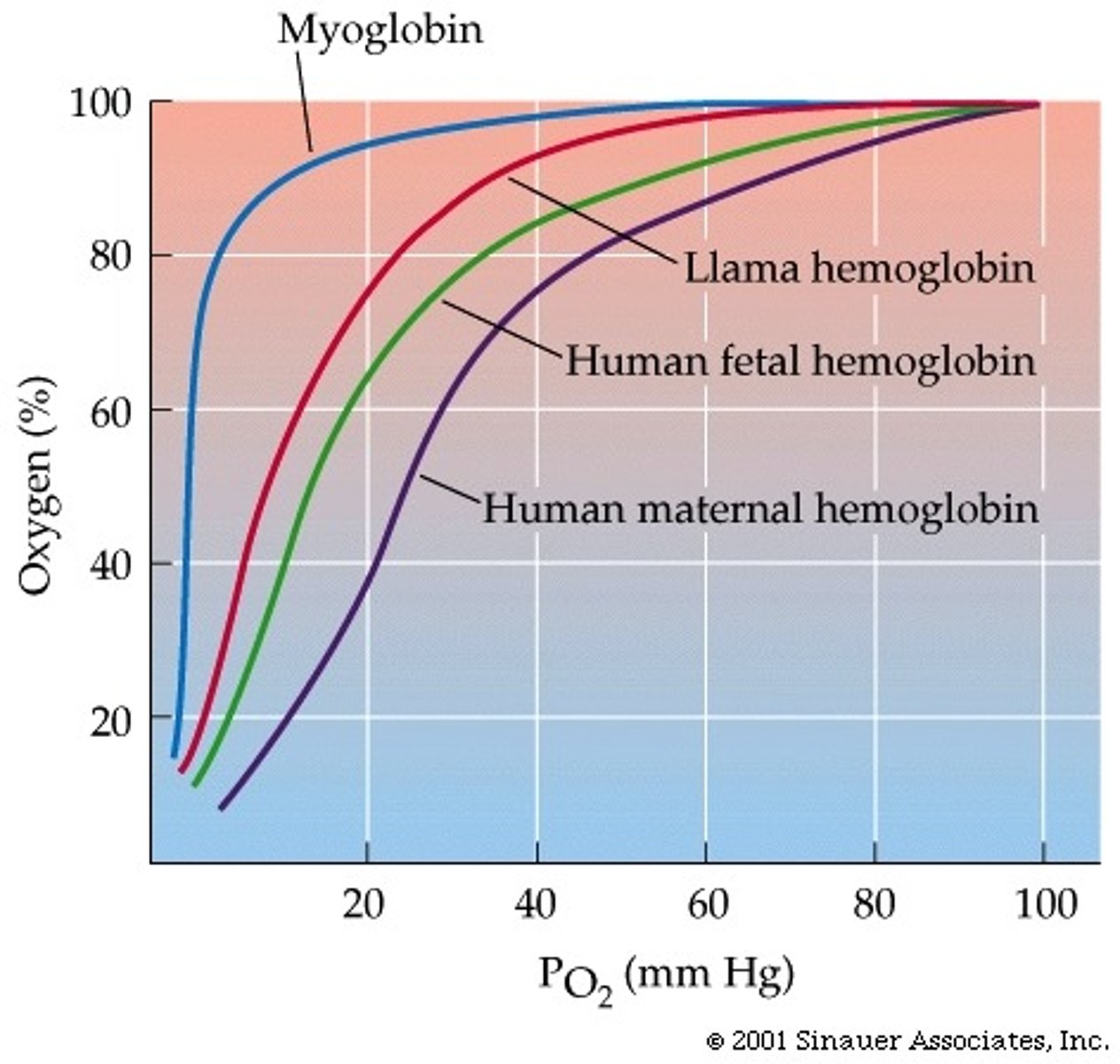
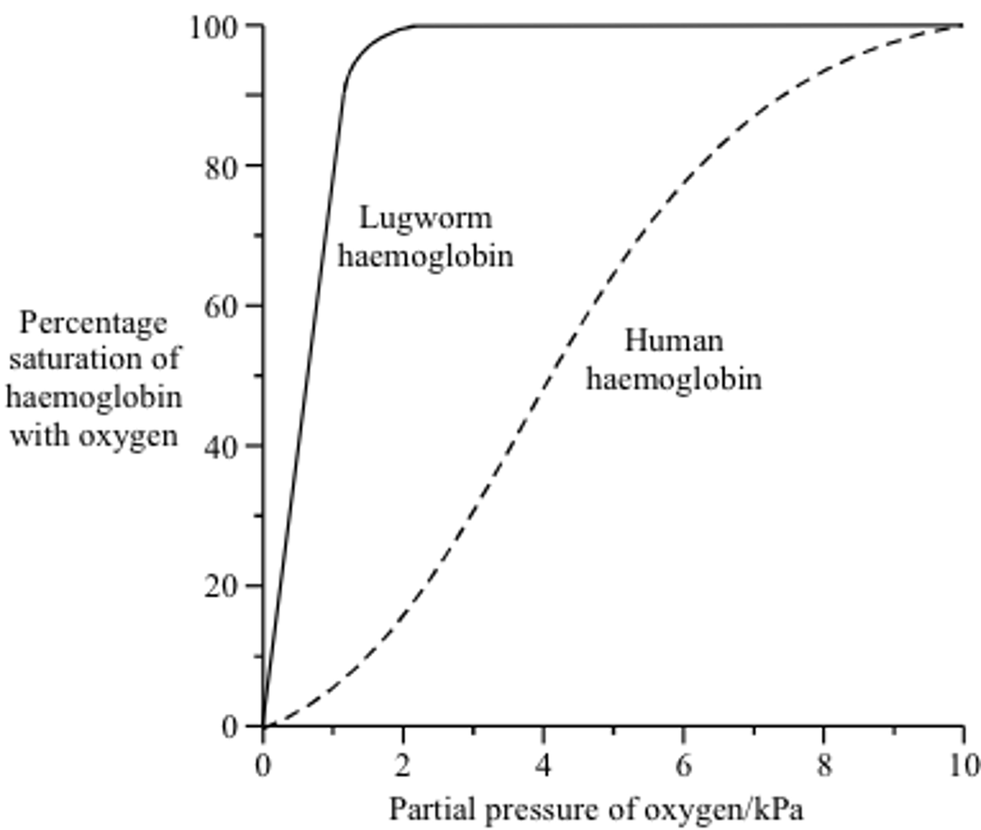
explain how organisms can be adapted to their environment by having a type of haemoglobin with higher affinity for oxygen + give examples
- if the curve is shifted to the left, that type of haemoglobin has a higher affinity for oxygen
- more oxygen will associate with Hb more readily at gas exchange surfaces where pO2 is lower
- this will be useful for organisms in low oxygen environments
eg. high altitudes (llamas), underground (lugworms) or foetuses
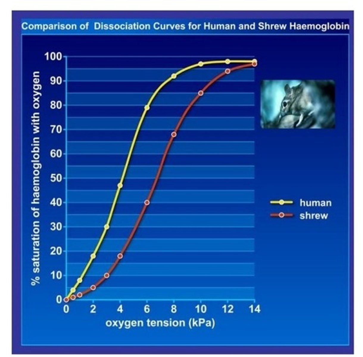
explain how organisms can be adapted to their environment by having a type of haemoglobin with lower affinity for oxygen + give examples
- if the curve is shifted to the right, that type of haemoglobin has a lower affinity for oxygen
- more oxygen will dissociate from Hb more readily at respiring tissue where more oxygen is needed
- this will be useful for organisms who may be small or very active so will have high respiratory/metabolic rates
eg. shrew
type of circulatory system in mammals
𝐜𝐥𝐨𝐬𝐞𝐝, 𝐝𝐨𝐮𝐛𝐥𝐞 circulatory system
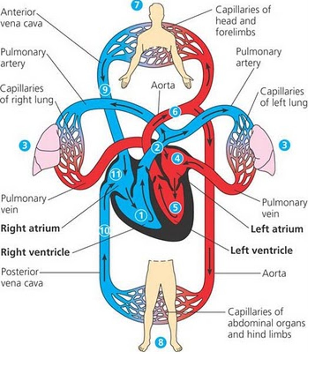
meaning of a closed double circulatory system
- blood is confined to vessels
- blood passes through the heart twice for each complete circuit around the body
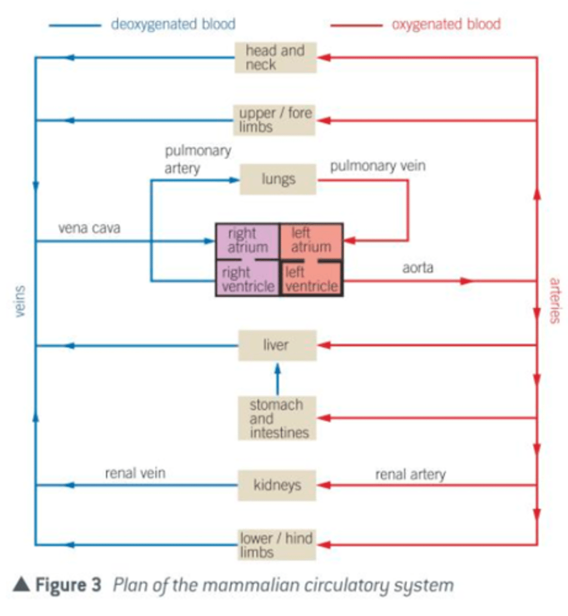
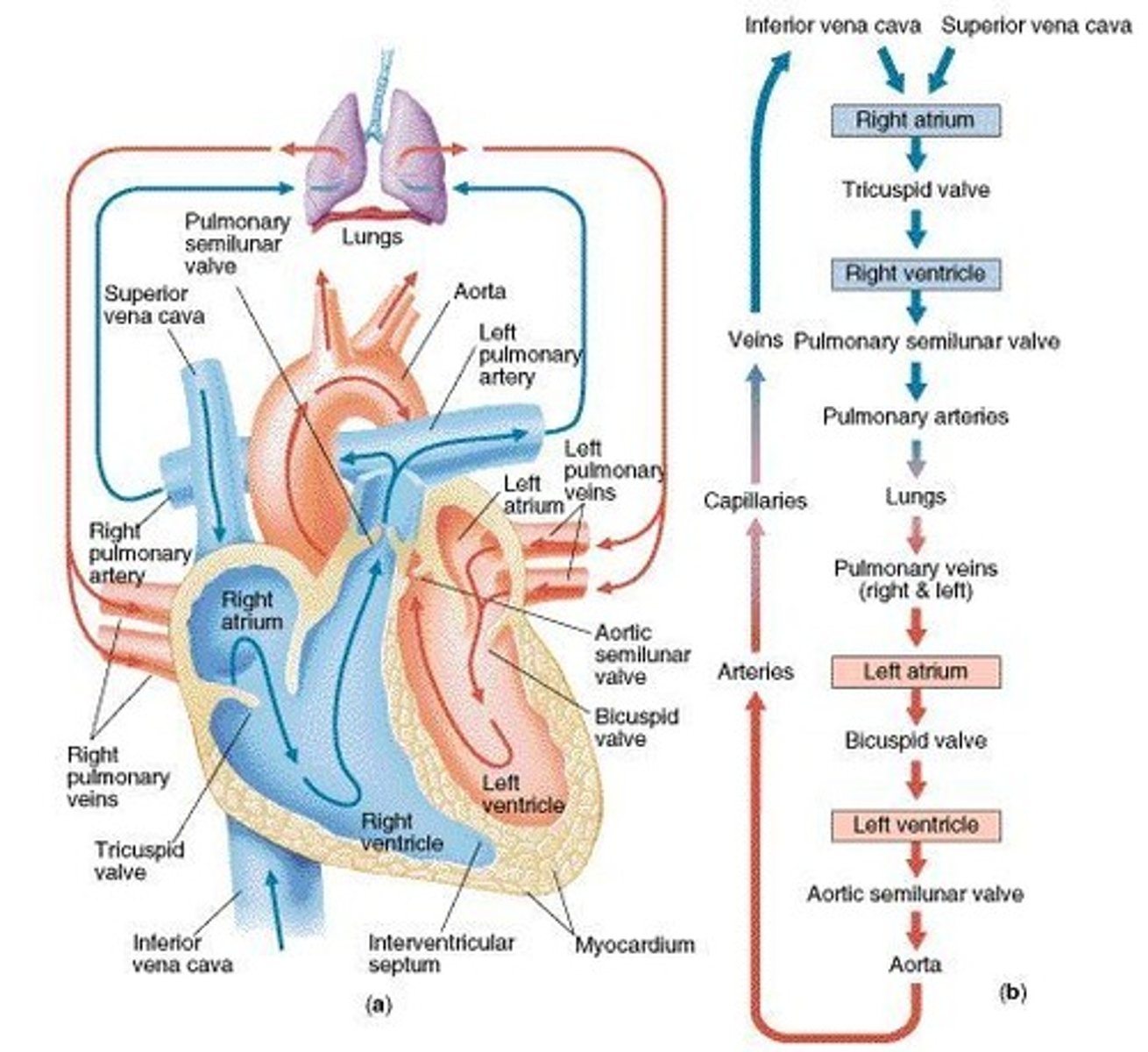
describe the general pattern of blood circulation
- deoxygenated blood in right side of heart pumped to lungs and oxygenated blood returns to left side
- oxygenated blood in left side of heart pumped to rest of body and deoxygenated blood returns to right side
what blood vessels carry blood away from the heart to the kidney?
renal arteries
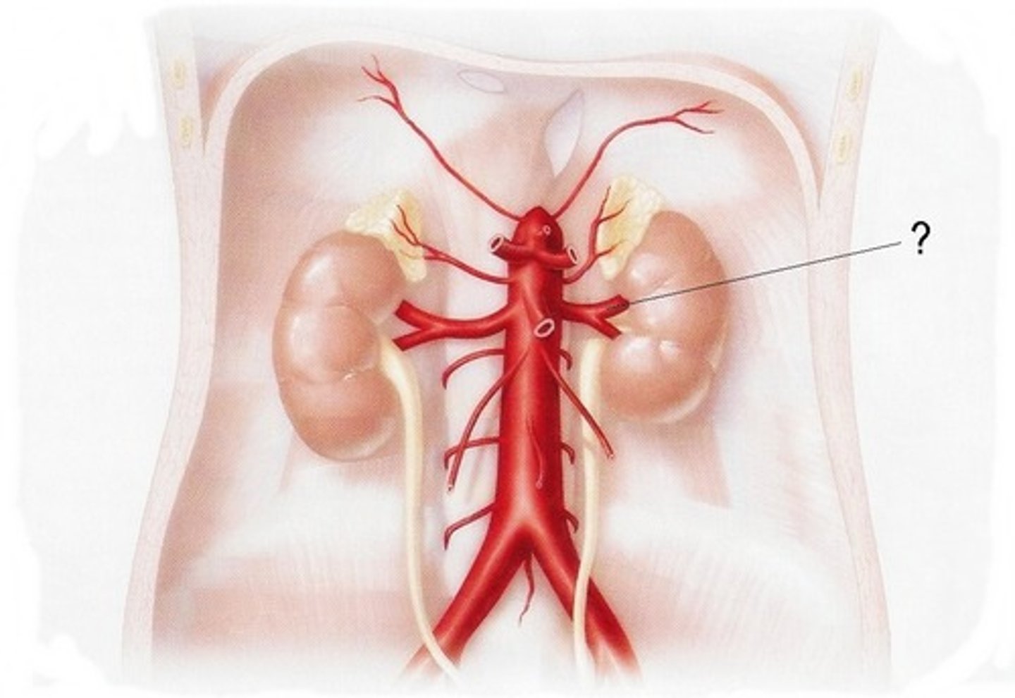
what blood vessels carry blood away from the kidneys back to the heart?
renal veins

2 reasons for the importance of a double circulatory system
1. prevents mixing of oxygenated and deoxygenated blood so blood pumped to body is fully saturated with oxygen for aerobic respiration/ increases efficiency of oxygen transport
2. allows blood to be pumped to body at a higher pressure so substances are delivered to and removed from cells faster
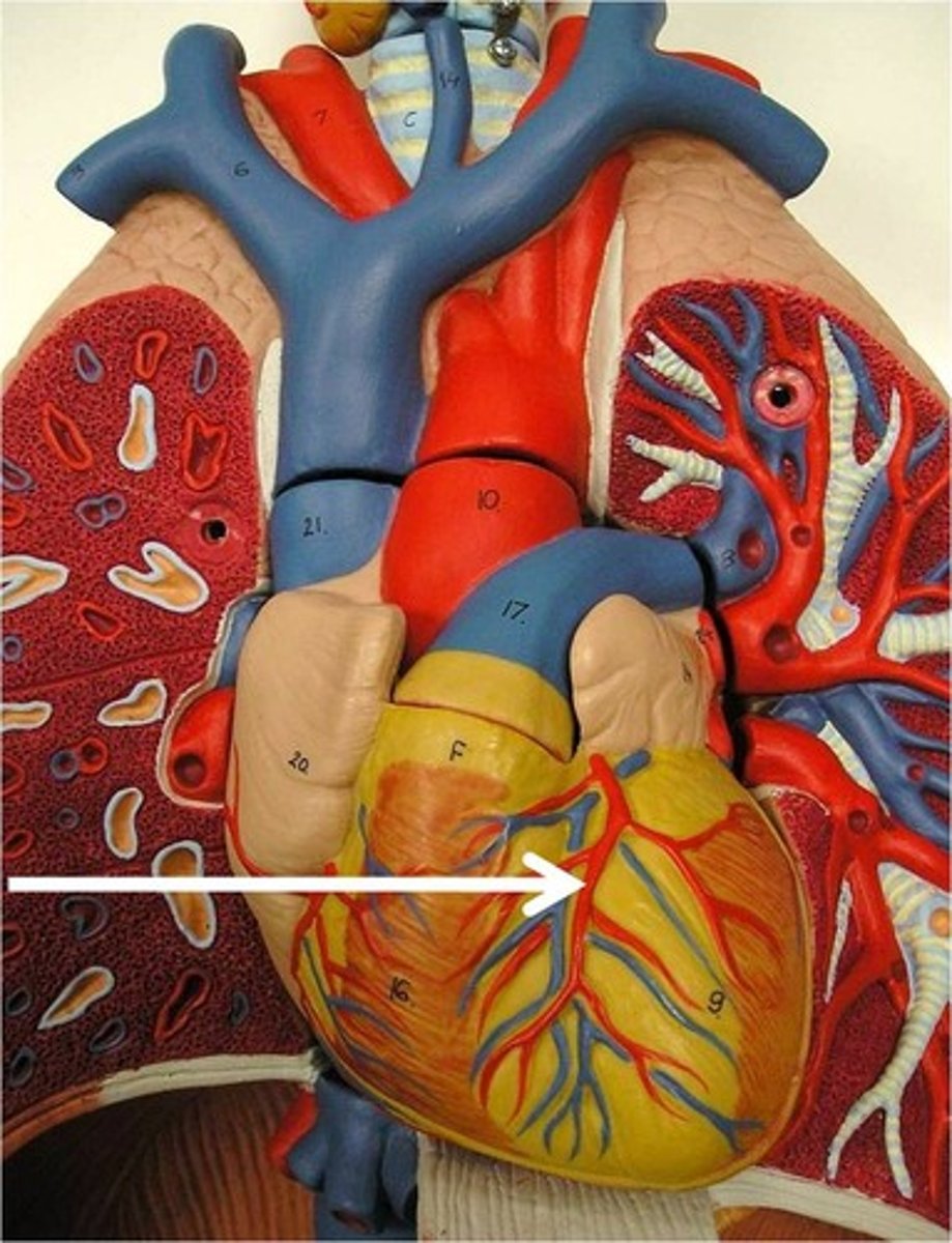
what blood vessels carry oxygenated blood to the heart muscle? where are they located?
coronary arteries, located on the surface of the heart branching from the aorta
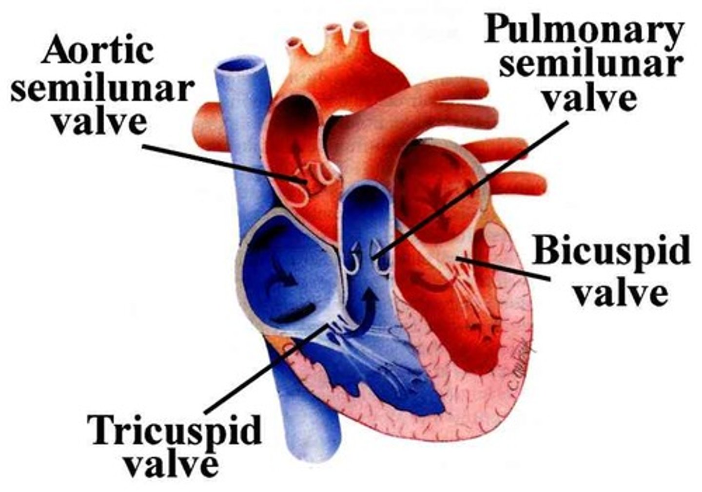
describe movement of blood through the heart
- deoxygenated blood flows from the vena cava into the right atrium, and then through the atrioventricular valve into the right ventricle which pumps it through semi-lunar valve to the lungs via the pulmonary artery for oxygenation.
- oxygenated blood returns to the left atrium via pulmonary vein and then through the atrioventricular valve into the left ventricle where it is pumped through the aorta through the aortic valve to the rest of the body
what ensures the unidirectional flow of blood in the heart? how?
- Valves which are flaps of tissue in the heart and large veins that prevent backflow of blood (remember to specify from where to where)
- only open one way which is when there is higher pressure behind the valve
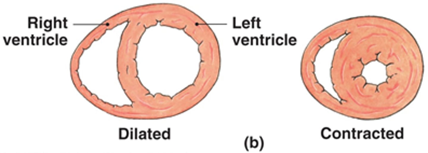
why is the wall of the left ventricle thicker than that of the right?
thicker muscles contract with greater force so higher pressure is generated to pump blood around the entire body
cardiac cycle steps
A complete heartbeat consisting of atrial systole, ventricular systole and diastole
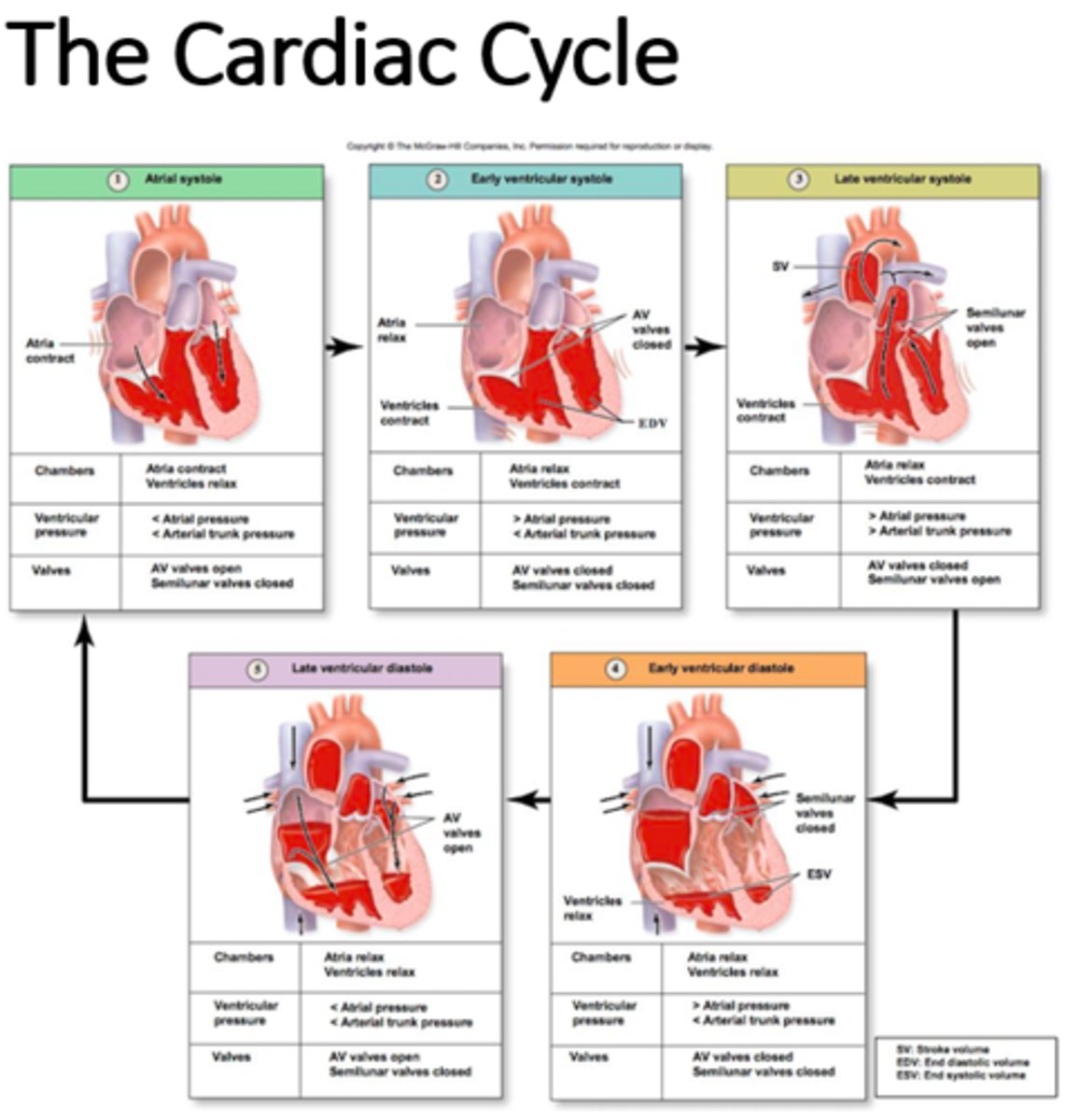
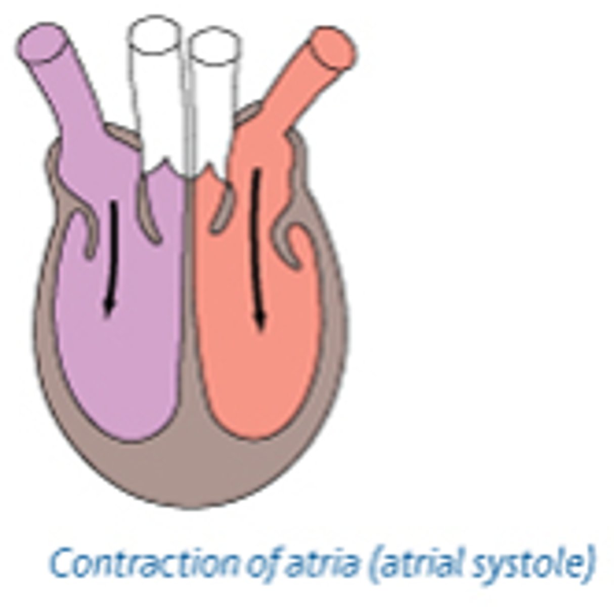
atrial systole
1. both atria contract - volume decreases, pressure increases
2. AV valves remain open as pressure in atria exceeds pressure in ventricles
3. SL valves remain shut as pressure in arteries exceeds pressure in ventricles
4. blood is therefore pushed from atria into ventricles
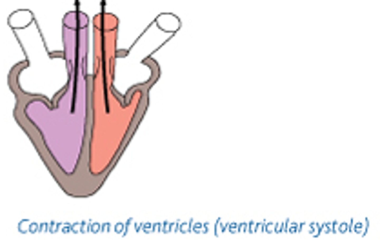
ventricular systole
1. after a short delay to allow ventricles to fill with blood, they contract - volume decreases, pressure increases
2. AV valves shut when pressure in ventricles exceeds pressure in atria
3. SL valves open when pressure in ventricles exceeds pressure in arteries
4. blood is therefore pushed out from ventricles through the arteries out of the heart
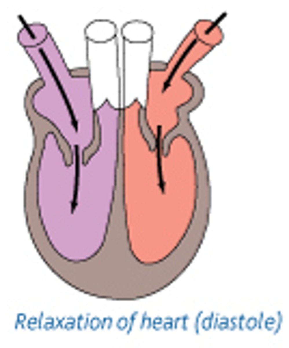
diastole
1. atria and ventricles relax - volume increases, pressure decreases
2. SL valves shut when pressure in arteries exceeds pressure in ventricles
3. AV valves open when pressure in atria exceeds pressure in ventricles
4. blood therefore passively fills the atria and to the ventricles from the veins
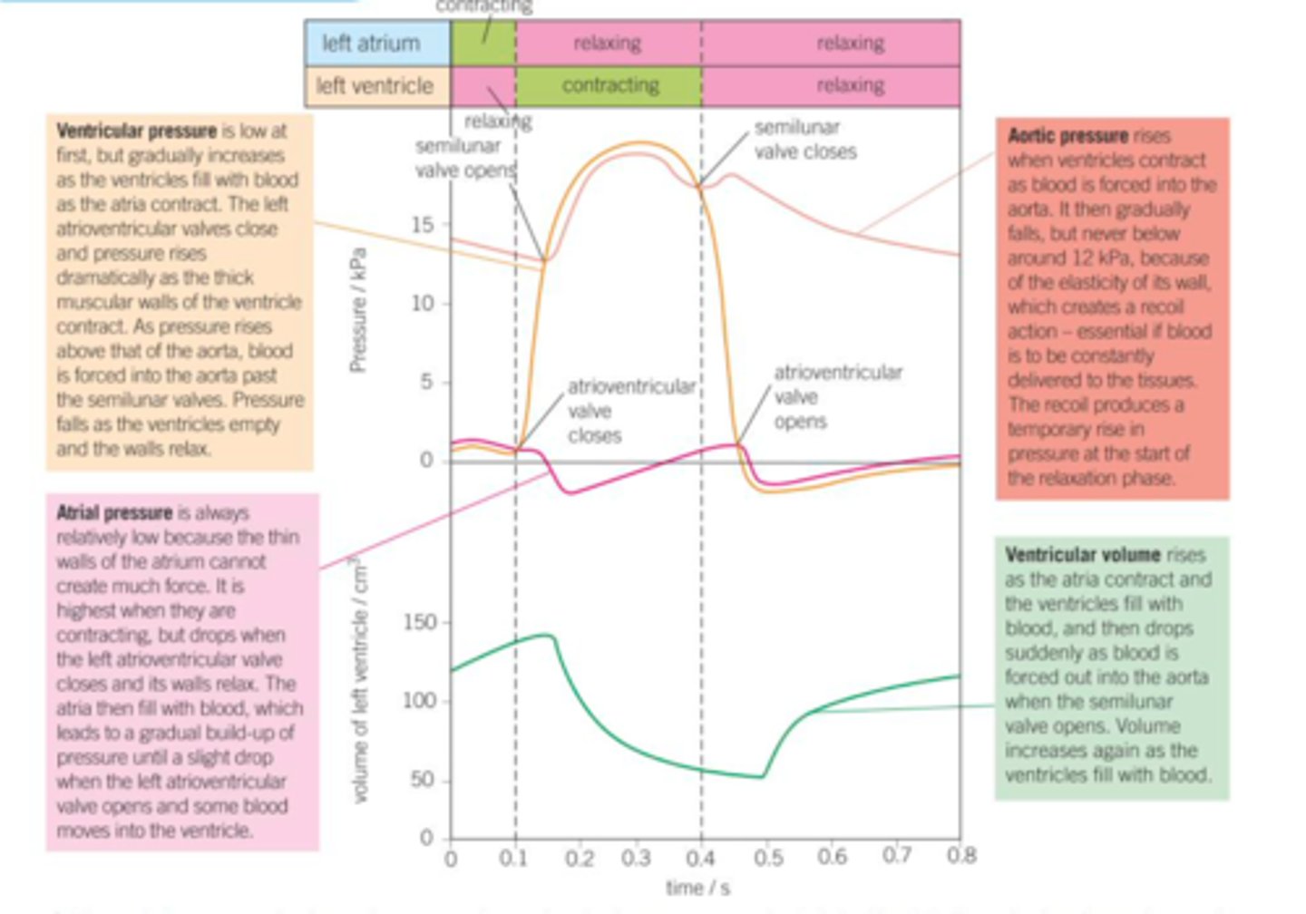
interpret graph showing cardiac cycle (4 points)
1. pressure in ventricle higher than in atria so AV valves close, preventing backflow of blood from ventricles to atria
2. pressure in ventricle higher than in artery so SL valves open, so blood flows from ventricle to artery.
3. pressure in artery higher than in ventricle so SL valves close, preventing backflow of blood from artery to ventricle.
4. pressure in atria higher than in ventricle so AV valves open, so blood flows from atria to ventricles.
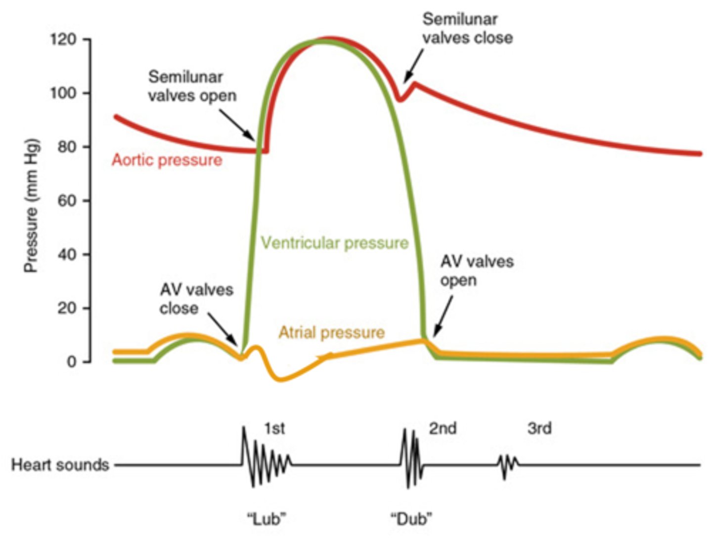
explain changes in ventricular volume in cardiac cycle
- ventricular volume rises as atria contract and the ventricles fill with blood
- volume drops suddenly as blood is forced out into the arteries when the SL valves open
- volume increases again as ventricles fill with blood
cardiac output
heart rate (bpm) x stroke volume

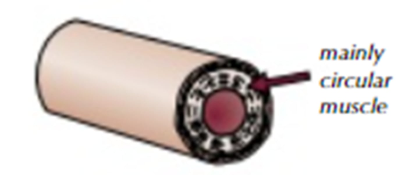
function of arteries
carry blood away from the heart at high pressure
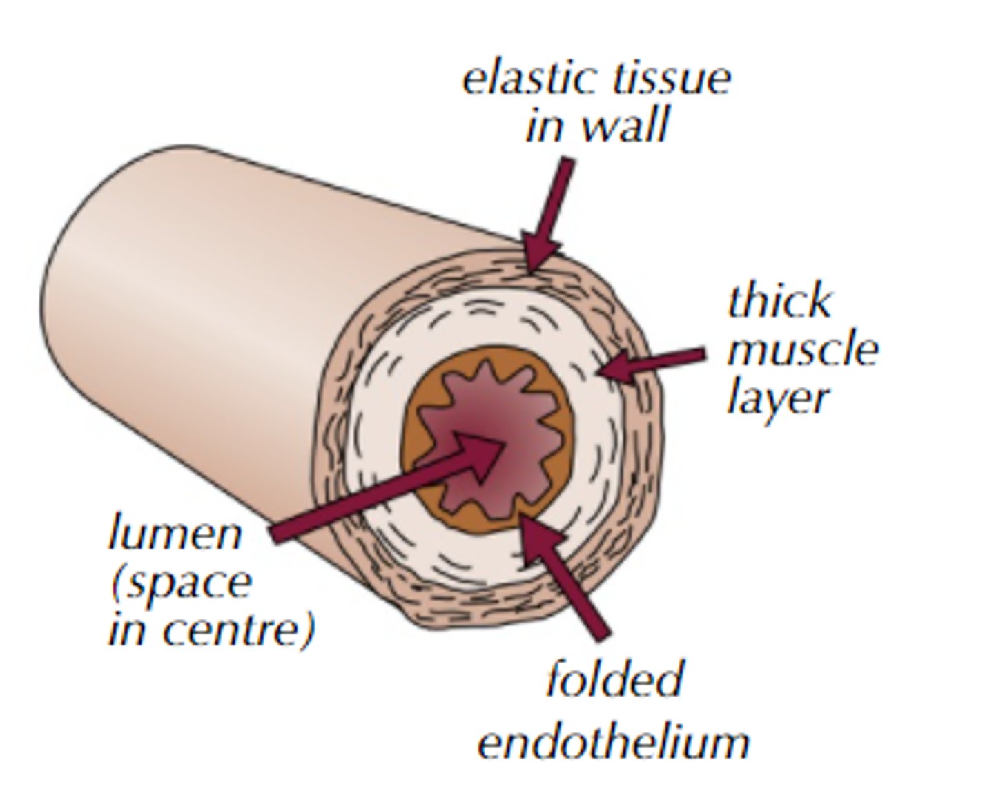
how are arteries adapted to their function? Explain 5 ways
1. Thick wall - withstand high pressure and prevent bursting
2. Thick smooth muscle - can contract and control/maintain blood pressure
3. Thick elastic tissue - can stretch and recoil as ventricles contract and relax to reduce pressure surges and maintain blood pressure
4. Narrow lumen - increases blood pressure
5. Smooth folded endothelium - reduced friction
function of arterioles
division of arteries to smaller vessels which transport and direct blood, controlling blood flow to capillaries/tissues
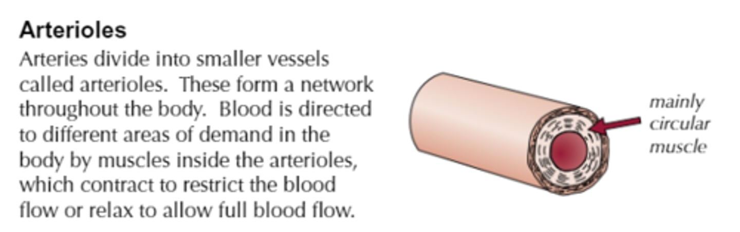
how are arterioles adapted to their function? Explain 2 ways
1. Thicker smooth muscle than arteries so they can:
- contract which narrows lumen (vasoconstriction) and reduces blood flow to capillaries
- relax which widens lumen (vasodilation) and increases blood flow to capillaries
2. Thinner elastic layer than arteries because there are lower pressure surges further from the heart
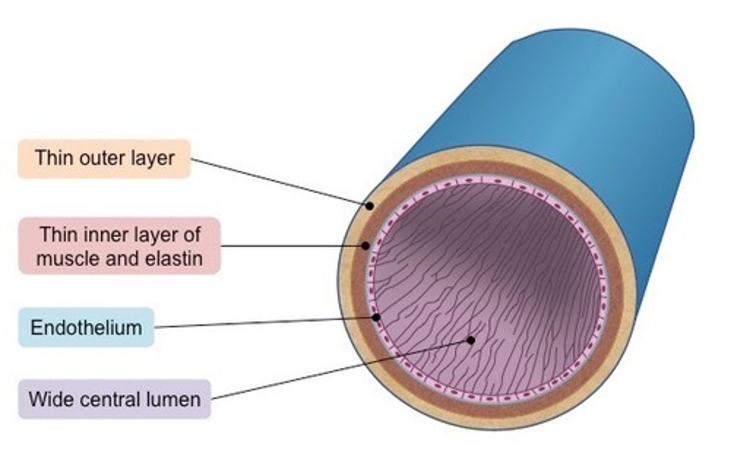
function of veins
return blood to the heart at lower pressure
how are veins adapted to their function? Explain 3 ways
1. Wider lumen - less resistance to blood flow
2. Thinner/ less muscle and elastic tissue - blood is being carried at a lower pressure
3. Valves - prevent backflow of blood
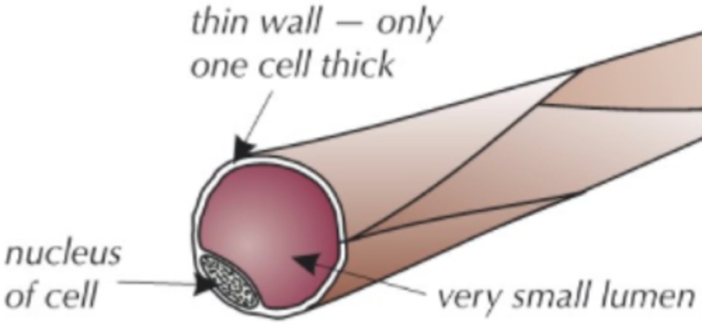
function of capillaries
allows efficient exchange of gases, nutrients, wastes, hormones, etc. between blood and tissue fluid
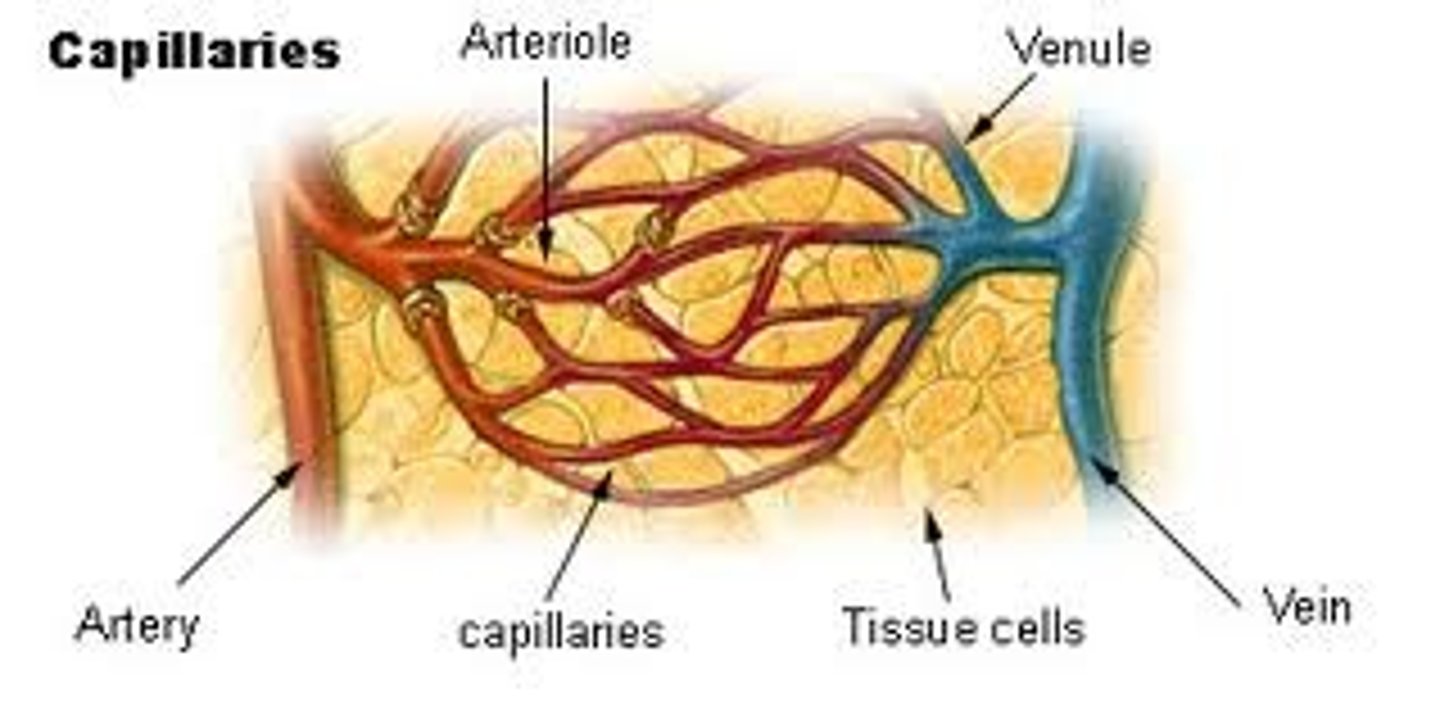
how are capillaries adapted to their function? Explain 4 ways
1. endothelial cells are only one cell thick - reduces length of diffusion pathway
2. capillary bed is a large network of branched capillaries - increased surface area for diffusion
3. narrow lumen - reduces rate of blood flow so more time for diffusion
4. permeable (pores in walls between cells) - allows larger substances through
what is tissue fluid?
- A watery substance contains all cells containing glucose, amino acids, ions, oxygen, and other nutrients.
- site of exchange between blood and cells by removing carbon
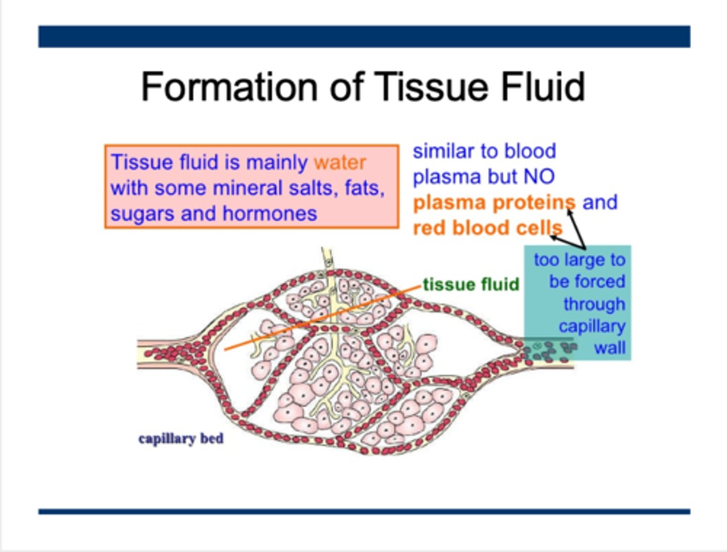
explain formation of tissue fluid
at arteriole end of capillaries...
1. contraction of ventricles produces higher hydrostatic pressure inside capillaries than in tissue fluid
2. this forces water and any other dissolved substances out of capillaries
3. large plasma proteins remain in capillaries (ultrafiltration)
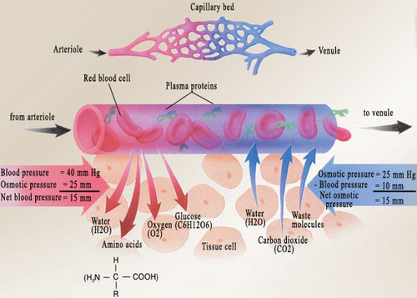
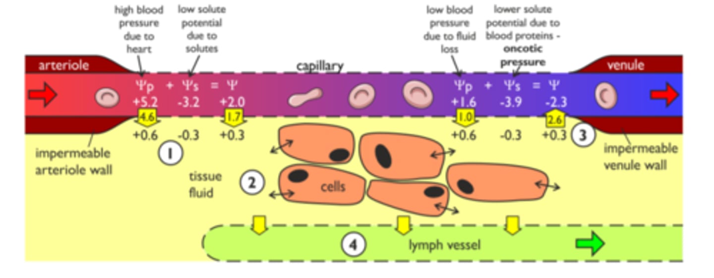
explain return of tissue fluid to circulatory system
at venule end of capillaries...
1. hydrostatic pressure decreases as fluid leaves capillaries
2. increasing concentration of plasma proteins as water is lost
3. water potential in capillaries is lower than that of tissue fluid so water enters capillaries down Ψ gradient via osmosis
4. excess water is taken up by lymph vessels and returned through veins
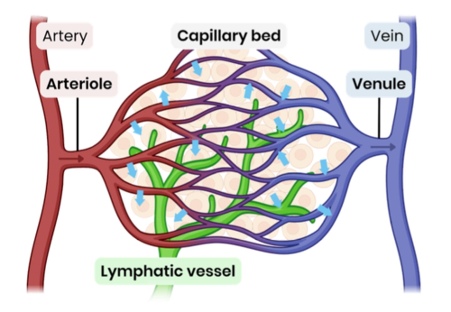
lymphatic system
not all tissue fluid can return to capillaries; remainder is carried in this network of vessels through which lymph drains from the tissues into the blood.
suggest two causes of excess tissue fluid accumulation
high blood pressure
low conc. of proteins in blood plasma
high salt conc. in tissue fluid
how will high blood pressure cause excess tissue fluid accumulation?
- high blood pressure = high hydrostatic pressure
- increased outward pressure at arteriole end and increased inward pressure at venule end
- more fluid forced out at arteriole end and less reabsorbed at venule end
- lymphatic system isn't able to drain excess tissue fluid fast enough
how will low conc. of proteins in blood plasma or
high salt conc. in tissue fluid cause excess tissue fluid accumulation?
- reduces water potential gradient
- less water reabsorbed at venule end by osmosis
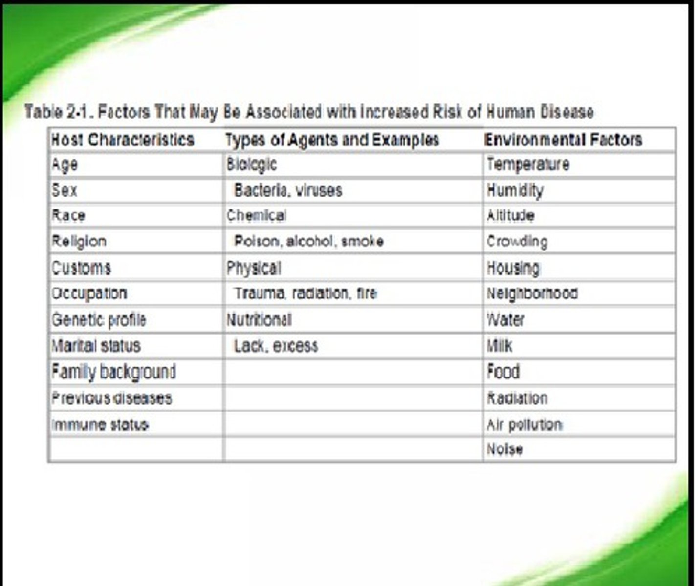
what is a risk factor?
any aspect of an individual's lifestyle (body or environment) that increases the likelihood of developing a disease or injury