IB Biology SL 2025 Exam - Unit 4 Study Guide
1/78
Earn XP
Description and Tags
Covering topics from Unit 4 that could be on the 2025 test!
Name | Mastery | Learn | Test | Matching | Spaced |
|---|
No study sessions yet.
79 Terms
What is the cell theory?
All living organisms are composed of cells
Cells are the basic unit of structure & function
All cells come from pre-existing cells
What are cells?
Fundamental units of life, they are capable of living on their own and make up all living organisms and tissues of the body
Developments in Microscopy
Improved light microscopes in the second half of the 19th century allowed discovery of bacteria and other unicellular organisms
Electron microscopes enabled scientists to discover the complexity of organs and find chromosomes
Common structures in all cells
Plasma Membrane
Cytoplasm
DNA
Prokaryote Cell Structure (mostly small in size)
Has a plasma membrane, sometimes having a cell wall (made of peptidoglycan) to further protect the cell
Inside is only filled with cytoplasm, no nucleus
70S ribosomes
Usually only a single molecule of DNA that forms a loop or circle`
Eukaryote Cell Structure (compartmentalized)
Plasma membrane
Nucleus (holds the cell’s chromosomes), DNA is typically linear
80S Ribosomes
Mitochondria
Processes of life in unicellular organisms
Homeostasis, Metabolism, Nutrition, Excretion, Growth, Response to Stimuli, Reproduction
Differences in eukaryotic cell structure between animals, fungi, and plants
Only plastids exist in plant cells
Only cell walls exist in plant and fungi cells
Small, temporary vacuoles in animal cells, whereas large, permanent vacuole in plant and fungi cells
Centrioles construct spindle to move chromosomes in mitosis in animal cells and 9+2 microtubules in cilia and flagella
Cilia and flagella are present in many animal cells
Atypical cell structure in eukaryotes
Red blood cells - do not have a nucleus
Phloem sieve tube elements - dividing walls between adjacent cells are sieve-like. Nucleus and most other cell contents break down, but plasma membrane remains
Skeletal muscle - some large multinucleate structures are formed when groups of cells fuse together (syncytium)
Aseptate fungal hyphae - nucleus divides repeatedly without any subsequent cell division, resulting in a multinucleate structure
What are organelles?
Discrete structures in cells that are adapted to perform one or more vital functions
Advantage of the separation of the nucleus and cytoplasm into separate compartments
Keeping chromosomes inside the nucleus safeguards the DNA
mRNA can be modified after transcription to prepare it for translation
Advantages of compartmentalization in the cytoplasm of cells
Enzymes and substrates for a particular process can be concentrated more
Substances that could cause damage to the cell can be kept inside the membrane of an organelle
Conditions such as pH can be maintained at an ideal level for particular processes
Organelles with their contents can be moved around
Larger area of membrane available for processes that happen within or across membranes
Totipotent Stem Cells
Typically are early-stage embryos, and are capable of differentiating into any cell type
Pluripotent Stem Cells
Embryonic stem cells that have gradually committed to developing on certain pathways. It is still capable of differentiating into a range of cell types, but not every type
Multipotent Stem Cells
Stem cells that can still differentiate into several types of mature cell
Hematopoietic stem cells are multipotent
Surface area-to-volume ratios and constraints on cell size
If the ratio is too small, substances will not enter the cell as quickly as they are required, waste products will accumulate, and the cell could overheat
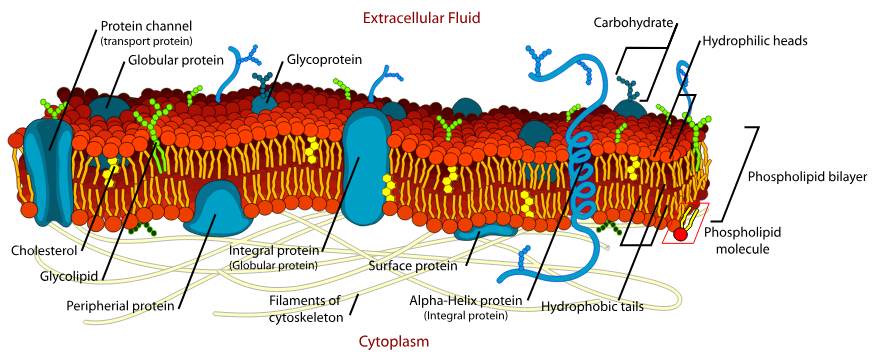
Lipid bilayers as the basis of cell membranes
Bilayer of phospholipids and other amphipathic molecules forms a continuous sheet that controls the passage of substances
Lipid bilayers as barriers
Membrane core has a low permeability to all hydrophilic particles
Solutes nearest to membrane surface might penetrate between the hydrophilic phosphate heads, but if they reach the hydrophobic core they will be drawn back to the aqueous solution outside
Larger the molecule size, lower the permeability
Simple Diffusion
Particles passing between phospholipids in the membrane, but can only happen if the bilayer is permeable to the particles
Particles will always move from higher to lower concentrations
Integral Proteins in Membranes
Hydrophobic and are embedded on the hydrocarbon chains in the center of the membrane
Many integral proteins are transmembrane proteins
Peripheral Proteins in Membranes
Hydrophilic and mainly attached to the surface of integral proteins
Osmosis
The net movement of water from a lower solute concentration to a higher solute concentration
Water, despite being hydrophilic, is small enough to pass through the phospholipid bilayer
Aquaporins
Water channels that can greatly increase the membrane permeability to water, allowing for more water to pass through the bilayer
At its narrowest point, the channel is only slightly wider than water molecules, so therefore they must pass through in a single file
Channel Proteins
Integral, transmembrane protein that connects the cytoplasm to the aqueous solution outside the cell
Ions and polar molecules typically pass through these channel proteins, typically down the concentration gradient
Some channels can be opened or closed, allowing for control of permeability of certain substances
Facilitated Diffusion
Channel proteins are required for the movement to occur
No energy is expended by the cell to cause this movement
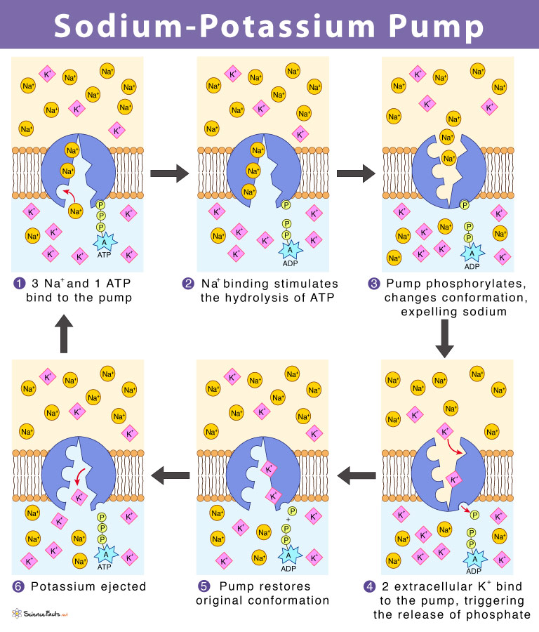
Active Transport
Process where molecules need energy (ATP) to move across the membrane
Pump Proteins
Integral membrane proteins that only moves particles in one direction, typically against the concentration gradient (low to high)
Have two conformations: one for transporting the particle from one side to a central chamber, and another to bring across an ion to the opposite side
Selectivity in membrane permeability
Allows the passage of particular particles, but not others
Facilitated diffusion and active transport are examples of selective permeability
Glycoproteins
Conjugated proteins with carbohydrate as the non-polypeptide component
Component of plasma membrane of cells, with protein part embedded in the membrane and the carbohydrate part projecting out
Are responsible for cell-to-cell recognition
Glycolipids
Molecules consisting of carbohydrates linked to lipids
Carbohydrate part is usually a single monosaccharide or a short chain between 2-4 sugar units
Lipid part contains one or two hydrocarbon chains to fit in the hydrophobic core of membranes
Help the immune system to distinguish between self and non-self cells
Solvation with water as the solvent
Solvation is the combination of a solvent with the molecules and ions of a solute
Polar solutes dissolve due to attractions of the partially positive and negative charges on the water molecules
When this happens, water molecules form shells around many types of ion and charged molecule
Hypertonic
A solution that has a higher concentration of solutes
Hypotonic
A solution that has a lower concentration of solutes
Isotonic
A solution that is equal in the concentration of solutes on both sides
Water movement by osmosis into or out of cells
Water will always move from the hypotonic solution to the hypertonic solution to try and balance out the concentration gradient
There is always movement of water molecules, but if it’s in a isotonic solution, there is an equal amount of water going in and out
Effects of water movement on cells that lack a cell wall
If the cells that don’t have a cell wall are exposed to hypotonic solutions, the cells will easily burst
If the cells that don’t have a cell wall are exposed to hypertonic solutions, the cells will shrink and form indentations (crenations)
Effects of water movement on cells with a cell wall
Cells with a cell wall that are bathed in hypotonic solutions will expand and become turgid. This is the normal state of plant cells
Cells with a cell wall that are bathed in hypertonic solutions will shrink and become flaccid. If this happens, sometimes the plasma membrane will pull away from the cell wall (plasmolysis)
Medical applications of isotonic solutions
Isotonic sodium chloride solution is used (called normal saline), contains 9g of NaCl per cubic decimeter of solution
It can be safely introduced to a patient’s blood system
Used to rinse wounds and skin abrasions
Used to keep areas of damaged skin moistened prior to skin grafts
Used as the basis for eye drops
Frozen to the consistency of slush for cooling hearts, kidney, and other donor organs
Fluorescent Stains & Immunofluorescence
Fluorescent stains are used for substances to absorb light and re-emit it at a longer wavelength to make cells easier to identify
Immunofluorescence is a development of fluorescent staining. Antibodies that bind to particular chemicals are linked to fluorescent markers of different markers. Then, a multicolored fluorescent image can be produced showing where the chemicals are located
Freeze-Fracture Electron Microscopy
Sample is plunged into liquified propane at -190C to freeze the sample
Steel blade is used to fracture the sample
A vapor of platinum or carbon is fired onto the fracture surface at an angle of 35 degrees to form a coating, creating a replica
The replica can be analyzed through an electron microscope
Cryogenic Electron Microscopy
Thin layer of pure protein solution is applied to the grid and flash-frozen to create smooth vitreous ice
Grid with frozen protein solution is placed in an electron microscope and detectors record the pattern of electrons transmitted by individual protein molecules, producing many different patterns
Computational algorithms will combine patterns to produce a 3D image of the protein molecules
Plasma Membrane
Outer boundary of the cell and encloses all of its contents
Controls entry and exit of substances
Prevents entry of unwanted or toxic substances
Allows the cell to maintain concentrations of substances that are very different from those in the surrounding environment
Lysis
When the plasma membrane of the cell bursts due to excess pressure or by viruses
When this happens, the cell will always die
Autolysis
When the cell carries out the lysis itself
Cytoplasm
Water is main component of cytoplasm and there are many substances dissolved or suspended in the water
Enzymes in cytoplasm catalyze a multitude of chemical reactions (called the metabolism, which provides a cell with energy, produces proteins and other substances to make up the cell structure)
DNA
Contains the information needed for a cell to carry out all its functions
Many genes carry information for making proteins, with some of them used for growth and repair and others acting as enzymes
Nucleus
Has a double membrane with pores in it that holds chromosomes, which consist of one long DNA molecule wound around the outside of proteins (histones)
Plastids
Family of organelles with two outer membranes and internal membrane sacs
Cell Wall
A rigid layer outside the plasma membrane to strengthen and protect the cell
Vacuole
Flexible fluid-filled compartment surrounded by a single membrane
Centrioles
Cylindrical organelles that organize the assembly of structures composed of microtubules
Undulipodia
Cilia and flagella used to generate movement of a cell or movement of fluid adjacent to a cell
Rough Endoplasmic Reticulum
Consists of flattened membrane sacs called cisternae
80S ribosomes attach to the outside of cisternae
Synthesizes protein for secretion from the cell, which passes to the cisternae and moved by vesicles to the golgi apparatus
Smooth Endoplasmic Reticulum
Consists of a branched network of tubular membranes, does not have ribosomes attached
Synthesizes lipids, phospholipids and steroids (one special one stores calcium ions in muscle)
Golgi Apparatus
Has cisternae, but not as long, often curved, don’t have ribosomes and have many vesicles nearby
Processes proteins brought in from vesicles from the rER
Most proteins are then carried in vesicles to the plasma membrane for secretion
Lysosomes
Formed from golgi vesicles and are spherical in shape
Contain high concentrations of protein
Contain digestive enzymes to break down ingested food in vesicles
Can break down organelles or cells
Mitochondria
Double membrane surrounds mitochondria, with inner membrane invaginated to form structures called cristae
Fluid inside is called the matrix
Shape of mitochondria is variable but usually spherical or ovoid
Produce ATP for the cell by aerobic cell respiration (fat can be digested here if it’s being used as an energy source)
Have their own DNA
Ribosomes
Appear as dark granules in the cytoplasm and not surrounded by a membrane, typically 80S ribosomes
Synthesize proteins, releasing it to work in the cytoplasm
Constructed in the nucleolus
Chloroplast
Double membrane surrounds the chloroplast with stacks of thylakoids inside
Shape of chloroplasts are typically spherical or ovoid
Produce glucose and other organic compounds by photosynthesis
Vesicles
Small vacuoles used to transport materials inside the cell
Cytoskeleton
Constructed from several types of protein fiber
Contains microtubules (made of tubulin) and microfilaments (actin)
Shape is dynamic since microtubules and microfilaments can be constructed and taken apart
Organelles with No Membranes
Ribosomes, Centrioles, Microtubules, Proteasomes, Nucleoli
Organelles with One Membrane
Vesicles & Vacuoles, rER, sER, Golgi Apparatus, Lysosomes
Organelles with Two Membranes
Nuclei, Mitochondria, Chloroplasts, Amyloplasts, Chromoplasts
Stem Cell Niche in Skeletal Muscle
These niches remain inactive until there’s injury, in which the changes will cause the stem cells to proliferate and differentiate rapidly
Stem Cell Niche in Bone Marrow & Hair Follicles
Two areas where the microenvironment enables the stem cells to have continuous proliferation and differentiation
Sperm
50 micrometers in length
Narrow shape and small volume allow it to swim to the egg easily
Egg
110 micrometers in diameter and spherical in shape
Allows large quantities of food reserves to be stored in cytoplasm
Red Blood Cells
6 to 8 micrometers but indented on both sides and only about 1 micrometer thick in the middle
Small size and shape allow passage along narrow capillaries and gives a large surface area to load and unload oxygen
White Blood Cells
Only about 10 micrometers in diameter but can enlarge up to 30 micrometers if they are activated and become antibody-secreting plasma cells
Extra volume comes from cytoplasm with rER and Golgi apparatus for protein synthesis
Cerebellar Granule Cells
Cell body is only about 4 micrometers in diameter
Twin axons extend about 3 millimeters in the cerebellar cortex
Small volume allows cerebellum to accommodate 50 billion of them
Motor Neurons
Cell body is about 20 micrometers in diameter
Large size allows enough proteins to be synthesized to maintain the immensely long axon
Can extend to a meter or more to allow it to carry signals from the central nervous system to a distant muscle
Striated Muscle Fibers
Diameter from 20 to 100 micrometers and lengths that exceed 100 mm
Dimensions allow the fiber to exert greater force and contract by a greater length
Magnification Triangle
Actual Size * Magnification = Image Size
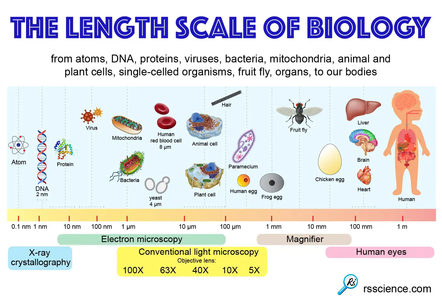
Cell Size Chart
Differentiation
Process during development whereby newly formed cells become more specialized and distinct from one another as they mature
Morphogens
The process of early embryo differentiation is driven by the release of specialized signaling molecules
They play a critical role in the formation of different layers of cells (ectoderm, mesoderm, endoderm) in the embryo
Fluid Mosaic Model
Active Transport Visual