Bite Wing Technique Chapter 21, Radiology 1
1/36
There's no tags or description
Looks like no tags are added yet.
Name | Mastery | Learn | Test | Matching | Spaced | Call with Kai |
|---|
No analytics yet
Send a link to your students to track their progress
37 Terms
What are bite-wings primarily for?
Used to examine the inter-proximal surfaces of teeth
Between two adjacent surfaces
interproximal
Inspect crowns of maxillary and mandibular on single image
interproximal exam
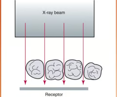
Patient "bites" on tab/wing to stabilize receptor
Bite Wing Receptor
Supporting bone around roots of teeth
Alveolar Bone
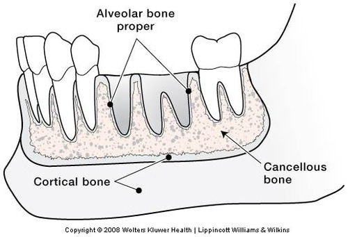
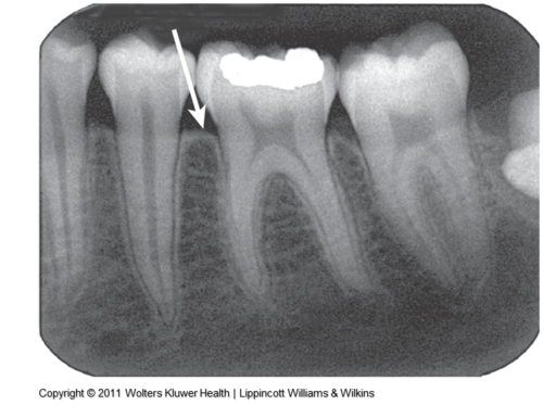
Coronal portion of alveolar bone found between teeth (AKA alveolar crest)
Crestal Bone
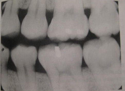
Where adjacent tooth surfaces contact each other
Contact Areas
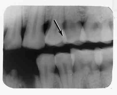
On image, appears as thin radiolucent lines between adjacent tooth surfaces
Opened Contacts/ black or radiolucent
Where contacts of two teeth are superimposed
Overlapped (closed) Contacts
closed contacts
it will look like a cloud of radiopaque (very white)
Vertical and horizontal bite-wings? What are the differences?
Horizontal Bitewing- the bite-wing receptor is placed in the mouth with the long portion of the receptor in a horizontal direction.
Vertical Bitewing- The bitewing receptor is placed in the mouth with the long portion of the receptor in a vertical direction.
A bite wing image includes
The crowns of maxillary and mandibular teeth, interproximal areas, and areas of crestal bone on the same image
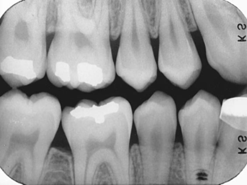
Bite wings are usually used to
detect interproximal caries
Principles of Bite-wing Technique
-Receptor placed parallel to crowns of both maxillary and mandibular teeth
The receptor is stabilized by a bite block or beam alignment device or bite-wing tab
When using a bite-wing tab on film or PSP, the central ray of
beam is directed through contacts of teeth using a vertical
angulation of +10___degrees
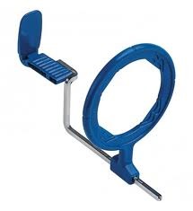
Beam alignment device: Rinn XCP (you will be trained on this
equipment)
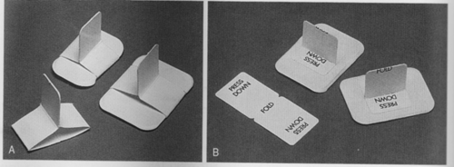
Bite-wing tabs: We have them in clinic (hard to find good ones for digital sensors)----Wingers work well (pink plastic and
DISPOSABLE)
Beam alignment device
used to help the radiographer position the PID in relationship to the tooth and film
It is not always possible to use a beam alignment device to expose a bite wing image especially in children
Therefore the dental practitioner must be familiar with the original bite wing technique of using a tab attached to the receptor for use with such patients.
bite-wing tab
A heavy paperboard tab or loop that is fitted around an intraoral receptor and is used to stabilize the receptor during the procedure.
Size 0
Child/Pedo (posterior teeth in primary dentitions—horiz placement)
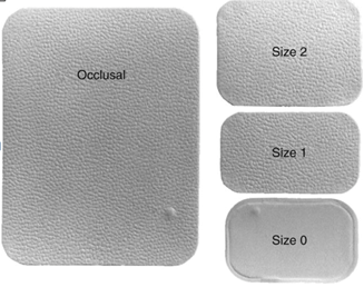
Size 1
Vertical bite-wing in anterior for adult patient (this is NOT in your book!) or bitewing for pedo patient that has 6 year molars/mixed dentition---Vert BW in ant. for adults are very difficult to eliminate overlap and not recommended any longer
Size 2
Recommended for posterior adult bite-wings (horiz/vert)
Size 3
Long, narrow bite-wing for capturing premolar AND molar region (not recommended due to excessive overlap, only used horizontally in past)
-only for bitewing
Angulation
The alignment of the central x-ray beam in the horizontal and vertical planes.
Horizontal angulation
-Positioning of the central ray in a horizontal (side-to-side)
plane
-Correct horizontal angulation: Central ray directed
perpendicular to the curvature of the arch, through the contacts of the teeth—produces ___open________ contacts
-Incorrect horizontal angulation: "Overlapping"---> Cannot dx. caries
Vertical angulation
-Positioning of PID in a vertical or up-and-down plane
Positive or negative
Degrees measured on outside of tubehead

positive vertical angulation
PID ABOVE occlusal plane, central ray directed DOWNWARD
Negative vertical angulation
PID BELOW occlusal plane, central ray directed UPWARD
correct vertical angulation
+10 degrees (for film or phosphor plate use); usually for digital, receptor is more rigid, so may not need as much vertical angulation
incorrect vertical angulation
-Results in a distorted image
-If a negative vertical angulation is used, the occlusal surfaces of maxillary teeth are evident, and the apical regions of mandibular teeth are seen
- A bite-wing image exposed with an excessive negative vertical angulation is non diagnostic.
Rules for bite wing technique
1. receptor placement (to cover correct teeth)
2. receptor position (parallel to crowns & stable)
3. vertical angulation (central ray is +10 degrees)
4. horizontal angulation (central ray is directed through contact areas)
5. receptor exposure (x-ray beam must be centered)
Patient Preparation for bitewing images
1. explain to the patient
2. Adjust the chair so the patient is upright
3. adjust the headrest to support patient's head- the maxillary arch is parallel to floor and the midsaggital plane is perp to the floor
4. place and secure lead apron with a thyroid collar
5. remove patient eyeglasses- retainer
Equipment prep
1. Set up kV and mA and time
2. if beam alignment device is needed, open the package and assemble the device
3. if a bite wing tab is used then attach the tab to the white side of the film or the correct side of the receptor
Exposure sequence
Bite-wings are usually LAST in a FMX ( book refers to as CMS), expose premolar view then molar view; repeat on opposite side of mouth
Vertical bite wings
7 films - 3 anterior and 4 posterior
THESE ARE NOT USED AS MUCH NOW
What is the proper placement for the molar and premolar bite wing?
premolar bitewing- step one position PID, place one finger parallel to the lower premolars and place the opening of PID parallel to the finger
Step 2- Place receptor and hold tab against teeth while patient bites
Step 3 stand behind the PID, and look down the PID If you see the receptor a cone cut will result.
Molar bitewing- step 1- place finger parallel to lower molars and place opening of PID parallel to finger
Step 2- place receptor and hold tab against teeth while patient bites
Step 3- stand behind the PID, and look down the PID If you see the receptor a cone cut will result.
Edentulous spaces
Teeth no longer present
Modification in placement may be necessary---> Use cotton roll to support in edentulous area or a different beam alignment device
Bony growths
Torus/Tori Don't place receptor on tori, benefit to use beam alignment device instead of tab

Horizontal bite wings are standard but vertical bite wings are for people with
perio disease