Ch 2 Chest
 Call Kai
 Learn
Learn Practice Test
 Spaced Repetition
 Match
Match Flashcards
 Knowt Play
1/54

There's no tags or description
Looks like no tags are added yet.
Name | Mastery | Learn | Test | Matching | Spaced | Call with Kai | Chat |
|---|
No analytics yet
Send a link to your students to track their progress
55 Terms
Chest X-ray Overview
chest is the most common radiographic procedure
reasons?
trouble breathing, chest pain, trauma
the chest/thorax is divided into what 3 parts?
bony thorax
respiratory system
mediastinum
Bony Thorax
what does this provide? what does the framework include?
provides protection to respiratory + circulatory system
framework:
- sternum = 3 parts
a) manubrium = superior portion
b) body = center
c) xiphoid process = inferior
- 2 clavicles - 2 scapulae - 12 pairs of ribs
- 12 thoracic vertebrae
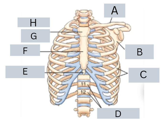
label parts
A - clavicle (2)
B - scapula (2)
C - ribs (12 pairs)
D - thoracic vertebrae (12)
E - xiphoid process (inferior)
F - body (center)
G - manubrium (superior)
H - sternum (3 parts, G/F/E, top to bottom)

What are the 3 positioning landmarks for chest x-ray?
vertebra prominens
spinous process of C7, may be easier to find if neck is flexed, good for PA chest positioning
jugular notch
deep notch on superior portion of sternum, good for AP chest positioning
xiphoid process
corresponds to level of T9-T10 + anterior portion of diaphragm, not reliable due to body habitus
Respiratory System
purpose?
what are the 4 respiratory system divisions?
p = exchange of gas bt air we breath + bloodstream
1) pharynx 2) trachea 3) bronchi 4) lungs
what is also part of the respiratory system, but not in the divisions?
diaphragm
primary, dome shaped, muscle
each half is a hemidiaphragm
creates pressure differences that allow for breathing
separates the thoracic + abdominal cavities
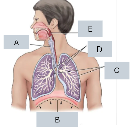
label system
A - trachea
B - diaphragm (right/left hemi diaphragms)
C - lungs
D - bronchi
E - pharynx
pharynx + oral cavity
description and purpose?
p = passageway for food, fluid, air + part of respiratory AND digestive system
pharynx = approx 5 inches long, contains 3 parts
a) nasopharynx b) oropharynx c) laryngopharynx
oral cavity = hard and soft palate form roof of mouth, posterior aspect of soft palate is the uvula
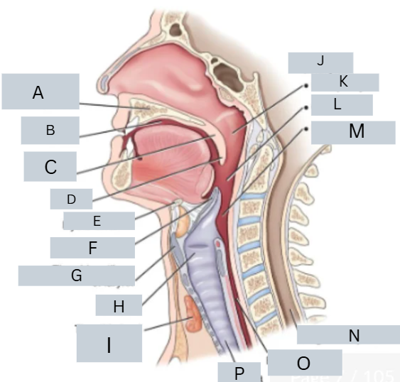
label parts
A = hard palate B = oral cavity
C = soft palate D = uvula E = hyoid bone
F = epiglottis G = thyroid cartilage of larynx
H = larynx I = thyroid gland
J = pharynx K = nasopharynx
L = oropharynx M = laryngopharynx
N = region of spinal cord
O = esophagus P = trachea
larynx
purpose? description? prominence?
purpose = organ of voice, protects airway
descript = approx. 1.5 - 2 inches, anterior portion of neck, suspended from hyoid bone, ranges from C3 to C6
laryngeal prominence = anterior projection of thyroid cartilage, located at C4 to C5

label parts
A = hyoid bone B = thyroid cartilage
C = cricoid cartilage D = epiglottis
E = true vocal cords F = trachea
G = level of C3 H = larynx
I = level of C6 J = trachea
K = epiglottis L = hyoid bone
M = laryngeal prominence (level of C5)
N = thyroid cartilage
O = cricoid cartilage
trachea
purpose? description? what type of rings?
p = connects larynx to main bronchi + provides structural rigidity
d = fibrous muscular tube, approx. ¾ inch diameter + 4 ½ inch long, extends from C6 - T4/T5
cartilage rings = 16-20 C shaped rings in anterior wall + provide rigidity to prevent collapse of trachea

label parts
A = larynx B = level of C6
C = trachea D = level of T4/T5
E = right bronchi (3) F = left bronchi (2)
G = region of thymus gland
H = parathyroid glands
I = thyroid gland J = thyroid cartilage
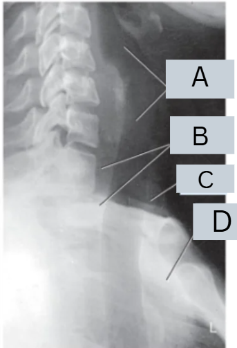
label image
A = trachea + larynx
B = esophagus
C = thyroid gland region
D = thymus gland region
trachea glands
what are the 3 glands? describe?
1) thyroid gland = located anteriorly in neck, below thyroid cartilage, very radiosensitive
2) parathyroid glands = embedded in posterior surface of each lobe of thyroid gland, assists in blood functions
3) thymus gland = inferior to thyroid gland + anterior and superior to heart
right and left bronchi
purpose? description?
p = allows air to enter each lobe of lung
d = right primary bronchus is wider + shorter than left
carina splits the trachea + primary bronchi
R bronchus contains 3 lobes, left contains 2 lobes
bronchi divide into bronchioles, bronchioles divide into alveoli
lungs
purpose? description?
p =
d = composed of parenchyma
right lung has 3 lobes: superior, middle, inferior which is divided by 2 deep fissures
left has 2 lobes: superior, inferior which is divided by 1 deep fissure

label parts
A = pleura B = parietal
C = pleural cavity D = pulmonary visceral
E = fissures F = right lung
G = left lung H = fissure
I = superior lobe J = inferior lobe
K = inferior lobe L = middle lobe
M = superior lobe
what are the 3 ‘pleuras’?
parietal pleura = outer layer of pleural sac
pulmonary/visceral pleura = inner layer pleura, covering surface of lungs + dipping into fissures
pleural cavity = potential space bt the double walled pleura
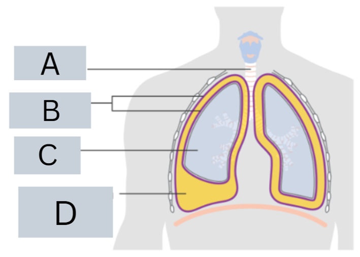
label parts
A = trachea (windpipe)
B = pleura (lung lining)
C = lung
D = pleural effusion (fluid bt pleural space)

what are the lung pathology for L and R?
L = pneumothorax
R = pleural effusion hemothorax

name position, projection, diagnosis

label chest radiograph
what projection?
PA
A = trachea
B = apex of lung
C = carina
D = base of lung
E = diaphragm
F = costophrenic angle of lung
G = hilum

label chest radiograph
what position
L lateral
A = apex of lung
B = upper lobe of lung (superior)
C = hilum
D = lower lob of lung (inferior)
E = base
F = diaphragm
what are the four important structures in the mediastinum
thymus gland, heart + great vessels + trachea + esophagus

label parts
A = esophagus
B = superior vena cava
C = ascending aorta
D = inferior vena cava
E = right F = abdominal aorta
G = left H = heart
I = pulmonary artery J = arch of aorta
K = thymus gland L = thyroid gland
M = trachea
thymus gland
purpose? description?
p = role in development of immune system
d = located behind upper sternum, prominent in infancy + decreases in size through aging, typically only seen on small children x=rays
heart and great vessels
what houses them?
what are the 3 sections of aorta?
pericardial sac = houses the heart + roots of great vessels
great vessels = inferior vena cava, superior vena cava, aorta, large pulmonary arteries + veins
aorta = largest artery in body, ¾ to 1 in diameter = ascending aorta, arch of aorta, descending aorta

label radiograph
A = superior vena cava
B = right atrium
C = inferior vena cava
D = left ventricle
E = left atrial appendage
F = main pulmonary artery
G = aorta

determine body habitus
L = PA chest for a sthenic patient
R = PA chest for hyposthenic patient
what are the 4 breathing movements/techniques?
1) suspended inspiration = take in deep breath + hold it - make exposure [chest, abdomen]
2) suspended expiration = take deep breath in, blow it all out, stop breathing - make exposure
3) suspended respiration = stop breathing, don’t breathe - make exposure once breathing has stopped
4) orthostatic = breath normally but don’t move - make exposure
what is the general rule for a full inspiration x-ray?
for average adult
need MINIMUM of 10 pairs of ribs seen on full inspiration x-ray

which is expiration and which is inspiration?
L = inspiration
R = expiration
what are technical considerations/selections for chest x-ray?
kVp = high (long scale, low contrast)
grid is utilized
mAs = high mA w short exposure time (s)
for centering for chest, you want to place top of IR where?
top of IR 1 ½ to 2 inches above shoulders
3-4 inches from jugular notch landing on T7
or
7-8 inches from C7 landing on T7
PA technical factors
IR, grid, kVp rang, SID, collimation, respiration
what about lateral technical factors?
IR = 14×17”, portrait or landscape (mainly L)
grid = yes kVp = 110-125
SID = 72”
Collimation = 4 sided to area of lung fluid
respiration = 2 deep breaths, hold on 2nd breath
LATERAL = everything the same except it is NOT landscape the IR is in portrait
criteria for PA chest structures on radiograph
both lungs
costophrenic angles + apices
hilum markings
heart + great vessels
minimum of 10 pairs ribs
long scale contrast
NO rotation (look at clavicles)
label radiograh
(pa chest struct)
criteria for Lateral chest structures
both lungs
costophrenic angles + apices
centered hilar region
long-scale contrast
no rotation
label radiograph
(lateral chest structure)
AP chest topography
which positioning landmark do you use?
jugular notch = 3-4 inches below notch
AP chest - supine, upright, semierect
what is the angle of the CR
angle caudal 5 inches
criteria of AP chest in supine, upright, semierect
both lungs
costophrenic angles + apices
air filled trachea from T1 down
hilum markings
heart + great vessels = will be MORE enlarged than PA projection
8-9 (NOT TEN) pairs of ribs
long scale contrast
no rotation
technical factors of AP pedi chest
IR, grid, kVp range, SID, collimation, respiration
what about for lateral positioning?
IR = vary based on patient size
grid = NO kVp = 75-85 SID = 50-60”
collimation = close to outer chest margins
respiration = exposure on full inhale if possible
LATERAL = all same except for kVp 80-55
AP pedi chest
patient position, CR, parent instructions
position = utilize parent help, tech don’t hold unless necessary, supine w arms + legs extended, place patient in middle of IR w shoulders 2 inches below top of IR [lateral arms are extended above head]
CR = centered to MSP at level of midthorax (approx. mammillary/nipple line)
parent = remove chest clothing, wear lead apron + gloves, position will not obstruct image, hold patient’s legs + arms
criteria for AP pedi chest
limited signs of motion
entire lungs
9-10 rib pairs
chin elevated
no rotation
criteria for lateral pedi chest
limited signs of motion
entire lungs
9-10 rib pairs
chin elevated
no rotation
technical factors for lateral decubitus chest
IR, grid, kVp range, SID, collimation, respiration
IR = 14×17” landscape grid = yes
kVp = 110-125 SID = 72”
collimation = 4 sides to lung field
respiration = exposure on 2nd full inspiration
lateral decubitus chest
patient position, CR, extras
p place radiolucent material under patient, true lateral on R/L for 5 min prior to exposure, arms raised above head, chin extended back
CR = horizontal CR directed at T7
extra = mark the film up, use decub marker if available
lateral decubitus chest criteria
true lateral position
entire lungs
arms not in field
markers placed correctly
no motion
AP lordotic chest tech factors
IR, grid, kVp, SID, collimation, respiration
IR = 14×17 landscape/portait
grid = yes kVp = 110-125 SID = 72
collimation = 4 sides to lung field
respiration = expose on 2nd full inspiration
AP lordotic chest
patient position, CR
p = stands roughly 1ft away IR, leaning shoulders/neck/back of head against IR
hands on hips, palms rolled out
shoulders rolled forward, center MSP to CR and center of IR
CR = midsternum, 3-4” below jugular notch
semiaxial AP lordotic chest
patient position, CR
p = lies supine, shoulders rolled forward + arms positioned for lordotic position
CR = midsternum (3-4” below jugular notch) + angled 15-20” cephalic
AP lordotic chest criteria
entire lung field
unobstructed apices
clavicles nearly horizontal + superior to apices
sternal end of clavicle equidistant
no motion