PBL Monday
 Call Kai
 Learn
Learn Practice Test
 Spaced Repetition
 Match
Match Flashcards
 Knowt Play
1/394

There's no tags or description
Looks like no tags are added yet.
Name | Mastery | Learn | Test | Matching | Spaced | Call with Kai | Chat |
|---|
No analytics yet
Send a link to your students to track their progress
395 Terms
what are the 2 main blood grouping systems?
- ABO
- Rhesus
what are the 4 different groups in the ABO grouping system?
- A
- B
- AB
- O
what are the 2 antigens of the ABO grouping system?
- A antigens
- B antigens
what antigens does type A have?
A antigens only
what antigens does type B have?
B antigens only
what antigens does type AB have?
A and B antigens
what antigens does type O have?
neither A or B antigens
what else contributes to the ABO grouping system?
antibodies:
- anti-A antibodies
- anti-B antibodies
what type of antibodies are anti-A and anti-B antibodies?
IgM
what antibodies does type A have?
anti-B antibodies
what antibodies does type B have?
anti-A antibodies
what antibodies does type AB have?
neither antibody
what antibodies does type O have?
both anti-A and anti-B antibodies
which blood type is the universal donor and why?
type O because it has no antigens on the surface that could interfere with other antibodies
which blood type is the universal receiver and why?
type AB because it does not contain any anti-A or anti-B antibodies that could interfere with antigens
how many different Rhesus (Rh) antigens are there?
5 clinically significant ones
what are the 5 clinically significant antigens?
- D
- C
- c
- E
- e
which is the main Rh antigen we focus on and why?
- Rh D
- because is it the most immunogenic, causes the greatest immune response
- most likely to cause a transfusion reaction
how do we determine whether someone is Rhesus positive or negative?
depends on whether they have Rh D antigen on surface of erythrocytes
when would someone be Rhesus positive?
if they have Rh D antigens
when would someone be Rhesus negative?
if they lack Rh D antigens
which blood can Rh+ people receive?
Rh+ and Rh-
which blood can Rh- people receive?
Rh- only
do all Rh- people have anti-D antibodies?
no, only when they have been exposed to Rh+ erythrocytes
when can the Rhesus grouping system cause problems in pregnancy?
if mother is Rh- and foetus is Rh+
what happens if foetus is Rh+ and mother is Rh-?
- presence of Rh D antigens stimulates production of anti-D antibodies
- maternal anti-D antibodies recognise and destroy foetal Rh D antigens - haemolysis
what is this called?
haemolytic disease of the new-born (HDN)
why is it not a large problem in nulliparous mothers?
the anti-D antibodies are mostly IgM which cannot cross the placenta
how do we prevent haemolytic disease of the new-born (HDN)?
administration of anti-D Ig prophylaxis
why is the Rhesus factor grouping system a large problem in multiparous mothers?
IgG anti-D anitbodies will be produced, which can cross the placenta
how does anti-D Ig prophylaxis work?
destroys feotal RBCs that leak into maternal circulation
what is the pericardium?
a fibro-serous, fluid-filled sac that surrounds the heart and roots of the great vessels
what are the 2 layers of the pericardium?
- fibrous layer
- serous layer
what is the fibrous pericardium?
- continuous with the central tendon of the diaphragm
- made up of tough connective tissue
what is the purpose of the fibrous pericardium?
it is non-distensible so prevents rapid overfilling of the heart
what 2 layers is the serous pericardium divided into?
- outer parietal layer
- internal visceral layer (epicardium)
what does the serous pericardium consist of?
single sheet of epithelial cells called the mesothelium
what lies between the outer parietal layer and internal visceral layer?
the pericardial cavity which contains some lubricating serous fluid
what is the purpose of the serous fluid?
minimises the friction generated by the heart as it contracts
what are all the layers from most outer to inner?
Fart Police Smell Villians:
Fibrous layer of pericardium
Parietal outer layer of serous pericardium
Serous fluid
Visceral inner layer of serous pericardium
give the 4 functions of the pericardium
- fixes the heart in the mediastinum
- prevents overfilling of the heart via the fibrous pericardium
- lubrication via the serous pericardium
- protection from infection as the fibrous pericardium acts as a physical barrier between heart and other organs
what nerve supplies the pericardium?
- phrenic nerve supplies the fibrous pericardium
- right and left vagus nerves supply serous pericardium
what arteries supply the pericardium?
- right and left internal thoracic arteries
- intercostal and superior phrenic arteries (inferior aspect)
- mediastinal branches of aorta (posterior aspect)
- epicardium by coronary arteries
what are the 5 surfaces of the heart?
anterior
posterior
inferior
right pulmonary
left pulmonary
what makes the anterior surface of heart?
right ventricle
what makes posterior surface of heart?
left atrium
what makes inferior surface of heart?
left and right ventricle
what makes right pulmonary surface?
right atrium
what makes the left pulmonary surface?
left ventricle
what are the 4 borders of the heart?
right border
left border
inferior border
superior border
what makes the right border?
right atrium
what makes the left border?
left ventricle (and some left atrium)
what makes inferior border?
left and right ventricles
what makes superior border?
right and left atrium and great vessels
what forms the sulci of the heart?
the four chamber divisions create grooves on the surface of the heart
what are the three sulci?
coronary sulcus
anterior interventricular sulcus
posterior interventricular sulcus
where is the coronary sulcus?
runs transversely where the atria divide form the ventricles
where are the anterior and posterior interventricular sulci?
they run vertically on anterior and posterior sides of heart, where the right and left ventricles separate
what are the pericardial sinuses?
passageways formed by the pericardium folds around the great vessels
what are the 2 pericardial sinuses?
oblique pericardial sinus - posterior surface
transverse pericardial sinus - superior surface
what is the definition of starlings law?
the ability of the heart to change its force of contraction and therefore stroke volume in response to changes in venous return
what happens to the stroke volume as the central venous pressure increases?
also increases
how does the stroke volume increase with venous pressure?
- the heart fills and stretches more
- this creates more regions of overlap for the actin-myosin cross-bridges to form
- allows for a greater force of contraction
is there a relationship between the force of a muscle fibre and the degree to which the muscle fibre is stretched?
yes - the more it is stretched the greater the force, until it reaches the optimal muscle fibre length
what happens if the preload is too high?
the optimal fibre length is surpassed which causes a decrease in contractility and stroke volume
what is on the x axis of the Starling curve?
left ventricular end diastolic pressure (LVEDP) in mmHg
what is on the y axis of the Starling curve?
stroke volume in μm
what happens to the curve in increased inotropy?
shifts up
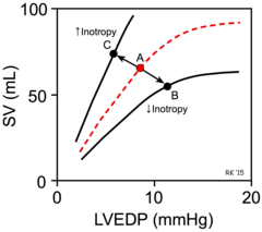
what is inotropy?
force of contraction
what happens to the curve in decreased inotropy?
shifts downs

what effect does the sympathetic nervous system have on contractility?
positive inotropic effect
how does the sympathetic nervous system have a positive inotropic effect?
noradrenaline acts on B1 adrenoreceptors
what effect does the parasympathetic nervous system have on contractility?
negative inotropic effect
how does the parasympathetic nervous system have a negative inotropic effect?
acetylcholine acts on M2 muscarinic receptors
what can affect stroke volume? (3)
- preload
- contractility
- afterload
what is stroke volume?
difference between end diastolic and end systolic volumes
what increases the preload? (2)
- fast filling time
- increased venous return
what does an increased preload do to stroke volume?
increases end diastolic volume - so increases stroke volume
what decreases preload? (7)
- high or low sodium
- high or low potassium
- low body temperature
- drugs
- decreased calcium ions
- hypoxia
- decreased thyroid hormones
what increases contractility? (5)
- sympathetic stimulation
- epinepherine and norepinephrine
- increased calcium ions
- thyroid hormones
- glucagon
what does increasing contractility do to stroke volume?
decreases end systolic volume - so increases stroke volume
what decreases contractility? (4)
- parasympathetic stimulation
- acetylcholine
- hypoxia
- hyperalkaemia
what increases afterload? (3)
- hypertension
- vasoconstriction
- atherosclerosis
what does an increase in afterload to do the stroke volume?
increases end systolic volume - so decreases stroke volume
what decreases afterload?
vasodilation
what in the biggest unit that makes up a muscle?
fasicle

what makes up the fasicles?
muscle fibres aka muscle cells aka myocytes

what makes up the muscle fibres/cells/myocytes?
myofibrils

what makes up the myofibrils?
myofilaments

what are the myofilaments?
actin and myosin
which out of actin and myosin is thinner or thicker?
actin - thin
myosin - thick
what is the sarcolemma?
plasma membrane of a muscle fibre/cell/myocyte
what is the myofibril organised into?
sarcomeres - short segments of myofibril
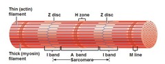
where does the sarcomere span to and from?
Z line to Z line
what is the Z line made of?
actin
what is the centre of the sarcomere called?
M line
what makes up the M line?
myosin
what is the A band?
length of the myosin

what is the I band?
actin only area

what is the H band?
myosin only
