Ch 2: Nucleic Acids
 Call Kai
 Learn
Learn Practice Test
 Spaced Repetition
 Match
Match Flashcards
 Knowt Play
1/33

There's no tags or description
Looks like no tags are added yet.
Name | Mastery | Learn | Test | Matching | Spaced | Call with Kai | Chat |
|---|
No analytics yet
Send a link to your students to track their progress
34 Terms
Nucleic Acids
nucleic acids are another class of biomolecules. They are biomolecules that are unbranched biopolymers, and what they do is that they store, transmit, & express genetic information. They will be classified as either DNA molecules or RNA molecules
EX: nucleic acids, proteins, lipids, carbohydrates etc.
DNA: deoxyribonucleic acid (DNA)
RNA: ribonucleic acid (RNA)

Nucleotides
(see image for the structure of a nucleotide) are monomeric units.
Nucleotides are the building blocks of DNA and RNA. Nucleotides are the monomeric units of nucleic acids.
Each nucleotide has 3 parts:
Phosphate + Sugar + Nitrogenous base
A nucleotide is a molecule that consists of a nitrogen containing base/heterocyclic structures (adenine). So the nitrogen bases are heterocyclic structures containing Carbon and Nitrogen.
Heterocyclic means relating to a ring-shaped chemical structure where at least one atom in the ring is not carbon.
Homocyclic is a cyclic chemical compound where the ring structure is made of atoms from a single element, usually carbon.
A sugar molecule that is covalently attached to the nitrogen base
phosphate there can be 1-3 phosphates attached.
As long as you have a nitrogen base, sugar and a phosphate, that constitutes a nucleotide.
1 phosphate: monophosphate
2 phosphates: diphosphate
3 phosphates: triphosphate
In purines, the number 9 Nitrogen is covalently bound to the 1 prime Carbon
In Pyrimidines, N1 position is bound to 1 prime Carbon
Third component:1, 2 or 3 phosphates.
Nucleic acids are polymers
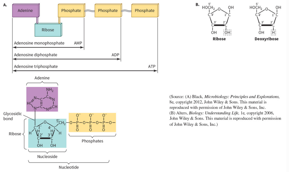
nucleoside
if you only have only the nitrogen base and the sugar. A nucleoside phosphate is a nucleotide. (remember the 3 components).

Monomeric units
A monomer unit is a small, simple molecule that chemically bonds with other similar molecules to form a larger chain called a polymer.
Monomeric units of nucleic masses are nucleotides
Monomeric units of proteins are amino acids
Monomeric units of polysaccharides are sugar molecules

Nitrogenous Bases
Nitrogenous bases define what the nucleotide is. There are 5 different commonly occurring nitrogen bases. Those nitrogen bases can be further classified as purines and pyrimidines.
Purines: the ones that have 2 cyclic components (bicyclic)
Pyrimidines: only have 1 cyclic structure

Purines are either
Adenine or guanine
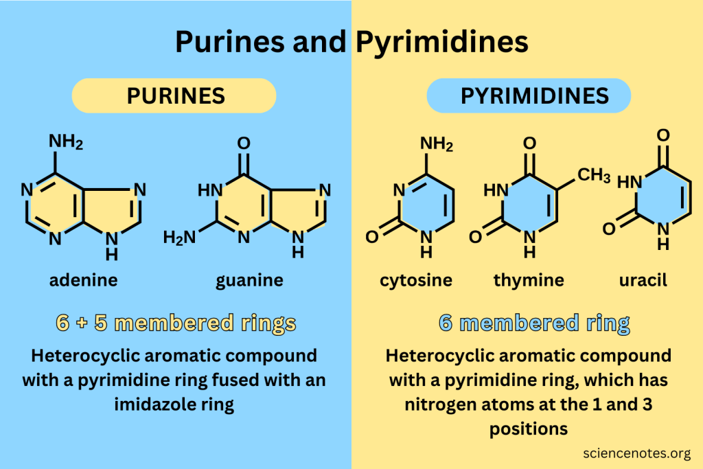
The Pyrimidines are
cytosine
thymine
uracil
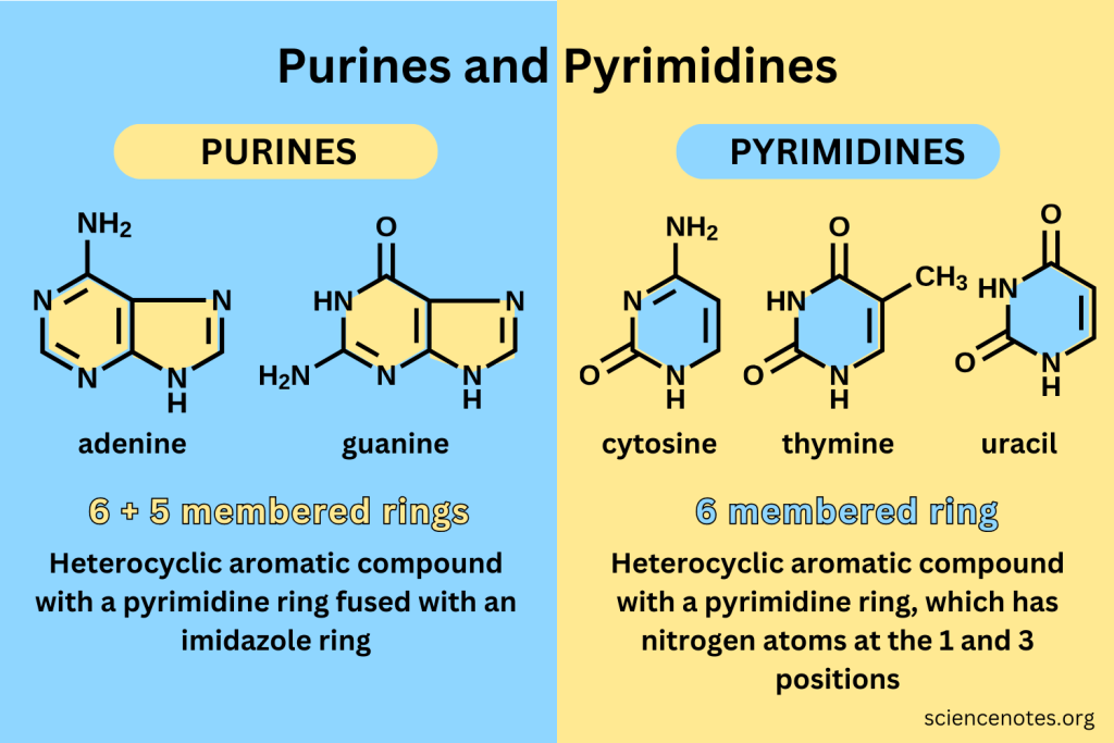
In DNA, the combination of Purines and Pyrimidines involve
adenine & thymine, guanine & cytosine
In RNA molecules, thymine is replaced with
uracil
The 4 bases that occur in RNA
Adenine
Uracil
Guanine
Cytosine

The 4 bases that occur in DNA
adenine
thymine
guanine
cytosine

the numbering system for these bases are
come back to this
Sugar components are going to be either
Ribose Sugar: a five Carbon sugar, it takes the cyclic furanose form of the sugar
Deoxyribose Sugar: In DNA, the sugar is a deoxyribose. In DNA there is not a hydroxyl group in the 2 prime position. In RNA, there is a hydroxyl.
(see image)
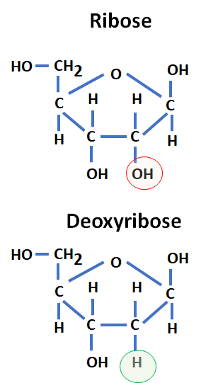
numbering system for sugars
those little ‘ are called primes. Count clockwise.

what occurs in nucleic acids/how you get them
Polymerize nucleotides. The polymerization occurs through the sugars and phosphates. That’s why nucleic acids are said to have a sugar phosphate backbone. The way the sugar backbone forms is that the first phosphate group (alpha phosphate) second phosphate (beta) third phosphate (gamma)
The way that nucleic acid molecule forms is that the alpha phosphate group that’s attached to the 5 prime carbon, gets attacked by the 3 prime hydroxyl group on a sugar molecule.
If we have a chain of nucleotides, the 3 prime hydroxyl group will attack the alpha phosphate with an incoming nucleotide, so what leaves is the 2 phosphates (vpi) the word for a molecule that contains 2 phosphates is pyrophosphate. and then you get the linkage between 3 prime hyxroxyl groupa nd incoming nucleotide.
When the next nucleotide, the nedt hydroxyl group will attack ther incoming nucleotide
What we see in a DNA molecule is a chain of sugars and phosphates and hanging off of the sugars are the various nitrogen bases. the nitrogen bases pair through hydrogen bonding to a nitrogen base on another strand of these sugar phosphates. This represents a dna molecule., the dna molecules consists of 2 polymers of these nucleotides running anti parrelel to each other. The hydrogen bonding occurs between an adinine nitrogen base on 1 of these strands with a thymine on another. so in dna, a always pairs with t, g always pairs with c
tthymine and adinenin base and how different bases can form from two different hydrogen bonds

In DNA, a always pairs with..
a always pairs with t, and g always pairs with c

Cytosine and Guanine form
3 hydrogen bonds can form from these Nitrogen bases
Watson-Crick base pairing
(occurs in DNA, see image)

Double stranded DNA twists into a helix structure
that helix structure of chromosomal DNA, characterized by 2 individual polynucleotide strands winding around each other (the central axis)

Weak forces, formation and stability of the double helix
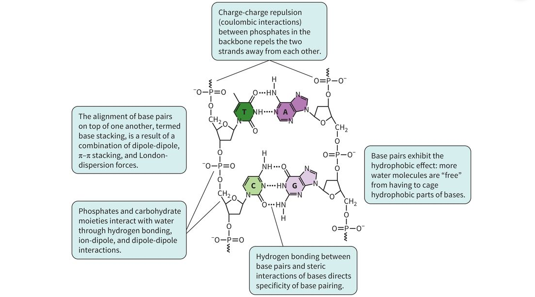
Forces That Stabilize the DNA Double Helix
There are strong covalent bonds within the DNA molecule, including those in the sugar-phosphate backbone and between the sugar and the nitrogenous base. However, the double-stranded structure of DNA is mainly stabilized by weaker interactions.
The two major weak forces that stabilize the DNA double helix are:
Hydrophobic interactions
Base-stacking interactions (van der Waals forces)
1. Hydrophobic Interactions
The nitrogenous bases are relatively nonpolar and hydrophobic. Because they do not interact well with water, they tend to avoid contact with water.
When the two DNA strands come together to form the double helix, the bases become buried inside the helix, away from the surrounding water.
This reduces the amount of water that needs to surround the hydrophobic bases, making the double-stranded structure more stable.
Simple idea:
The hydrophobic bases "hide" from water by being positioned inside the DNA double helix.
2. Base Stacking
The nitrogenous bases are arranged on top of one another inside the double helix. This is called base stacking.
The bases contain π-electron clouds, and when the bases stack closely together, they can interact through van der Waals forces and other favorable interactions.
These stacking interactions are an important source of stability for the DNA double helix.
Simple idea:
The bases stack like a pile of coins, creating stabilizing interactions between neighboring bases.
Hydrogen Bonds
Another important interaction is hydrogen bonding between complementary bases.
Adenine (A) pairs with thymine (T) through hydrogen bonds.
Guanine (G) pairs with cytosine (C) through hydrogen bonds.
These hydrogen bonds help hold the two DNA strands together. However, the overall stability of the double helix comes from multiple interactions working together, especially base stacking and hydrophobic interactions.
Electrostatic Repulsion and Its Stabilization
The phosphate groups in the DNA backbone carry a negative charge.
Because the two DNA strands have negatively charged phosphate groups, they naturally repel each other.
This repulsion can be reduced by positively charged ions, such as Mg²⁺, which help shield the negative charges.
In chromosomes, histone proteins also help with this. Histones are positively charged proteins that interact with the negatively charged DNA and help stabilize and package it.
Big Picture
The DNA double helix is stabilized by several interactions:
Hydrophobic interactions → keep the hydrophobic bases away from water.
Base stacking → bases stack on top of each other and interact through van der Waals and related forces.
Hydrogen bonds → connect complementary bases between the two strands.
Positive ions and histones → reduce repulsion between negatively charged phosphate groups.
The two major stabilizing forces to remember are: hydrophobic interactions and base stacking.

Urea & Guanidinium hydrochloride
are 2 compounds that are often used to help in the lab (not inside a living organism) to disrupt interactions between the 2 strands.
Urea is a highly soluble molecule that is able to form a lot of Hydrogen bonds. (both of them are really good at disrupting Hydrogen bonds, so other things can stabilize the DNA molecule.)

structure of RNA molecules
RNA Structure
RNA is typically a single-stranded nucleic acid. Like DNA, it has a sugar-phosphate backbone made up of nucleotides, with the nitrogenous bases extending from the backbone.
Unlike DNA, RNA usually does not form a long double-stranded structure. However, because RNA is single-stranded, it can fold back and pair with itself.
When an RNA strand folds back on itself, complementary bases can form Watson-Crick base pairs. These interactions create loops and other folded structures within the RNA molecule.
These folded structures are called secondary structural elements.
Primary vs. Secondary StructurePrimary Structure
The primary structure of a nucleic acid is the linear sequence of its nucleotides.
This applies to both DNA and RNA.
Simple idea:
Primary structure = the order of the nucleotides.
Secondary Structure
The secondary structure refers to the additional structures that form when different parts of the nucleic acid interact with each other.
These structures are stabilized largely by hydrogen bonding between complementary bases.
In DNA, this helps form the double helix.
In RNA, the single strand can fold back on itself, allowing bases within the same RNA molecule to pair with one another and create structures such as loops and stems.
Simple idea:
Secondary structure = the way the nucleotide sequence folds because bases can pair with each other.
RNA Base Pairing
In RNA, the standard Watson-Crick base pairs are:
Adenine (A) pairs with uracil (U)
Guanine (G) pairs with cytosine (C)
This is different from DNA, where adenine pairs with thymine (T).
Big Picture
RNA is usually single-stranded.
It has a sugar-phosphate backbone with bases attached.
RNA can fold back on itself.
Complementary bases can form Watson-Crick base pairs within the same RNA molecule.
These interactions create secondary structures in RNA, such as stems and loops.

different kinds of RNA
mRNA (messenger rna): involved in protein synthesis
tRNA (transfer rna): smaller adapter molecules that are involved in protein synthesis
rRNA(ribosomal rna): part of complex that occurs in protein synthesis in the ribosome

chemical stability of RNA vs DNA
1. DNA is more chemically stable than RNA
Both DNA and RNA are nucleic acids, but DNA is much more stable and tends to remain intact for longer periods of time.
The main reason RNA is less stable is because of a chemical difference in their sugars:
RNA has a 2′-OH (hydroxyl) group
DNA does not have a 2′-OH group; it has a hydrogen (H) instead
2. The 2′-OH group makes RNA easier to break down
The 2′-OH group in RNA can attack the phosphate backbone of the RNA molecule.
This can cause the sugar-phosphate bond to break through hydrolysis, resulting in the RNA strand being cleaved.
In simple terms:
RNA: 2′-OH → can attack the backbone → RNA can break apart more easily
DNA: no 2′-OH → cannot undergo this same reaction as easily → DNA is more stable
This is the major chemical reason why RNA is less stable than DNA.
3. Enzymes can also break down RNA
RNA can also be broken down by enzymes called RNases (ribonucleases).
RNases are enzymes that break down RNA by cleaving the phosphodiester bonds in the RNA backbone.
These enzymes are found throughout cells and are also common in the environment.
Different RNases have different functions and can break down RNA in specific ways.
4. Why don't RNases constantly destroy RNA inside our cells?
Our cells have strict regulation and control mechanisms for RNases.
RNases are not simply active everywhere all the time. Cells control:
Where RNases are located
When they are active
Which RNA molecules they target
How RNases themselves are regulated or inhibited
This allows cells to break down RNA when it is no longer needed while protecting RNA molecules that are still being used.
RNA is less stable than DNA mainly because RNA has a 2′-OH group. That hydroxyl group can participate in breaking the phosphodiester backbone. RNA is also actively degraded by RNases, which are highly regulated inside cells.
RNA has the 2′-OH → more reactive → less stable.
DNA lacks the 2′-OH → less reactive → more stable.


processes inside nucleic acid inside the cell
The central dogma describes how genetic information flows through a cell:
DNA → RNA → Protein
There are three major processes to know:
Replication: DNA → DNA
Transcription: DNA → RNA
Translation: RNA → Protein
1. Replication: DNA→ DNA
Replication is the process of making a copy of DNA.
Why does replication happen?
Replication occurs before a cell divides.
The goal is to make sure that each daughter cell receives an identical copy of the organism's genomic DNA.
How does replication
work?
DNA is normally double-stranded.
During replication:
The two DNA strands separate.
Each original strand acts as a template.
A new complementary strand is synthesized using each original strand as a guide.
This produces two DNA molecules.
Semi-conservative replication
Replication is called semi-conservative because each new DNA molecule contains:
One original (parental) strand
One newly synthesized strand
So:
Original DNA
→ separates into two strands
→ each strand gets a new complementary strand
→ 2 DNA molecules, each containing:
1 old strand + 1 new strand
New DNA is always synthesized in the 5′ → 3′ direction.
Why? New nucleotides are added to the 3′-OH group of the growing DNA strand.
The 3′-OH attacks the alpha phosphate of the incoming nucleotide, forming the new phosphodiester bond.
So remember: 3′-OH → attacks phosphate → new nucleotide is added This is why the new strand grows 5′ → 3′.
2. Transcription: DNA → RNA
Transcription is the process of using information stored in DNA to make an RNA molecule.
During transcription:
A section of DNA is used as a template.
The DNA sequence is read.
A complementary RNA strand is produced.
For example:
DNA template:
3′-TACG-5′
RNA:
5′-AUGC-3′
The RNA contains a sequence of bases that is complementary to the DNA template.
Transcription = making RNA from DNA, DNA → RNA = transcription
3. Translation: RNA → Protein
Translation is the process of using the information stored in RNA to make a protein.
The nucleotide sequence in RNA contains the instructions for determining the amino acid sequence of a protein.
So: RNA → amino acids → protein. Translation occurs using structures called ribosomes, which read the RNA sequence and help assemble the appropriate amino acids into a protein. Translation = making protein from RNA

What is replication? Why is it necessary?
The process of duplicating DNA before cell division. It is necessary so that each daughter cell receives a complete copy of the genome.
What does semi-conservative mean?
Each daughter DNA molecule contains:
1 parental strand + 1 newly synthesized strand
Which direction is new DNA synthesized?
5′ → 3′
Where are new nucleotides added? What chemical reaction forms the new bond?
To the 3′-OH of the growing strand. The 3′-OH attacks the alpha phosphate of the incoming nucleotide, forming a new phosphodiester bond.
The replication process in procaryotic cells is different then eukaryotic cells
Prokaryotic Cells
Prokaryotic cells do not have a membrane-bound nucleus or other membrane-bound organelles.
Examples include bacteria, which are usually unicellular organisms.
In prokaryotic cells:
DNA replication begins at one origin of replication.
Replication proceeds from this single starting point.
The original double-stranded DNA molecule is copied, producing two identical double-stranded DNA molecules.
When the bacterial cell divides, each daughter cell inherits one copy of the DNA molecule.
Eukaryotic Cells
Eukaryotic cells have a nucleus and other membrane-bound organelles.
Examples include:
Human cells
Plant cells
Yeast cells
Yeast is an example of a unicellular eukaryotic organism.
In eukaryotic cells:
DNA replication begins at multiple origins of replication.
Replication happens at many locations along the DNA molecule at the same time.
This allows the large amount of DNA in eukaryotic cells to be copied efficiently.
Key Difference to Remember
Prokaryotes → one origin of replication
Eukaryotes → multiple origins of replication
Pro = one origin
Eu = multiple origins
The Transcription process
The transcription process involves using a DNA template as the information source for making an RNA product. (Process where by DNA template is used to make an rna product (any type of rna). Any time you are making an RNA molecule, you are using a DNA template. The direction of synthesis of these RNAs is in a 5 prime 3 prime direction.
Translation process
The process whereby messenger RNA serves as the template to inform the biochemical machinery inside a cell what order of amino acids there will be in the protein product.
Things like proteins are sometimes called gene products.
Are RNAs also gene products? YES
A gene product can be a protein & an RNA.
Both of them ultimately rely on a DNA molecule having information to tell them what they’re going to be.
(How) Manipulating & Using Nucleic Acids in a laboratory is possible
There are several important techniques used to study and manipulate nucleic acids, including DNA and RNA.
1. Nucleic Acid Sequencing
Sequencing is the process of determining the exact order of nucleotides in a nucleic acid molecule.
You can sequence:
DNA
RNA
Key Point: Sequencing = determining the exact nucleotide sequence.
DNA sequencing is an important tool for studying and manipulating DNA.
2. Polymerase Chain Reaction (PCR)
Polymerase Chain Reaction (PCR) is a technique used to rapidly amplify a specific piece of DNA.
In other words, PCR allows you to make many exact copies of a particular DNA sequence.
PCR was originally developed for DNA, but related techniques can also be used to work with RNA.
Key Point: PCR = make many copies of a specific DNA sequence.
3. Restriction Enzymes
Restriction enzymes are enzymes that cut DNA at specific nucleotide sequences.
They recognize a specific DNA sequence, often 5–6 nucleotides long.
They cut the sugar-phosphate backbone of the DNA.
Because they cut DNA at specific locations, they can be thought of as molecular scissors.
Where Do Restriction Enzymes Come From?
Restriction enzymes are naturally produced by bacteria.
Bacteria can be infected by viruses that inject viral DNA into the bacterial cell. Bacteria evolved restriction enzymes as a form of defense against these viruses.
The enzymes recognize specific sequences in the invading viral DNA and cut the DNA, helping protect the bacteria.
Scientists realized that if bacteria can use these enzymes to cut DNA at specific sequences, scientists can use the same enzymes to cut DNA in the laboratory.
Key Point: Restriction enzymes = cut DNA at specific sequences.
Heterologous Gene Expression
These techniques can be used together to produce a protein from one organism inside another organism.
Heterologous gene expression is when a gene from one organism is introduced into a different organism so that the second organism produces the protein encoded by that gene.
For example:
A specific human gene is identified.
The gene is copied and/or isolated.
The gene is placed into a DNA construct that allows it to be expressed.
The construct is introduced into bacteria.
The bacteria produce the human protein.
The protein can then be collected and purified.
This essentially turns the bacteria into a protein factory.
These techniques are useful in research and in the pharmaceutical industry.
Example: Insulin
Insulin is an example of a protein used as a medicine. People with diabetes may not produce enough insulin naturally. Before recombinant DNA technology, insulin used for treatment could be obtained from the pancreases of animals such as pigs. Scientists later developed methods to introduce the human insulin gene into bacteria. The bacteria can then produce human insulin, which can be collected and purified for use as a medicine. Large pharmaceutical facilities can grow bacteria in very large containers called fermentors, allowing them to produce large quantities of the protein.
Putting the Techniques Together
Scientists can use different techniques to manipulate DNA in many ways.
For example:
Sequencing → determine the exact DNA sequence
PCR → make many copies of a specific DNA sequence
Restriction enzymes → cut DNA at specific sequences
Heterologous gene expression → use a gene from one organism to produce its protein in another organism
PCR can also be used as part of methods that allow scientists to change specific nucleotides in a DNA sequence.
There are many different ways to manipulate DNA, but the important thing is to know the names of these techniques and what each one is used for.
Quick Study Guide
Technique | What it does |
|---|---|
Sequencing | Determines the exact nucleotide sequence |
PCR | Makes many copies of a specific DNA sequence |
Restriction enzymes | Cut DNA at specific sequences |
Heterologous gene expression | Produces a protein from one organism using another organism |
Remember:
Sequencing = Read it
PCR = Copy it
Restriction enzymes = Cut it
Heterologous expression = Make the protein
Formation of the Sugar-Phosphate Backbone
The sugar-phosphate backbone of DNA is formed when a new nucleotide is added to the growing DNA strand.
How the Bond Forms
The 3′ hydroxyl (OH) group on the sugar of the growing DNA strand attacks the alpha (α) phosphate of an incoming nucleotide triphosphate.
This reaction forms a covalent bond between:
The oxygen of the 3′ hydroxyl group on the existing sugar
The phosphate group of the incoming nucleotide
This creates the sugar-phosphate backbone and extends the DNA strand by one nucleotide.
Pyrophosphate
When the new nucleotide is added:
Pyrophosphate (PPi) is released.
Pyrophosphate is then hydrolyzed into two inorganic phosphate molecules (2 Pi).
This hydrolysis helps drive the reaction forward and makes nucleotide addition favorable.
Key Point to Remember:
3′ OH → attacks α-phosphate → phosphodiester bond forms → PPi is released → PPi is hydrolyzed to 2 Pi
The covalent bond formed between the 3′ oxygen of one sugar and the phosphate of the next nucleotide is called a phosphodiester bond.