Introduction to the Immune System - Slide Deck
 Call Kai
 Learn
Learn Practice Test
 Spaced Repetition
 Match
Match Flashcards
 Knowt Play
1/45

Earn XP
Description and Tags
Immuno Exam 1 - Basic Science Core
Name | Mastery | Learn | Test | Matching | Spaced | Call with Kai | Chat |
|---|
No analytics yet
Send a link to your students to track their progress
46 Terms
What Comprises the Immune System? (3 Groups)
1) Cells
2) Protein/Chemicals
3) Organs and Tissues
Basic Functions of the Immune System (3)
1) Identifies and eliminates pathogens
2) Promotes healing and tissue repair
3) Develops long-term immunity
Blood Composition (2)
1) Plasma
2) Formed Elements
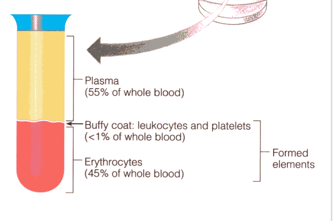
How is blood seperated
With a Centrifuge
A Centrifuge spins blood to separate it
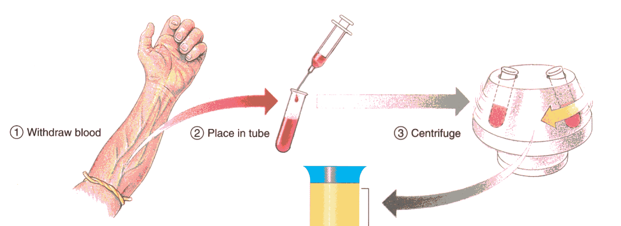
What are the Formed Elements
1) RBCs (erythrocytes)
2) WBCs (leukocytes),
3)) platelets (thrombocytes)
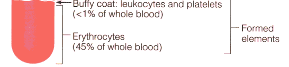
The plasma contains: (3)
1) H2O
2) Solutes
3) Plasma proteins
Serum
is plasma minus its clotting factors.
Plasma Proteins (3)
1) Albumins
2) Fibrinogens
3) Globulins
Where is Red Bone Marrow Found? (3)
1) Flat bones
(sternum, ribs & cranial bones)
2) Irregular Bones
(coxal bones, vertebrae (singular – vertebra) and scapulae (singular – scapula)
3) The epiphyses (singular – epiphysis) of long bones such as the femur and humerus
Gross Anatomy of Long Bones
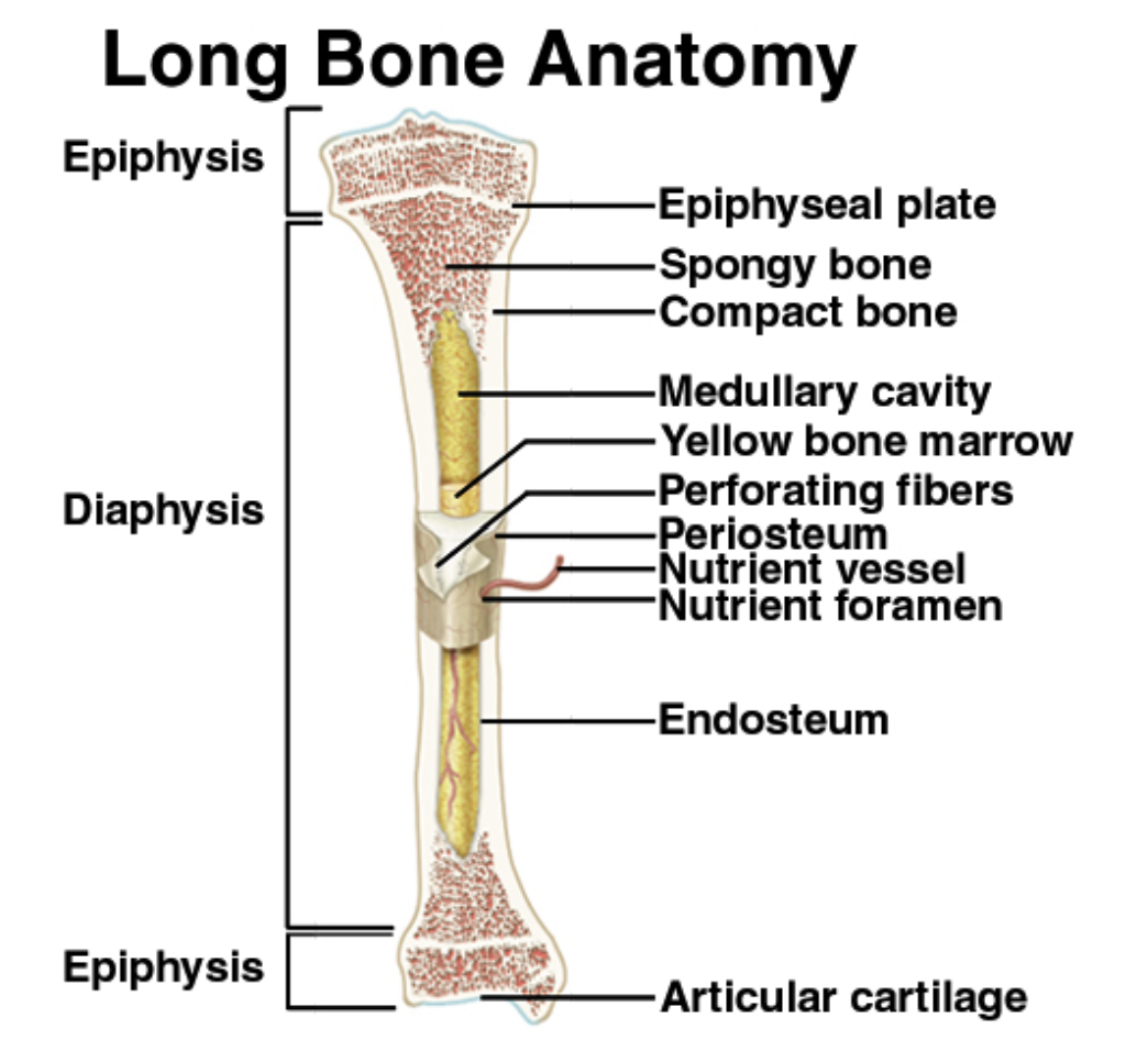
Where is Yellow Marrow found?
Medullary cavity
In cases of severe blood loss, the body can convert yellow marrow back to red marrow to increase blood cell production.
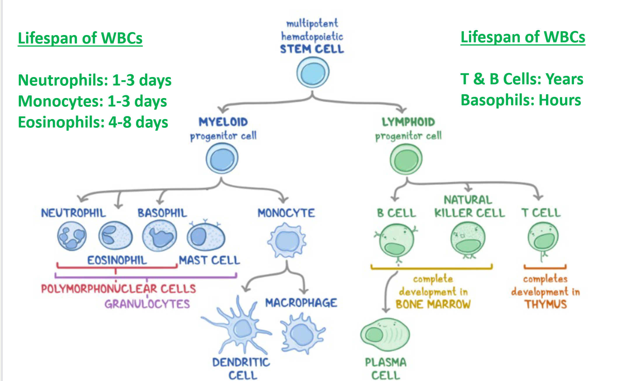
Lifespan of WBCs (5 Types)
Neutrophils: 1-3 days
Monocytes: 1-3 days
Eosinophils: 4-8 days
T & B Cells: Years
Basophils: Hours
Process of making White Blood Cells
Multipotent Hematopoietic Cells → Myeloid → Speciallized WBC (Except T & B Cells)
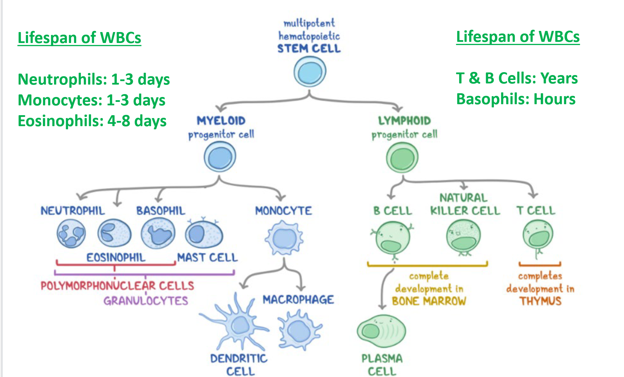
Process of making T, B & Killer Cells
Multipotent Hematopoietic Cells → Lymphoid→ T, B & Killer Cells
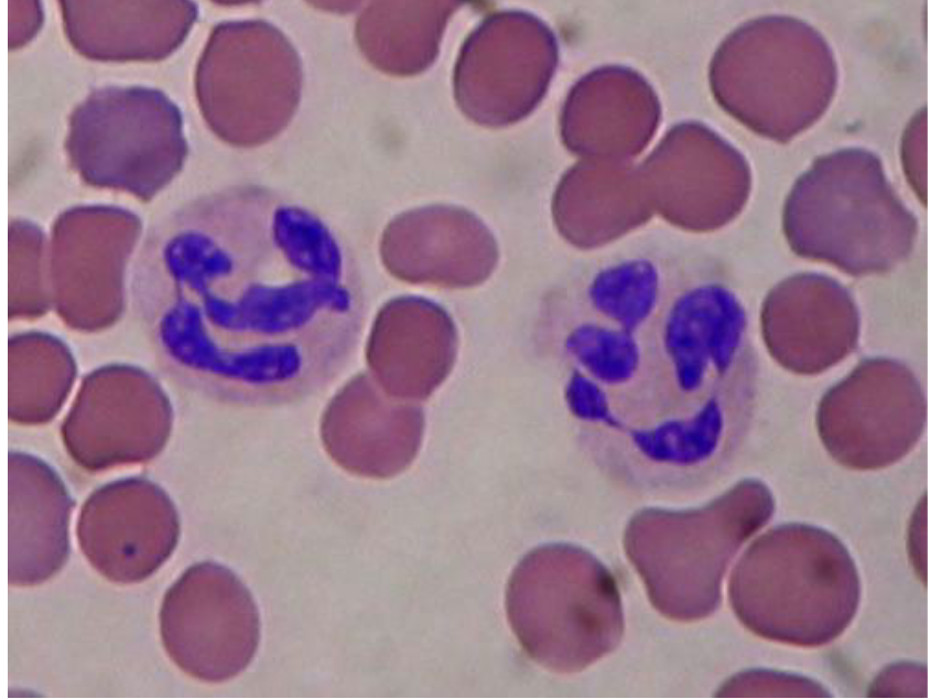
Neutrophils (3)
Nucleus typically divided into 3-5 lobes which are connected by thin strands of chromatin
Most Common white blood Cell (~60-70%)
Function is primary defense against bacteria and fungi
Large numbers of neutrophils will invade tissues where an infection has occurred
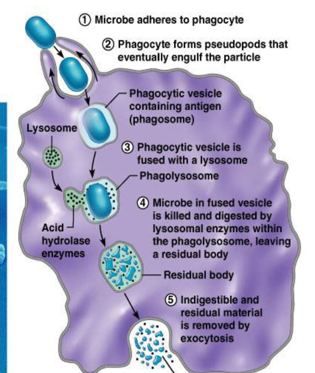
Neutrophils Phagocytosis (5 Steps)
A neutrophil uses its membrane to make a pseudopod to ingest pathogens.
A lysosome creates particles to destroy the pathogen.
After it is destroyed, the debris is called the residual body, which is then dumped before another cell is eaten.
Eosinophils
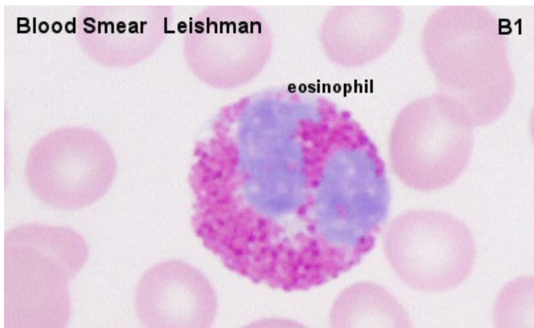
Nucleus typically divided into 2 lobes
Almost all of the cytoplasm appears filled with granules
Granules are not neutral but stain red or pink
Main function → defense against parasitic infections
Basophils
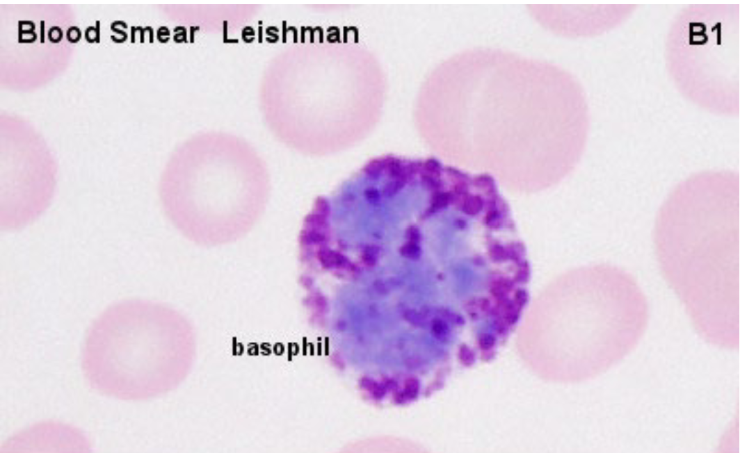
Nucleus usually has 2-3 lobes
The lobes are typically not as well defined (May be “S” Shaped)
The granules of basophils are usually stained deeply bluish or reddish-violet
Less Granules
Stores lots of Histamines
HISTAMINE
causes some of the symptoms of an allergic reaction
Found in Basophils
Monocytes
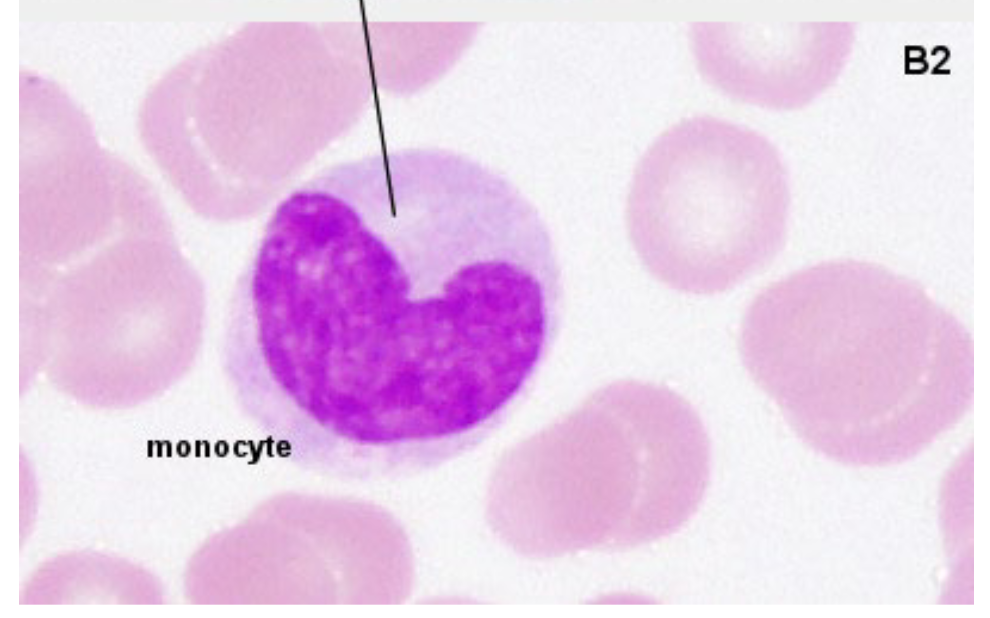
C Shaped (Kidney Bean Shape)
Few to no granules
become MACROPHAGES when they migrate to tissue
Monocytes can also become DENTRITIC CELLS
Macrophages
They are formed by Monocytes when they migrate to tissues
Macrophages kill intracellular microorganisms (certain bacteria, fungi) similar to neutrophils (i.e., phagocytosis)
Macrophages also promote inflammation and participate in wound healing and tissue repair
Dendritic Cells
Specialized antigen-presenting cells crucial for activating the adaptive immune response (more on this in a future lecture)
Also formed by monocytes
Lymphocytes
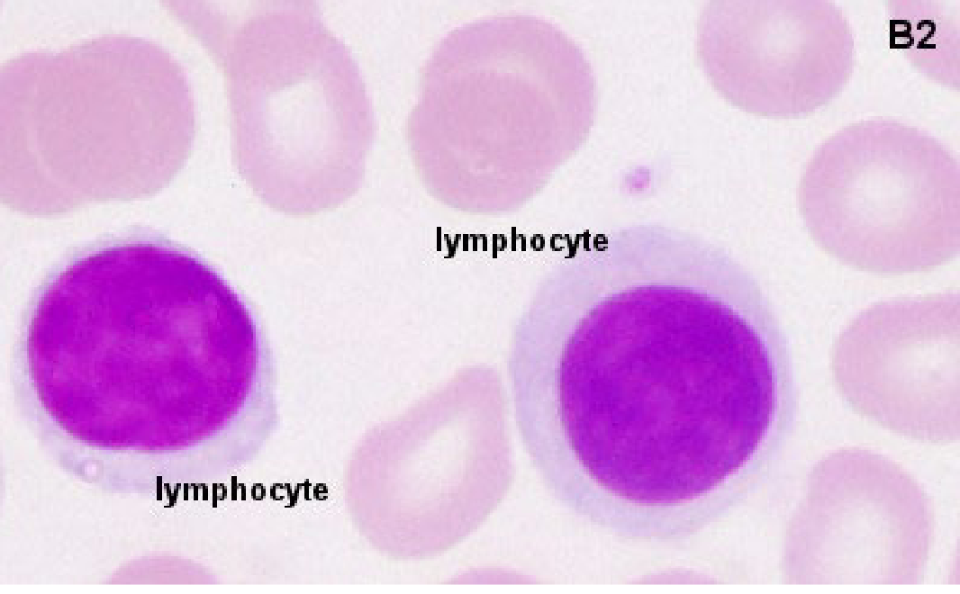
Lymphocytes are generally the smallest of all the WBCs → usually a tad larger than RBCs
Round nucleus & very little cytoplasm
Few to no granules
Two main types → B and T lymphocytes (indistinguishable microscopically)
Third Type is Killer T Cells
The function of T cells and B cells is to identify “non-self” antigens, particularly on viruses
B lymphocytes (2)
Acts as antigen-presenting cells
When a B lymphocyte encounters a specific antigen, it can become activated and either differentiate into a PLASMA CELL or MEMORY B CELL.
Plasma cells
Plasma cells produce large quantities of ANTIBODIES which then attack bacteria and viruses.
Type of B lymphocyte
T Lymphocytes
HELPER T CELLS or CD4 + CELLS (Which disease is characterized by low counts of these cells?), produce chemicals that direct the immune response.
While other T cells, called CYTOTOXIC T CELLS or CD8 + CELLS, produce enzymes which induce the death of pathogen infected cells.
Memory B Cells
Following activation, B and T lymphocytes can “remember” the antigens
they have encountered. Throughout the lifetime of an individual, they will
recall each specific pathogen encountered, and are able to mount a strong
and rapid response if the pathogen is ever detected again
Natural killer cells
a type of lymphocyte that play a major role in the rejection of tumors and cells infected by viruses.
They kill cells by releasing small cytoplasmic granules of proteins called perforin and granzyme that cause the target cell to die.
Complete Blood Count (CBC)
used as a broad screening test to check for such disorders as anemia, infection, and many other diseases.
It is actually a panel of tests that examines different parts of the blood
Tests We need to know about CBC (2)
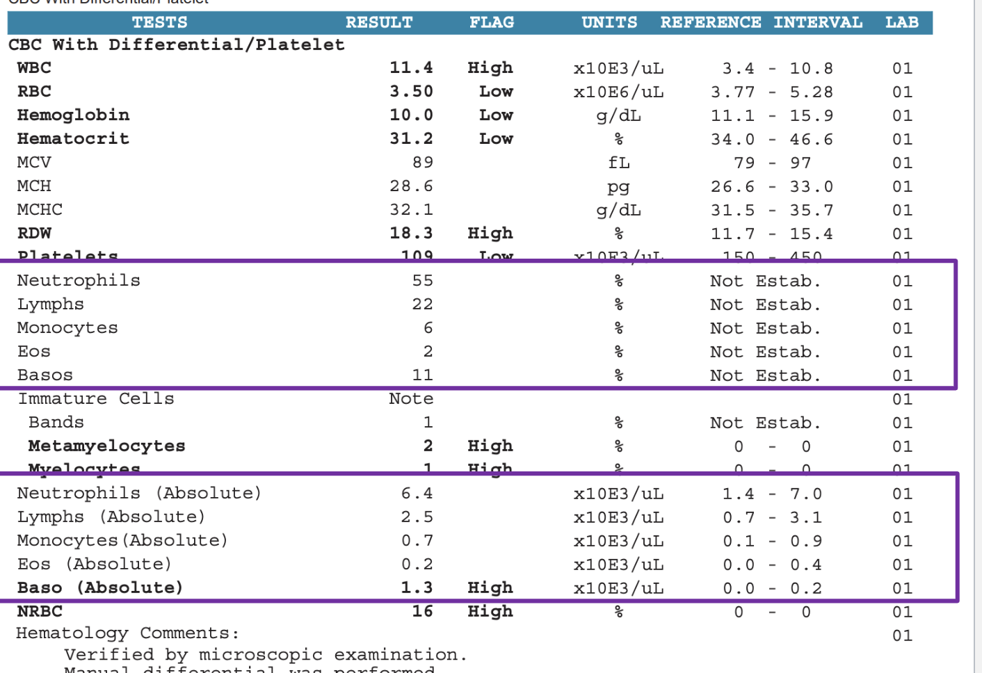
White Blood Cell Count
WBC Differential Count
White Blood Cell Count
Count of the actual number of white blood cells per volume of blood
May be increased (LEUKOCYTOSIS) with infections, inflammation,
cancer, leukemia
May be decreased (LEUKOPENIA) with some medications, bone
marrow failure, chemotherapy, and congenital marrow aplasia
(marrow doesn't develop normally)
Normal is (3.4 to 10.5)

LEUKOCYTOSIS vs LEUKOPENIA
LEUKOCYTOSIS (too much WBC)
LEUKOPENIA (too little WBC)
WBC Differential Count
Counts the percentage of white blood cells in blood, 5 types tested
Try to narrow down why specifically the total white blood cells is high
Include band Neutrophils (Immature Neutrophils)
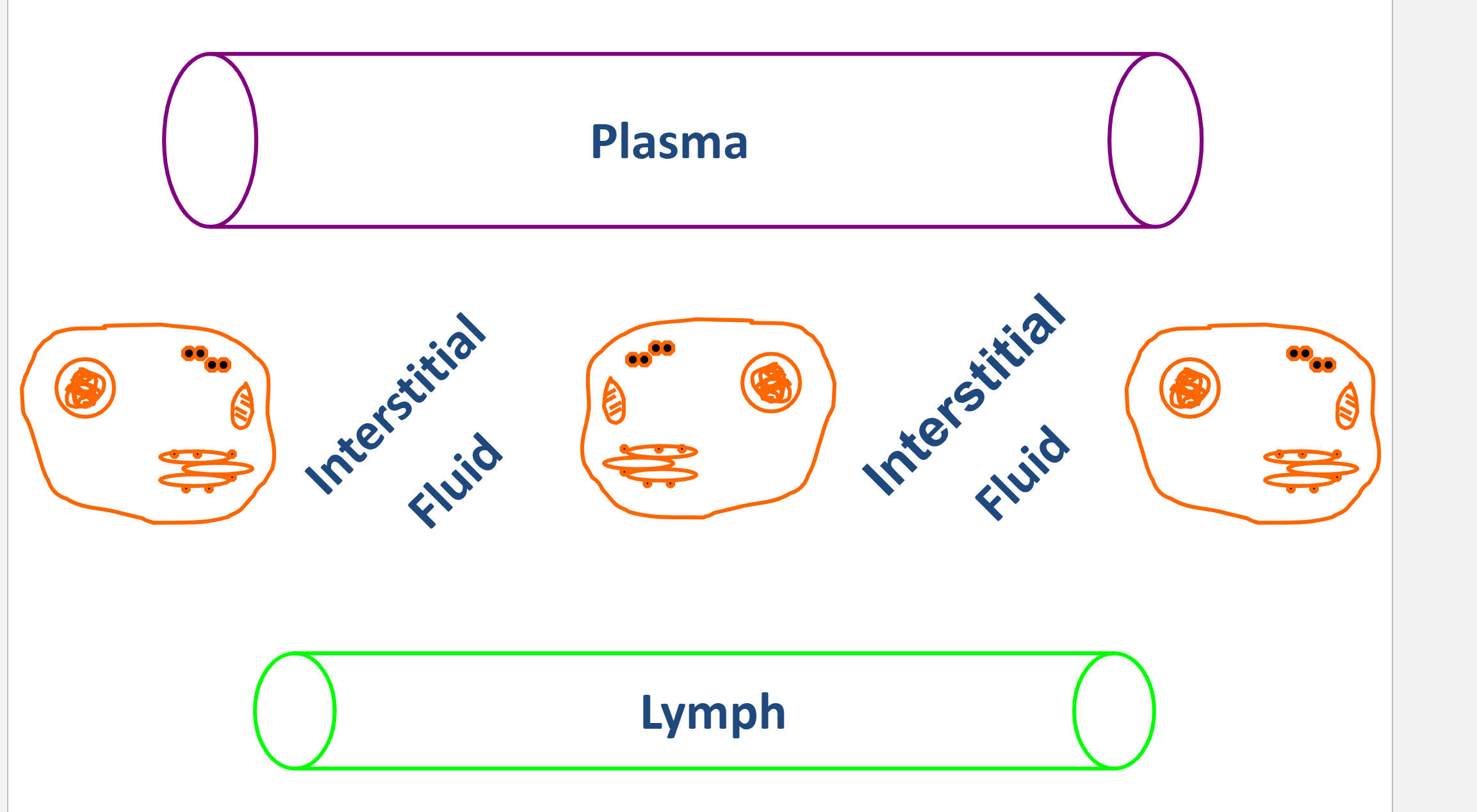
Lymph
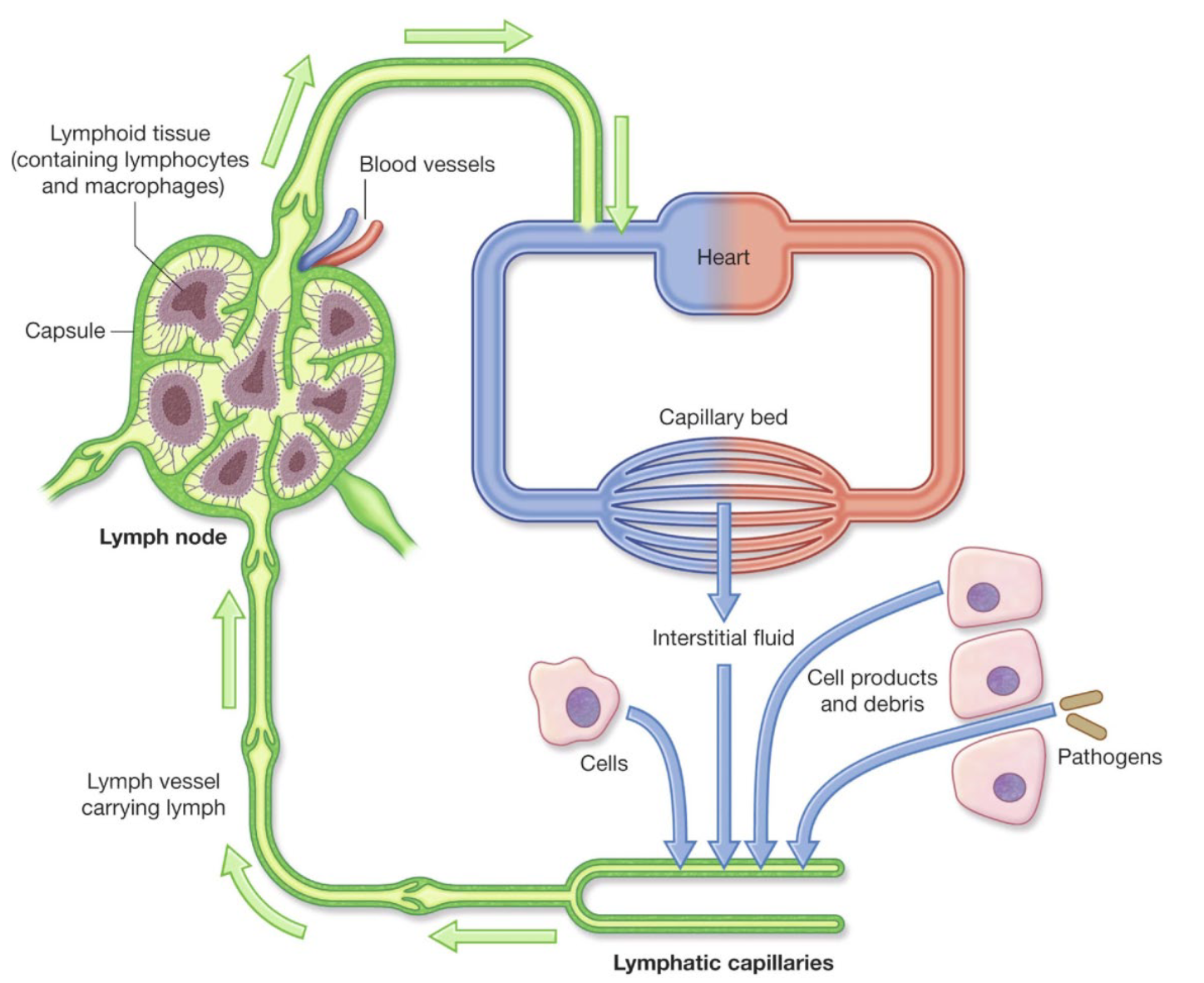
Lymph is a fluid that is:
created in the interstitial spaces of tissues (found in spaces between cells)
carried away by lymphatic vessels
put back into the circulation through the blood
Mixes with the blood
Fluid Exchange Between Compartments (Lymph)
Lymph is leftover liquid and nutrients dropped by the blood, which is eventually brought back to the blood
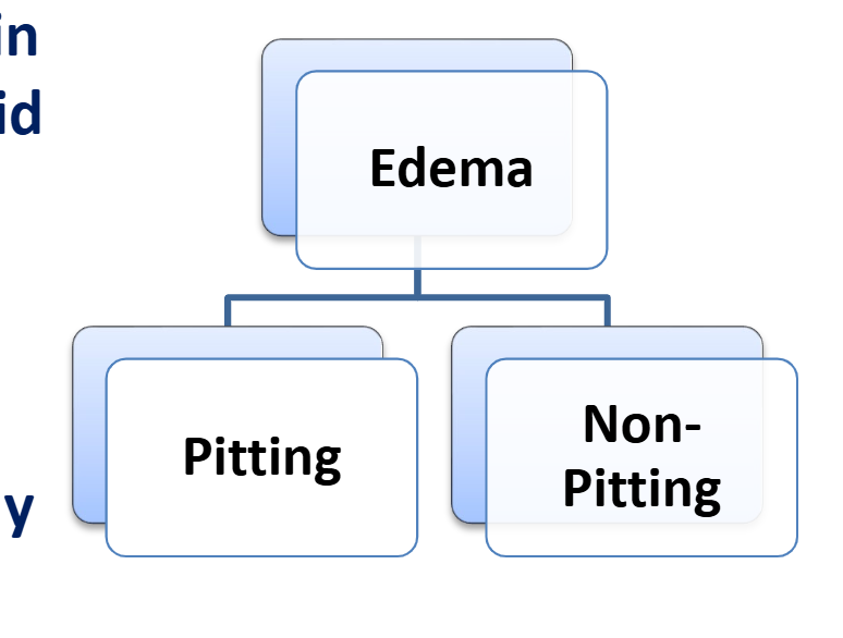
Edema (2 Types)
Definition: palpable swelling due to expansion of extracellular fluid
Two Types:
Pitted
Non Pitted
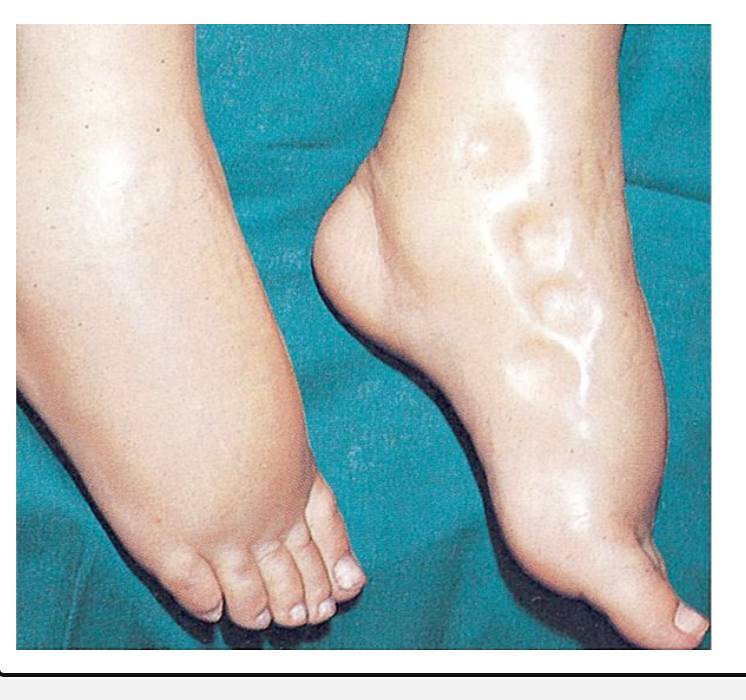
Pitted Edema
A finger pressed into the skin over an accumulation of fluid resulting in a temporary depression in the skin
Normal skin and subcutaneous tissues quickly rebound when the pressure is released.
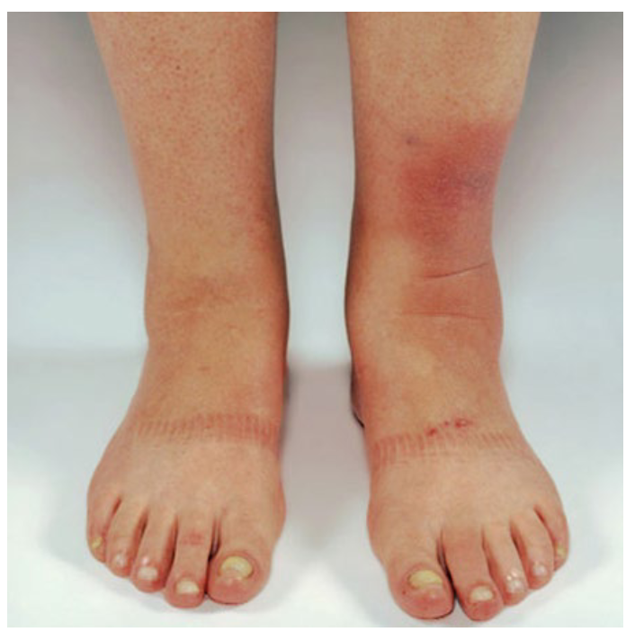
Non-pitted Edema
Excess fluid in the interstitium becomes infiltrated with protein or other material so that it cannot move freely within the tissue spaces.
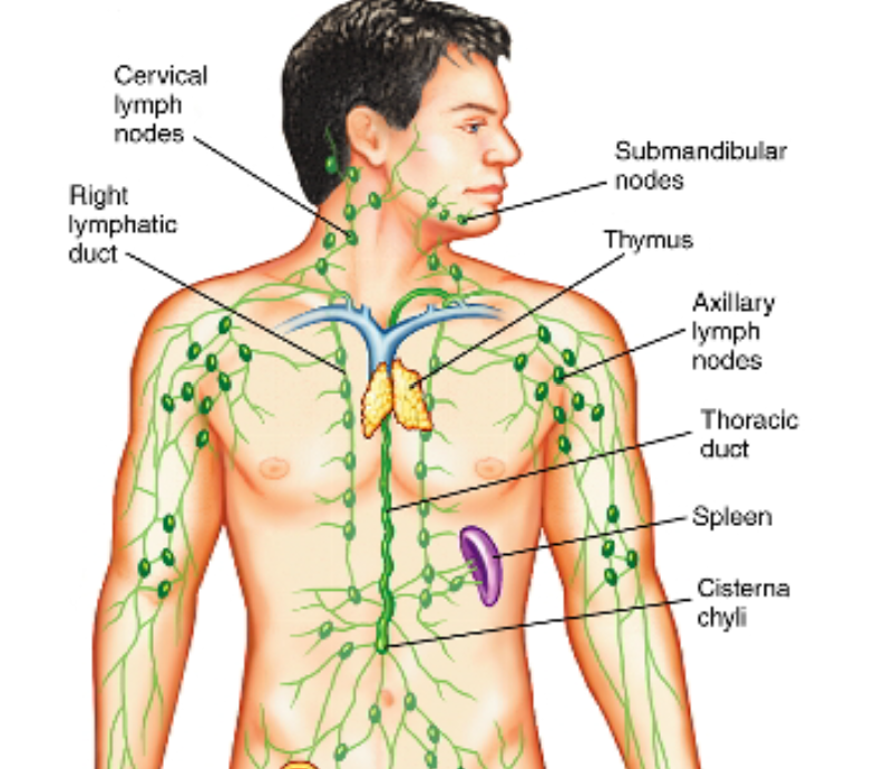
Lymphatic Vessels
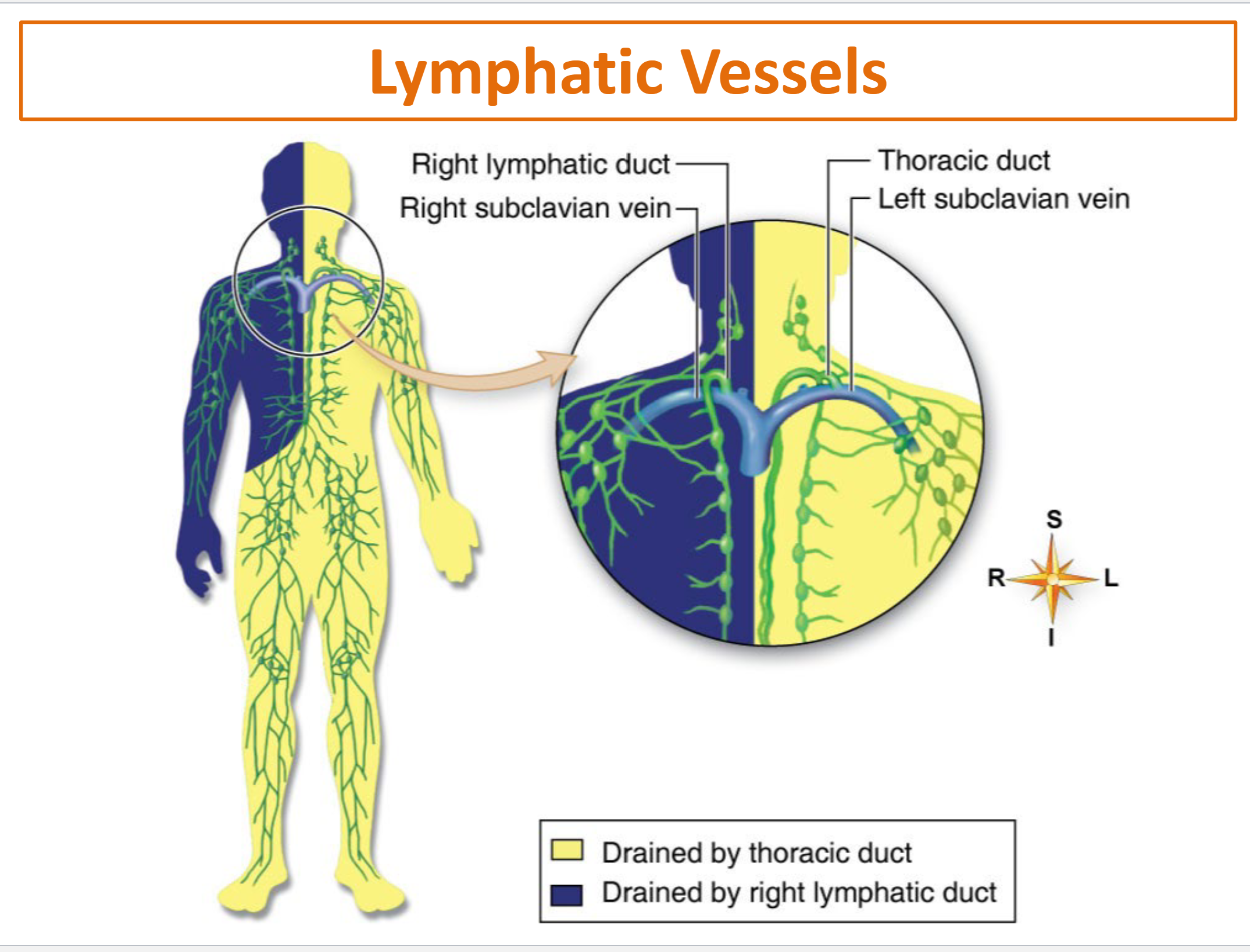
Lymphatic Capillaries → Lymphatic Venules & Veins → Lymphatic Ducts (Right Lymphatic Duct & Thoracic Duct)
Lymph Nodes
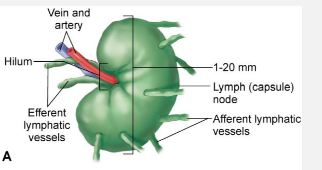
Lymph nodes are “beads” or “swellings” along certain lymphatic vessels.
They are FILTERS!
Most extensive in the breast, neck, axilla, abdomen, thorax, pelvis & groin
Found in the Lymphatic Venules & Veins
How many lymphatic ducts are there?
2 (the right and left)
Both drain into the subclavian veins (right and left)
Thymus Gland
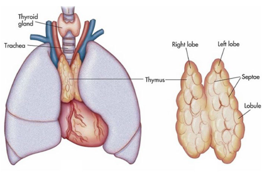
Located in the upper chest in front of the ascending aorta and behind the sternum
Active during childhood and adolescence; gradually shrinks in adults
T lymphocytes “mature” in the thymus after produced in the bone marrow
The thymus “teaches” the T cells how to recognize self vs. non-self antigens
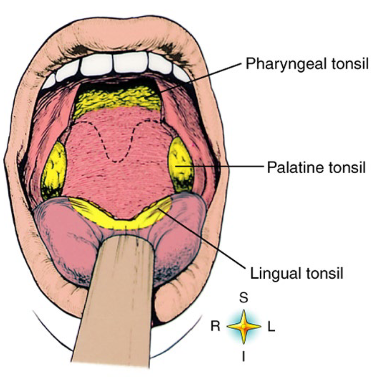
Tonsils
Tonsils are essentially clusters of WBCs hanging out in the pharynx.
There are 3 types:
Pharyngeal (adenoids)
Palatine
Lingual
Spleen
Located in LUQ
Removes dead/deteriorating RBCs, WBCs, and platelets from the circulation
Can store or “pool” blood, which can be released into the circulation when needed (e.g., hemorrhage) Particularly pools lymphocytes, where they can be presented with antigens and produce antibodies Often injured in abdominal trauma
Splenectomy = removal of spleen
Splenectomy
Removal of spleen
Mucosa associated lymphoid tissue (MALT) (GI Tract)
Small concentrations of lymphoid tissue the walls of tissue throughout the body.
MALT is populated by T lymphocytes, B lymphocytes and macrophages, each of which are well situated to encounter pathogens trying to enter the body.