MCAT Biology Pt. 1
1/152
There's no tags or description
Looks like no tags are added yet.
Name | Mastery | Learn | Test | Matching | Spaced | Call with Kai |
|---|
No study sessions yet.
153 Terms
Cell Theory
all living things are composed of cells
the cell is the basic functional unit of life
cells arise only from preexisting cells
cells carry genetic information in the form of deoxyribonucleic acid (DNA), This genetic material is passed on from parent to daughter cell.
Cell Theory vs Viruses
Viruses: not living bc violate third and fourth tenents of cell theory (unable to reproduce on own)
Eukaryotic cells
Unicellular or multicellular
Contain true nucleus enclosed in membrane
Prokaryotic cells
Single celled
Do not contain nucleus
Membrane Bound Organelles
most cells have membrane enclosing semifluid cytosol where organelles are suspended
membrane bound allow for compartmentalization of functions
cytosol: allow for diffusion of molecules throughout the cell
Nucleus
surrounded by nuclear membrane or envelope
nuclear pores: allow for selective two-way exchange material between cytoplasm and nucleus
Coding:
Genes: coding regions of DNA
Histones: organizing proteins where DNA wrapped around
Chromosomes: liner strands of DNA
Nucleolus: subsection of DNA where rRNA is synthesized (25% of volume of nucleus, darker spot)
Mitochondria functions
power plants of the cell
semi-autonomous: own genes and replicated independently of nucleus via binary fission
cytoplasmic / extranuclear inheritance: transmission of genetic material independent of nucleus (b/c engulfing of aerobic prokaryote by anaerobic prokaryote)
in charge of kicking-off process called apoptosis via releasing enzymes from electron transport chain
Mitochondria parts
Outer membrane: barrier between cytosol and inner environment of mitochondrion
Inner membrane: arranged into infoldings called cristae, contain molecules and enzymes for electron transport chain
Intermembrane space: between inner and outer membrane
Mitochondrial matrix: space inside inner membrane
Lysosomes
membrane bound structures
hydrolytic enzymes for breaking down many different substrates (endocytosis and cellular waste products)
endosomes: transport, package and sort cell material traveling to and from membrane → also HELP transport material to lysosomal pathway for degredation
autolysis: destruction of cellular components and cell via hydrolytic enzymes released from lysosomes → apoptosis
Endoplasmic Reticulum (ER) structure
interconnected membranes contiguous with nuclear envelope
double membrane
Rough Endoplasmic Reticulum
studded with ribosomes
translation of proteins that will be secreted
Smooth Endoplasmic Reticulum
lack ribosome
use for lipid synthesis (like phospholipids in cell membrane)
detoxification of certain drug and poison
Golgi Apparatus
stacked membrane-bound sacks
modify proteins made in ER
add groups like carbohydrates, phosphates and sulfates
introduce signal sequences which direct delivery of product to specific cellular location
repackaged in vesicles afterwards → exocytosis if exiting cell
Peroxisomes
contain hydrogen peroxide
break down long chain fatty acids via beta-oxidation
synthesis of phospholipids
also contain some enzymes invovled in pentose phosphate pathway
Cytoskeleton
provide structure to cell and help to maintain its shape
Cytoskeleton - Microfilaments
made up of solid polymerized rods of actin
organized into bundles and networks and resistant to both compression and fracture → protection of cell
also use ATP for movement by interacting with myosin
ex. muscle contraction
Cytoskeleton - Microfilaments: Cytokinesis
cytokinesis: division of materials between daughter cells
cleavage furrow formed from microfilaments → ring at site of division between two new daughter cells
actin filaments here contract and ring becomes smaller to separate daughter cells
Cytoskeleton - Microtubules
hollow polymers of tubulin protein
radiate throughout cell
primary pathways for carrying vesicles
use motor proteins like kinesin and dynein to carry vesicles
Cytoskeleton - Microtubules: Cilia and Flagella
cilia: projection from cell involved in moving materials along surface of cell
flagella: structures involved in moving of cell itself
9+2 structure (9 pair of microtubule forming in outer ring+ 2 pair in center)
structure for both cilia and flagella
Cytoskeleton - Microtubules: Centrosome
found in region of cell called centrosome
nine triplets of microtubules with a hollow center
during mitosis:
centriole go to opposite poles of dividing cell and organize mitotic spindle
microtubule attaching to chromosomes via complexes called kinetochores
pulls sister chromatids apart
Cytoskeleton - Intermediate Filaments
diverse group of filamentous proteins
include keratin, desmine, vimentin, lamins
involved in cell-cell adhesion and maintenance of overall integrity of cytoskeleton
able to withstand tremendous amount of tension
also helps anchor other organelles including nucleus
Epithelial Tissue
cover body and line cavities, means of protection for pathogen invasion and desiccation
underlying layer if basement membrane (connective tissue)
in most organs, epithelial cells constitute parenchyma: functional parts of organ
polarized: one side faces lumen (inside of organ/tube) or outside world, while other side interact with underlying blood vessel and structural cells
Types of Epithelial Cells
Simple epithelia: one layer of cells
stratified epithelial: multiple layers
Pseudostratified epithelia: appear to have multiple layers due to differences in cell height but only one layer
cuboidal cells: cube-shaepd
columnar cells: llng adn thin
Squamous cells: flat and scale-like
Connective tissue
supports body and provide framework for epithelial cells to carry out functions
main contributor to stroma: support structure
bone, cartilage, tendons, ligaments, adipose tissue and blood
most cells in connective tissues secrete materials like collagen and elastin to form extracellular matrix
Nucleoid region
region where single circular molecule of DNA for prokaryote is concentrated
Prokaryote: Archaea
single celled organisms that visually similar to bacteria but with genes and metabolic pathways more similar to eukaryotes than to bacteria
extremophiles: most commonly isolated from harsh environment with hight temp, high salinity or no light but actually found in human body too
use alternative sources of energy: photosynthetic, chemosynthetic
Archaea and Eukarya Common Origin
translation with methionine
contain similar RNA polymerases
associate DNA with histones
BUT archaea has single circular chromosome
Prokaryote: Bacteria
cell membrane and cytoplasm
some have flagella or fimbriae (like cilia)
Prokaryote: Bacteria - Targeting
Can target via differences in flagella from eukarya vs bacteria
differences in ribosomes (bacteria ribosomes smaller than eukarya)
Prokaryote: Bacteria and Eukarya Relationships
mutualistic symbiotes: both humans and bacteria benefit from relationship
parasites: provide no advantage or benefit to host, but rather cause disease
Shapes of Bacteria
Cocci: spherical bacteria like strep pyogenes
Bacilli: rod-shaped bacteria like e. coli
Spirilli: spiral-shaped bacteria like treponema pallidum
Aerobes vs Anaerobes
obligate aerobes: require oxygen for metabolism
anaerobes: cellular metabolism that does not require oxygen
obligate anaerobes: anaerobes tha cannot survive in oxygen-containing environment
facultative anaerobes: can toggle between oxygen for aerobic metabolism or anaerobic metabolism if no oxygen present
aerotolerant anaerobes: unable to use oxygen for metabolism but not harmed by presence in environment
Prokaryotic Cell structure: Cell Wall
outer barrier of cell
next layer is cell membrane (plasma membrane)
envelope: cell wall + cell membrane
provide structure and control movement of solute in and out of bacterium
Prokaryotic Cell structure: Cell Wall - Gram Positive and Gram Negative
Gram positive: thick layer of peptidoglycan (polymeric substance made from amino acids and sugars)
provide protection from host organism’s immune system
contain lipoteichoic acid too
Gram negative: thin layer of peptidoglycan
periplasmic space: between peptidoglycan walls and cell membrane
also contain outer membranes with phospholipids and polysaccharides
Prokaryotic Cell structure: Flagella
Chemotaxis: ability of cell to detect chemical stimuli and move away from or toward them
composed of:
filament: hollow, helical struucture with flagellin
basal body: complex structure that anchors flagellum to cytoplasmic membrane and motor of flagellum
hook: connects filament and basal body
Prokaryotic Cell structure: Other Organelles
plasmids: DNA acquired fomr external sources carried on these smaller circular structures
cell membrane site of electron transport chain and generation of ATP
primitive cytoskeleton
Binary Fission
form of asexual reproduction in prokaryotes
circular chromosome attach to cell wall and replicate while cell continues to grow in size and begin to grow inward to make two identical daughter cells
faster than mitosis
Genetic Recombination
Plasmids: extrachromosomal (extragenomic) material
often carry genes impart some benefit to bacterium, like antibiotic reistance or virulence factors: increase pathogenicity
episomes: subset of plasmids capable of integrating into genome of bacterium
Bacterial Transformation
results from integration of foreign genetic material into host genome
comes from other bacteria that upon lysing spill contents into vicinity of bacterium capable of transformation
most gram-negative rods able to do this
Bacterial Conjugation
bacterial form of mating (sexual reproduction)
two cells form conjugation bridge: facilitates transfer of genetic material
formed from sex pili found on donor male
to form pilus, bacteria must have plasmids known as sex factors that contain necessary genes, like F (fertility) factor in E. Coli
Hfr (high frequency of recombination): sex factor plasmid being integrated into host genome → able to transfer entire host genome into other bacteria
unidirectional transfer: donor male (+) to recipient female (-)
Bacterial Transduction
only genetic recombination process requiring vector: virus carries genetic material from one bacterium to another
Viruses are obligate intracellular pathogens, cannot reproduce outside host cell
bacteriophages: viruses that infect bacteria
may accidentally incorporate segment of host DNA during assembly
when infecting other bacteria may put in that segment of host DNA
Bacterial Transposons
genetic elements capable of inserting and removing themselves from genome
both in prokaryotes and eukaryotes
may disrupt gene if inserted in coding region of gene
Bcterial Growth
Lag phase: bacteria first adapt to new local conditions
Exponential/log phase: bacteria exponentially increase once they adapt to local conditions
Stationary phase: when reduction of resources slows reproduction
Death phase: bacteria exceeded ability of environment to support number of bacteria
Viral Structure
Capsid: protein coat of virus
Envelope: surrounding capsid, composed of phospholipids and virus-specific proteins
sensitive to heat, detergents and desiccation, so enveloped viruses easier to kill
Virions: viral progeny released to infect additional cells
Viral structure - Bacteriophages
tail sheath: syringe, injecting genetic material into bacterium
tail fibers: help bacteriophage recognize and connect to correct host cel
Viral genomes - Single Stranded RNA viruses
Positive sense single stranded RNA viruses: genome directly translated to functional proteins by ribosomes of host cell (like mRNA)
Negative-sense single-stranded RNA viruses: template for synthesis to complementary strand → template for protein synthesis
Must have RNA replicase to ensure complementary strand is synthesized
Retroviruses: enveloped, single-stranded RNA viruses
Two identical RNA molecules
carry reverse transcriptase: synthesized DNA from single-strand DNA → integrate into host cell genome and replicate and transcribed like host cell’s own DNA
Viral Life Cycle
infection: virus binds to correct receptor on cell and enveloped viruses fuse with cell membrane
translation of viral genetic material: translocation of genetic material to correct location in cell → translate into protein with host ribosomes, tRNA, amino acids and enzymes with capsids
progeny release: cell death → spill viral progeny, or host cell lyse, or virus fuse with plasma membrane (extrusion) to keep host cell alive and allow continued use by virus (productive cycle)
Viral Life Cycle - Bacteriophages
Lytic cycle: cell lyses, virulent: viruses in lytic state
Lysogenic cycle: virus does not lyse bacterium, but integrate into host genome as provirus or prophage, replicated as bacterium reproduces but environmental factors may cause provirus to leave genome and revert to lytic cycle
superinfection: simultaneous infection with other phages; infection with one strain of phage makes bacterium less susceptible to this
Prions
infectious proteins, nonliving things
trigger misfoldings of other protieins (a helical to b pleated sheet)
protein aggregates form → interfere with cell function
Viroids
small pathogens with very short circular single-stranded RNA that infect plants
bind to large number of RNA sequences and silent genes on plant genome
also present for humans too
Diploid vs Haploid Cells
diploid (2n): 2 copies of each chromosome, autosomal cells = 46
haploid (n): 1 copy of each chromosome, germ cells = 23
Cell Cycle
cell cycle: specific series of phases during which cell grows, synthesizes DNA and divides
interphase: G1, S, G2, longest part of cell cycle
chromosomes not visible with light microscopy because in less condensed form known as chromatin
G0 stage: cell simply living nand carriying out its functions without preparation ofr division
Cell Cycle: G1 Stage: Presynthetic Gap
cells make organelles for energy and protein production while also increasing their size
passage into next stage governed by restriction point: need to contain proper complement of DNA
main protein in control is p53
Cell Cycle: S Stage: Synthesis of DNA
cell replicates genetic material so daughter cell have identical copies
after replication, two identical chromatids bound at specialized region known as centromere
still only have 46 chromosomes, even though 92 chromatids are present
double content, but content stay same
Cell Cycle: G2 Stage: Postsynthetic Gap
cell passes through another quality control checkpoint:
cell checks to ensure enough organelles and cytoplasm for two daughter cells
make sure that DNA replication proceeded correctly
main protein in control is p53
Cell Cycle: M Stage: Mitosis
mitosis + cytokinesis
occur in somatic cells (not involved in sexual reproduction)
Molecules responsible for cell cycle
Cyclin-dependent kinases (CDK): CDK require presence of right cyclins
During cell cycle, concentrations of specific cyclins increase and decrease during specific stages
cyclin binds to CDK → CDK-cyclin complex → phosphorylate transcription factor → promote transcription of genes required for next stage of cell cycle
Cancer
cell cycle control becomes deranged and damaged cells are able to undergo mitosis
mutation of gene producing p53 → not able to stop and repair damaged DNA
tumors: cancer cells that undergo rapid cell division
metastasis: distant spread of cancerous cells through bloodstream or lymphatic systems
Mitosis: Prophase
condensation of chromatin into chromosomes
centrosome: region outside of nucleus where paired cylindrical organelles help divide DNA → form spindle fibers made up of microtubules
asters: anchor centrioles to cell membrane
Kinetochores: appear at centrosome on centromeres that attach specific fibers of spindle apparatus (kinetochore fibers)

Mitosis: Metaphase
centriole pairs at opposite ends of cell
kinetichore fibers and fibers of spindle apparatus interact to align chromosomes at metaphase plate (equatorial plate)
Mitosis: Anaphase
centromeres split so chromatid has own distinct centromere
pulled towards opposite poles of cell by shortening of kinetochore fibers
Mitosis: Telophase and Cytokinesis
Telophase:
reverse of prophase
spindle apparatus disappears
nuclear membrane reform around each set of chromosomes
nucleoli reappear
chromosomes uncoil
Cytokinesis:
separation of cytoplasm and organelles
Mitosis
occur in gametocytes (germ cells) and results in up to four nonidentical sex cells (gametes)
Meiosis 1: homologous chromosomes separated, generating haploid daughter cells (reductional division)
Meiosis 2: separation of sister chromatids without change in ploidy (equational division)
Meiosis: Prophase I
chromatin condense into chromosomes
spindle apparatus form
nucleoli and nuclear membrane disappear
homologous chromosomes come together and intertwine in process called synapsis
each chromosome has two sister chromatids, so synaptic pair has tetrad (four chromosomes)
all held together by synaptonemal complex
recombination: chromatids of homologous chromosomes may break at point of contact (chiasma/chiasmata) and exchange equivalent pieces of DNA via crossing over
explains Mendel’s second law of independent assortment: inheritance of one allele has no effect on likelihood of inheriting certain alleles for other genes
Meiosis: Metaphase I
homologous pairs (tetrads) align at metaphase plate
held by one spindle fiber
Meiosis: Anaphase I
homologous pairs separate (father vs mother) and pulled to opposite poles of cell (disjunction)
accounts for Mendel’s first law of segregation: during disjunction, each chromosome of paternal origin separate from homologue of maternal origin, and either chromosome can end of up either daughter cell → distribution of homologous chromosomes is random with respect to parental origin (segregation)
Meiosis: Telophase I
nuclear membrane forms around new nucleus → cells are now haploid (n=23)
may have interkinesis: short rest period during which chromosomes partially uncoil
Meiosis: Prophase II
nuclear envelope dissolves
nucleoli disappear
centrioles migrate to opposite poles
spindle apparatus begins to form
Meiosis: Metaphase II
chromosomes line up on metaphase plate
Meiosis: Anaphase II
centromeres divide, separating chromosomes into sister chromatids
chromatids pulled to opposite poles by spindle fibers
Meiosis: Telophase II
nuclear membrane forms around each new nucleus
two daughter cells formed
four haploid daughter cells produced by gametocyte
Chromosomal Sex - X Chromosome
X chromosome: sizeable amount of genetic information
mutations in these genes → sex-linked (X-linked) disorders
hemizygous: males, many of genes on X chromosome because only have one copy so will necessarily express that allele of disease-causing allele on unpaired part of X chromosome
most x-linked disorders are recessively inherited
carriers: females with diseased allele on X chromosome but not exhibiting disease
Chromosomal Sex - Y Chromosome
Y chromosome: very little genetic information
SRY (sex-determining region Y): codes for transcription factor initiating testis differentiation
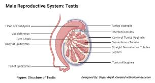
Male Reproductive Anatomy - Testes
primitive gonads → testes
seminiferous tubules: sperm produced, nourished by sertoli cells
interstitial cells of leydig: secrete testosterone and other male sex hormones (androgens)
testes located in scrotum, external pouch below penis (maintain 2 to 4 degrees celsius lower than body via muscle, ductus deferens)
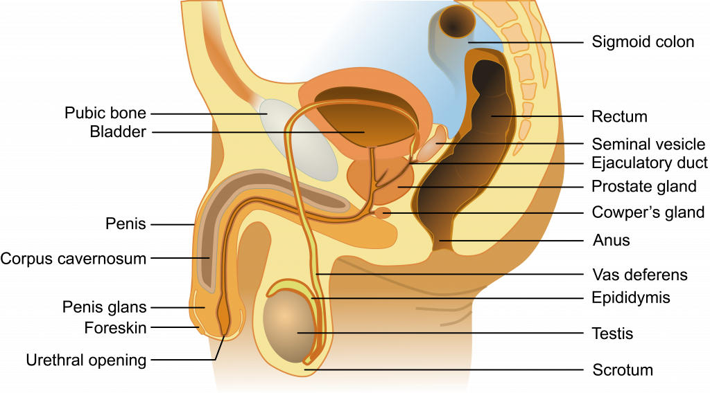
Male Reproductive Anatomy - Pathway of Sperm
sperm → epididymis until ejaculation → vas deferens → ejaculatory duct → urethra
Male Reproductive Anatomy - Semen
semen: sperm mixed with seminal fluid (produced by seminal vesicles, prostate gland and bulbourethral gland)
seminal vesicles: fructose to nourish sperm
seminal vesicle and prostate gland: fluid mildly alkaline properties so sperm survive in acidity of female reproductive tract
bulbourethral (cowper’s glands): clear viscous fluid that cleans out remnants of urine and lubricates urethra during sexual arousal
Spermatogenesis
formation of haploid sperm via meiosis
spermatogonia: diploid stem cells
diploid primary spermatocytes: after replicating genetic material (S stage)
haploid secondary spermatocytes: after first meiotic division
spermatids: after second meiotic division
spermatozoa: mature spermatids
Structure of Sperm
midpiece: ATP from fructose, filled with mitorchondria, generate energy for swimming through female reproductive tract to reach ovum in fallopian tubes
Acrosome: covers sperm head, derived from Golgi apparatus and necessary to penetrate ovum
head: contain genetic material
flagella: motility
Female Reproductive Anatomy:
gonads → ovaries: produce estrogen and progesterone
thousands of follicles: multilayers sacs that contain, nourish and protect immature ova (eggs)
one egg per month ovulated into peritoneal sac: lines abdominal cavity
egg → fallopian tube/oviduct lined with cilia to push egg forward → uterus
sperm go from vaginal canal → cervix → uterus
vulva: external parts of female genital organs
Oogenesis
production of female gametes
primary oocytes (2n): arrested in prophase I
after menarche (first menstrual cycle):
complete meiosis I → secondary oocyte (ample cytoplasm to one cell and polar body pretty empty) → wait until fertilization for remainder of meiosis II to complete
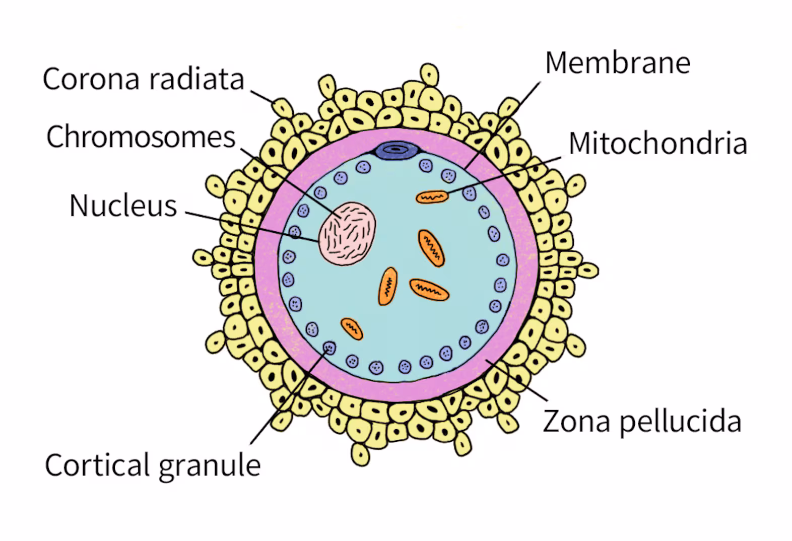
Oocyte Structure
Zona pellucida surround oocyte itself and acellular mixture to protect oocyte and contain compound necessary for sperm binding
corona radiata: lies outside zona pellucida and layer of cells that adheres to oocyte during ovulation
Sperm acrosomal enzymes → penetrates the two layers → trigger completion of meiosis II → forms another mature ovum and polar body
Ovum: contributes nearly everything to zygote (half of DNA, all of cytoplasm, organelles, mitochondria and RNA for early cellular processes)
Completion of meiosis II: haploid pronuclei of sperm and ovum join → diploid zygote
Male Sexual Development
Fetal period:
Y chromosome → production of androgens
Puberty:
Testosterone by testes rises a lot during puberty
sperm production:
FSH stimulate Sertoli cell and trigger sperm maturation
LH causes interstitial cells to produce testosterone
development of secondary sexual characteristics: facial and axillary hair, deepening of voice, increased muscle and bone mass
negative feedback to anterior pituitary gland: synthesize and release follicle-stimulating hormone (FSH) and luteinizing hormone (LH) and hypothalamus: restrict production of gonadotropin-releasing hormone (GnRH)
Female Sexual Development - Estrogen
secreted in response to FSH
development and maintenance of female reproductive system and female secondary sexual characteristics: breast growth, widening of hips, changes in fat distribution
in embryo: estrogen stimulate development of reproductive tract
in adult: estrogen → thickening of lining of uterus (endometrium) each month to prepare for implantation of zygote
Female Sexual Development - Progesterone
secreted by Corpus luteum: remains of ovarian follicle following ovulation in response to LH
development and maintenance of endometrium but not initial thickening of endometrium
by end of first trimester, progesterone is supplied by placenta, and corpus luteum atrophies and ceases to function
Menstrual cycle - Follicular Phase
begin when menstrual flow (shed uterine lining) begins
GnRH secretion from hypothalamus increased and decreased estrogen and progesterone toward end of cycle
increased secretion of FSH and LH → develop several ovarian follicle → produce estrogen → negative feedback effects → stimulate regrowth of endometrial lining, stimulating vascularization and glandurization of decidua
Menstrual cycle - Ovulation
estrogen later parodoxically result in positive feedback effects of GnRH, LH, and FSH → ovulation, release of ovum from ovary into abdominal/peritoneal cavity
Menstrual cycle - Luteal Phase
ruptured follicle forms corpus luteum → secretes progresterone → progesterone levels rise and estrogen levels remain high to maintain uterine lining for implantation → progesterone cause negative feedback on GnRH, FSH, and LH → prevent ovulatinon of multiple eggs
Menstrual cycle - Menstruation
no implantation → corpus luteum loses stimulation from LG, progesterone levels decline, uterine lining sloughed off → loss of estrogen and progesterone removes block on GnRH so next cycle can begin
Menstrual cycle - Pregnancy
fertilization → zygote become blastocyst that will implant on uterine lining and secrete human chorionic gonadotropin (hCG) analog of LH → maintain corpus luteum → critical during first trimester development to keep uterine lining in place → second trimester placenta make own progesterone and estrogen by itself so no more corpus luteum → negative feedback on GnRH secretion
Menstrual cycle - Menopause
ovaries less sensitive to FSH and LH → ovarian atrophy
estrogen and progesterone levels drop → endometrium also atrophies and menstruation stops
blood levels of FSH and LH rise because no more negative feedback
symptoms: flushing, hot flashes, bloating and headaches between 45-55
Fertilization
Secondary oocyte ovulated from follicle on day 14 of menstrual cycle in ampulla (widest part of fallopian tube)
Acrosomal apparatus (tube-like structure) after acrosomal reaction → cortical reaction (release of calcium ions) → fertilization membrane (now depolarized and impenetrable membrane)
Twins
Dizygotic (fraternal) twins: form from fertilization of two different eggs released during one ovulatory cycle by two different sperm
Monozygotic (identical) twins: single zygote splits into two
incomplete division → conjoined twins
monochorionic/monoamniotic: share same amnion and chorion
monochorionic/diamniotic: own amnion but share same chorion
dichorionic/diamniotic twin: have their own amnions and chorions
Cleavage
move to uterus for implantation → rapid mitotic cell divisions called cleavage → first cleavage makes embryo
Indeterminate cleavage: cells that can still develop into complete organisms
Determinate cleavage: result in cells with fates taht are already determined → committed to differentiating into a certain type of cell
Blastulation
Zygote → morula (solid mass of cells) → blastula
blastulation: forms blastula/blastocyst
fluid-filled inner cavity known as blastocoel
inner cell mass (protrudes into blastocoel): gives rise to organism
trophoblast: hollow surrounding of blastocoel, giving rise to chorion (extraembroynic membrane that develops into placenta) and placenta
Implantation
Blastula moves → uterus → burrow into endometrium
chorion → chorionic villi which are finger-like projections that penetrate endometrium → maternal-fetal gas exchange
umbilical cord - connects embryo to placenta via two arteries and one vein
yolk sac: supports embryo until placenta is functional, site of early blood cell development
allantois: extraembryonic membrane involved in early fluid exchange between embryo and yolk sac
amnion: surrounds allantois, thin, tough membrane filled with amniotic fluid that shock absorber during pregnancy
Gastrulation
generation of three distinct cell layers: endoderm, ectoderm, mesoderm
archenteron: membrane invagination into blastocoel → gut
blastopore: opening of archenteron → anus for deuterostomes, mouth for protosomes
Differentiation of cells
via selective transcription of genome (some genes transcribed)
induction: ability of one group of cells to influence fate of nearby cells, mediated by inducers (chemical substances) which diffuse from organizing cells to responsive cells
Neurulation
development of nervous system from ectoderm
notochord: rod of mesodermal cells, forms along long axis of organism like primitive spine (intervertebral discs)
overlying ectodermal cells slide inward to form neural folds → surround neural groove → grow toward each other until fuse into neural tube → CNS
neural crest cells: on tip of each neural fold → migrate outward to form PNS and other cell types in other tissues
ectoderm then covers all of this
Problems in Early Environment
Teratogens: substances that interfere with embryo development, causing defects or even death
maternal health also affects embryo development
Cell Specification/Determination
Specification: initial stage of cell specialization, where cell is reversibly designated as specific cell type
Determination: commitment of cell to particular function in the future
Due to mRNA and protein in parent cell being split differently to daughter cells
Morphogens: secretion of specific molecules form nearby cells that cause them to follow specific developmental pathway