Cont. Chest Anat, HERNIAS
 Call Kai
 Learn
Learn Practice Test
 Spaced Repetition
 Match
Match Flashcards
 Knowt Play
1/78

There's no tags or description
Looks like no tags are added yet.
Name | Mastery | Learn | Test | Matching | Spaced | Call with Kai | Chat |
|---|
No analytics yet
Send a link to your students to track their progress
79 Terms
Diastasis Recti
Increased distance between the rectus abdominus muscles caused by weakness in anterior abdominal wall
Linea alba is normally a narrow fibrous band in the midline abdomen that separates the two rectus abdominus muscles
Muscle weakness and thinning/stretching of the linea alba
This distance between the 2 muscles can increase with pregnancy, obesity and in newborns
It can be differentiated from ventral hernia b/c there is no fascial defect
Diastasis Recti presents as
A midline bulge that increases with increased abdominal pressure
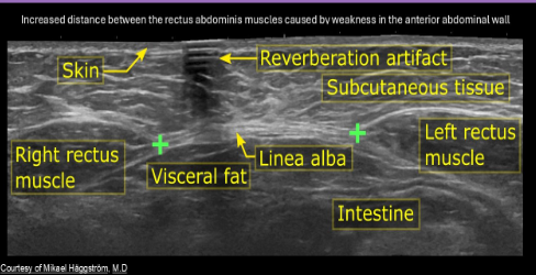
Diastasis Recti
Abdominal wall lipoma
Fatty tumor localized within the abdominal wall
Usually hypoechoic to isoechoic with surrounding tissues
Soft, compressible, movable
Usually removed for cosmetic purposes
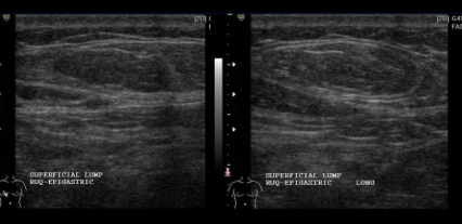
Abdominal wall lipoma
Other abdominal wall masses
Desmoid tumors
Endometriomas
Sarcoma
Mets
Desmoid tumors
Benign neoplasm
Usually found in the abdominal wall but can be intra- or extra-abdominal
Most commonly seen in women age 25-40yrs
Associated with pregnancy and previous abdominal surgery
Homogenous, isoechoic to hypoechoic mass
Endometriomas
Associated with cesarean section or laparotomies
Commonly mistaken for incisional hernia
Variable sonographic appearance
Important to determine if the mass has invaded the abdominal fascia
Hernia sac -
Has a neck, body, fundus and contents
Neck of hernia is in the _____
Orifice
A narrow, rigid or angled neck increases risk for:
Strangulation
Most hernias contain
Intraperitoneal or preperitoneal fat, but bowel can also protrude into sac
Pts with one hernia, usually have ________ and should be evaluated for other types of ipsilateral and contralateral groin
Multiple
Incarcerated hernias
Hernias that are nonreducible
Obstructed hernias
Contain incarcerated bowel loops that have become mechanically obstructed
Strangulated hernias
Contain incarcerated contents with compromised vascularity
Most significant complication of a hernia
Strangulation
Signs of hernia strangulation
Hyperechoic fat, thickened hernia sac, fluid in the sac, thickened bowel loops in the sac
Use color
Most common surgical procedures in the US
Herniorrhaphy
Hernia repair is usually performed using:
Mesh material
Abdominal wall or Ventral hernia are caused by
Chronically increased intraabdominal pressure or connective tissue weakness
Allows for protrusion of intestines → can cause intestine to continuously enter in and out
Linea abla hernias
Anterior abdominal wall hernias that penetrate through lina alba
Superior to umbilicus = epigastric
Inferior to umbilicus = hypogastric
Found fecentieters of the umbilicus
Linea alba
Thick layer of aponeurosis that separates the rectus abdominus muscles
Most common type of ventral hernia
Umbilical
Most common reason for missing an epigastric linea alba hernia
Examination for abdominal pain was performed with a low frequency transducer
Epigastric hernias usually have…
A narrow neck, are NOT reducible with increased risk for strangulation
Hypogastric hernias are usually found
Within a few centimeters of the umbilicus and have narrow necks, NOT reducible and are at increased risk for strangulation
Spigelian hernia
Nearly all these occur inferior to arcuate line, where spigelian fascia is penetrated by the inferior epigastric vessels
Associated with conditions that increase intraabdominal pressure
Spigelian hernia USA
Complex mass within the anterolateral aspect of the abdominal wall, could contain fluid or bowel loops or fluid
Mushroom or anvil shaped
Groin
Region of the ilioinguinal crease at the junction of the abdomen and the thigh and the adjacent areas immediately above and below
Groin hernia maneuvers
Quiet breathing - demonstrates bowel peristalsis
Valsalva maneuver (pt in supine position, hernia should increase in size with this)
Compression maneuver (assesses reducibility and tenderness in pts)
Upright position (demonstrates pooling of related peritoneal fluid)
Groin hernia with a broad fundus and narrow neck =
Usually NONreducible
A hernia with a broad neck and more narrow fundus =
Usually reducible
Inferior epigastric artery
Key landmark in distinguishing among the direct, indirect and spigelian hernias
The artery originates at the external iliac artery
Courses superiomedially, across the spigelian fascia and semilunar line
Distally it courses along the mid-posterior aspect of the rectus abdominus
Direct inguinal hernia =
Herniated structures medial to inferior epigastric artery
Indirect inguinal hernia =
Herniated structures lateral to the inferior epigastric artery
Most common type of hernia overall and most commonly in males
Indirect Inguinal hernia
Inguinal ligament
Extends from the anterior superior iliac spine to the pubic tubercle
Inguinal canal
Runs between the external oblique and the transversalis fascia, superficial to the inguinal ligament
Inguinal hernia
Weakness in pelvic floor muscles allows pelvic contents to drop into scrotal area
Hernia sac - composed of peritoneum
Indirect inguinal hernia
Due to migration of testis, spermatic cord, or round ligament penetration of the pelvis floor
Neck is SUPERIOR to IEA origin
Fundus is oriented horizontally and expands inferior and medial, passing superficial to the IEA origin
Anterior lateral to the spermatic cord
Indirect inguinal hernia is associated with
Abnormal movement of fat, bowel, or both through the deep inguinal ring and inguinal canal
Sliding inguinal hernia -
Wide neck, usually reproducible
Non-sliding hernia -
Narrow neck, usually Nonreproducible
Direct inguinal hernia
Intra-abdominal structures move anteriorly through the aponeurosis at the Hesselbach triangle
Neck lies inferior and medial to the IEA origin
Inguinal hernia sac lies posterior and medial to the spermatic cord in males, round ligament in females
Aponeuroses
Layers of fibrous tissue that connect sheet-like muscles needing a wide area of attachment
Hesselbach triangle
Bordered inferiorly by the inguinal ligament, medially by the lateral aspect of the rectus abdominus muscle, and superolaterally by the IEA
Amyand hernia
Rare inguinal hernia that contains appendix
Incisional hernia
Delayed complication of abdominal surgery
Occurs in first few months of surgery
More common with vertical incisions than with transverse incisions
Elderly, obese and malnourished pts have increased risk of an incisional hernia
Incisional hernia is associated with
Transverse rectus abdominus myocutaneous (TRAM) flap breast reconstruction surgery
Femoral hernia
Occur in areas where vessels penetrate the abdominal wall
Located below the inguinal ligament
Abnormal intraabdominal contents move inferiorly through the femoral canal
MORE COMMON IN WOMEN ON RIGHT
Hernia sac usually lies medial to the common femoral vein
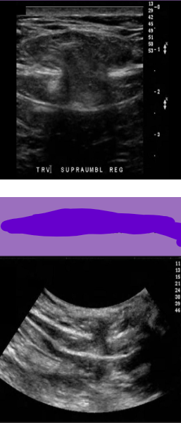
Abdominal wall hernia
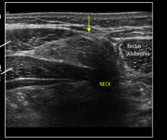
Spigelian hernia
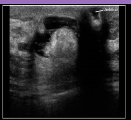
Umbilical hernia
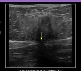
Epigastric hernia
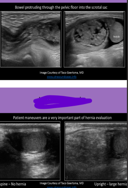
Inguinal hernia
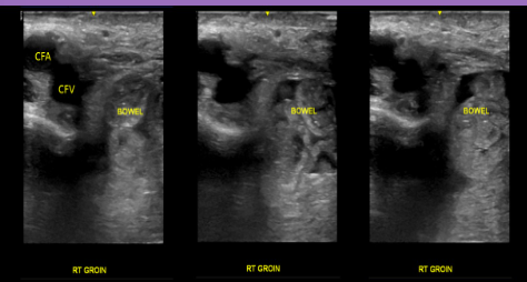
Femoral hernia
M-mode is commonly used to eval a pt for
Pneumothorax
What causes the diaphragm to appear highly echogenic on US evaluation?
Specular reflection
In cases of pneumothorax, what sign is identified on US evaluation?
Barcode sign
Transudate pleural effusion is related to:
CHF
Exudate pleural effusion is related to
Malignancy
Signs of pleural effusion
Diaphragm sign, displaced crus sign, bare area sign
Lactate dehydrogenase concentration can be used to differentiate:
Transudate from exudate effusion
US is used to eval the __________ for hernia formation
Anterior parietal peritoneum
The ___________ is called the foramen of Winslow and the __________ is called the morsion pouch
Right subhepatic space; Hepatorenal space
Liver disease causes a reduction in ________ levels which are associated with ascites formation
Albumin
The retroperitoneum is located between the:
Parietal peritoneum and the transversalis fascia
An enlarged, rounded lymph node with loss of hilar definition = _________
An enlarged oval lymph node with normal hilar characteristics = ___________
Malignancy; Infection
Which of the following can cause hydro, pedal edema, and scrotal swelling in a male pt?
Ormond disease
Which of the following are required when evaluating a pt with a suspected anterior wall hernia?
Ask them to perform the Valsalva
Scan them standing up
Obtain comparison views of unaffected side
All of the above
All of the above
Injury to the _________ is the most common cause of hematoma formation in the anterior abdominal wall
Inferior epigastric artery
Stretching of the linea alba during pregnancy results in what chronic disease
Diastasis recti
How can you differentiate diastasis recti from a ventral hernia?
Presence of a fascial defect
A narrow, rigid or angled hernia neck increases the risk of:
Strangulation
An incarcerated hernia cannot be:
Reduced
When reporting findings from an US eval for a hernia, what should be included?
Dynamic maneuvers used in exam, hernia contents, reducibility of hernia
Key landmark in distinguishing among direct and indirect Spigelian hernias?
Inferior epigastric artery
If a hernia does not increase in size with Valsalva strain, ______________ should be suspected
Strangulation, incarceration, or ischemia