Final Prep
 Call Kai
 Learn
Learn Practice Test
 Spaced Repetition
 Match
Match Flashcards
 Knowt Play
1/39

There's no tags or description
Looks like no tags are added yet.
Name | Mastery | Learn | Test | Matching | Spaced | Call with Kai | Chat |
|---|
No analytics yet
Send a link to your students to track their progress
40 Terms
Expected eye alignment values
Distance: ortho to 2^ xp, ortho vertically ± 0.25
Near: ortho to 6^ xp, ortho vertically ± 0.25
Advanced cover test results
Comitant= magnitude same in all 9 gazes (< 8^ difference) vs incomitant= not the same in all 9 gazes (>/= 8^ difference between gazes)
Mechanically restrictive incomitant deviations
Brown syndrome (SO catches), TED, Duane syndrome (lateral rectus innervated by CNIII/globe retraction), ocular myasthenia (ptosis)
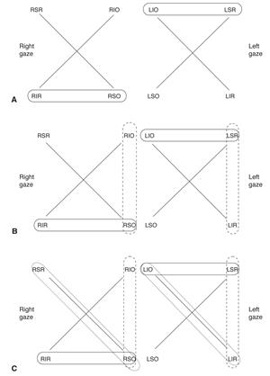
Park’s 3 step test
CT in primary, right/left and tilted right/left gaze, worse gaze= circled, find muscle with all 3
Duane’s Classifications
Maddox rod
Subjective, dissociative eye alignment test, doesn’t distinguish tropia vs phoria, line above dot= use BU prism to correct, L Hyper + visa versa, orient lines opposite intended image, NOT USED LATERALLY B/C NON-ACCOMMODATIVE TARGET, can be performed without correction if induced prism is suspected
W4D
Used with tropias/reduced stereo, will not pick up phoria b/c eyes never truly dissociated, only present in light= shallow suppression, present in dark= intense/deep suppression
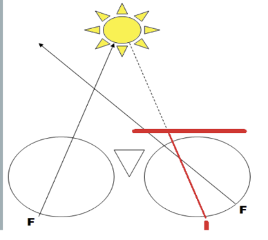
Lateral Maddox Rod: line to the right of light
RRUE: red right uncrossed eso, uncrossed diplopia, use BO prism (light on nasal retina of OD< perceived from temporal VF)
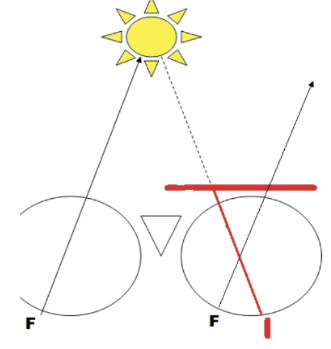
Lateral Maddox Rod: line to the left of light
RLCX: red left crossed exo, crossed diplopia, use BI prism to correct (light on temporal retina of OD, perceived from nasal VF)
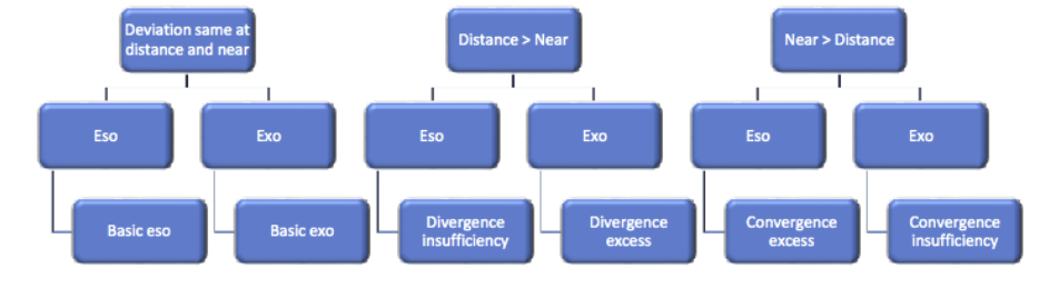
W4D: 5 dots, red to the left
Crossed diplopia, exotropia, temporal retina projects to nasal VF
W4D: 5 dots, red to the right
Uncrossed diplopia, esotropia, nasal retina projects to temporal VF
Sphygomomanometry
Indirect measure of blood pressure
Ascultatory gap
Temporary absence of sounds between phase 2 and 3 that causes underestimation of systolic pressure
Hypertensive retinopathy
Most common physical manifestation of hypertension, mild-severe, could be predictive of cardiovascular disease/stroke, disk edema common, emergent if systemic/ocular findings present
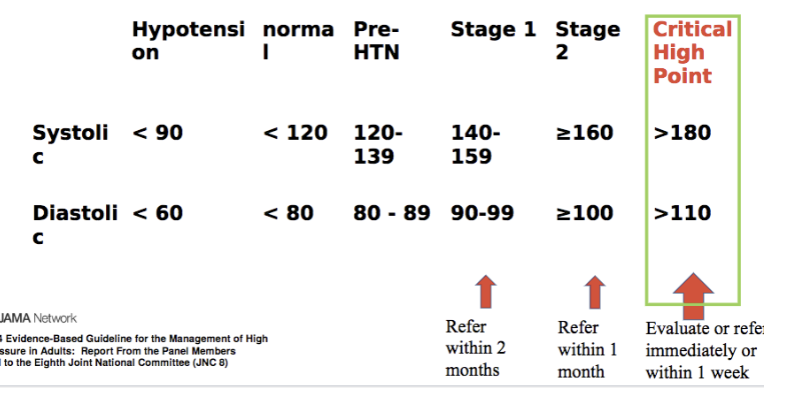
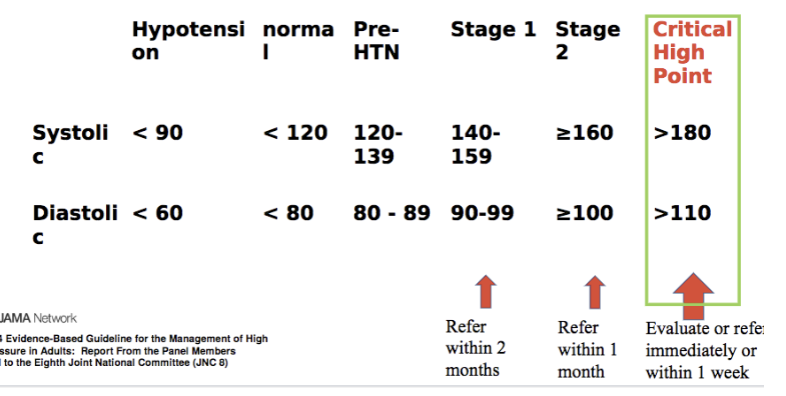
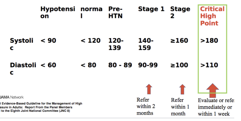
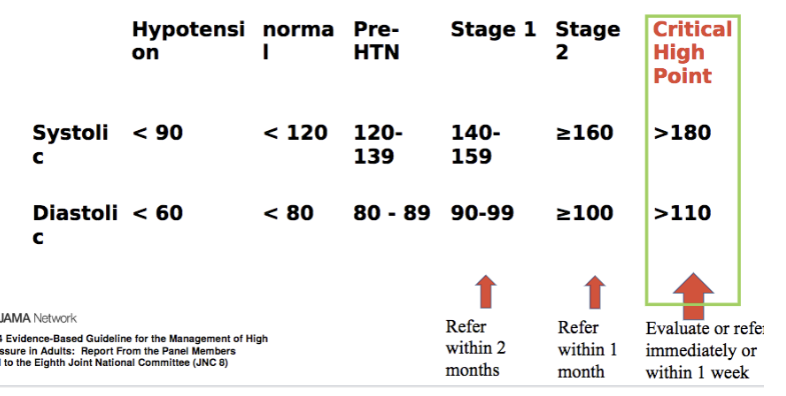
Hypotension numbers
Systolic < 90, diastolic < 60
Normal BP numbers
Systolic <120, Diastolic <80
Pre-HTN numbers
Systolic= 120-139, diastolic= 80-89
Stage 1 HTN numbers
Systolic= 140-159, diastolic= 90-99, refer within 2 months
Stage 2 HTN numbers
Systolic </= 160, diastolic </= 100, refer within 1 month
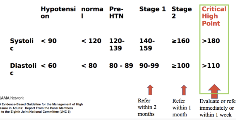
Critical high point HTN numbers
Systolic >180, Diastolic > 110, evaluate or refer immediately-within 1 week
BP considerations
Topical phenylephrine can alter BP (especially 10%), use alternatives/nasolacrimal occlusion to reduce systemic absorption
Korotkoff Sounds
Phase 1= systolic, appearance of clear tapping sounds, Phase 5= diastolic 2 (phase 4= diastolic 1/muffling), complete disappearance of sounds
False BP causes
High= cuff too small, arm too low, ascultatory gap measured for diastolic, Low= cuff too large, arm too high, deflating too rapidly, ascultatory gap measured for systolic, automated underestimate
Pt Education BP
Biggest impact on hypertension: weight loss/BMI under 24!!, DASH eating plan, dietary sodium reduction, 30 min/day physical activity, moderate alcohol consumption (1-2/day)
Causes of scissoring
Cataract or keratoconus, increase light slightly or focus on central reflex
Retinoscopy in Phoropter Set Up/Procedure
Set PD, confirm vertex distance, ensure sphere and cyl set to 0, dark room lighting, 20/400 RG E, both eyes unoccluded, confirm right eye cannot see the E but left eye can/check for against in both eyes and fog if needed, find principal meridians, neutralize most positive first, neutralize 2nd meridian with cylinder power, remove working distance, check monocular VAs
Posterior focal line vs anterior focal line
Sphere power, most plus meridian/axis, least plus meridian, cyl= difference between 2
Expected VA
1 VA line lower for each 0.25 D of spherical equivalent +1 line for oblique astigmatism
Impact of ±/cyl lenses
Plus lenses= shift interval of sturm anteriorly/towards the cornea, minus lenses= shift interval of sturm posteriorly/away from the cornea, minus cylinder lenses= impact only ONE focal line (focal line parallel to lens axis, orientation of axis marking on lens= focal line being altered)
Just noticeable difference
Denominator of VA/100, show patient choices that total JND (ex: pt is 20/100, JND= 1.00, show +0.50 and -0.50)
Monocular subjective
1st best sphere (3-4 above ret VA and walk down no lower than 2020, trying to place focal point/COLC on retina), JCC (power search/acceptance/axis refinement/power refinement/axis refinement again if >0.75 DC change, end w power, make sure to compensate sphere, goal= collapse interval of sturm), 2nd best sphere (no lower than 2020)
Focal lines during JCC
Flips will move AFL in one direction, PFL in one direction, and change size (NOT POSITION) of COLC. -0.25 moves focal lines of same orientation POSTERIORLY, +0.25 moves focal lines of same orientation ANTERIORLY
Power acceptance
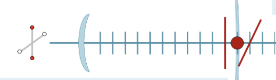
For every 2 white dots the patient rejects= add -0.25 D sphere compensation to place COLC back on retina
Power Search
Checking to see if cyl power was missed, check at 180, 045, 090, and 135, add -0.25 DC, then red/white dot preferences, don’t add more cyl here, endpoint= power rejected at every meridian or red/same
Axis Refinement
Chase the red, initially 15 degree rotations (exceptions= high cyl), repeat until reversal, bracket to 1 degree >/= 1.50 DC, small cyl= find middle of range
Power refinement
For every 2 white dots patient rejects add -0.25D sphere power to place COLC back on retina, if power refinement changes cyl by more than 0.75 DC you must return to axis refinement
Balance
Purpose= equalize stimulus to accommodate (not to balance VA/response), methods: bi-ocular: dissociated with prism (dissociated blur, red-green/duochrome, goodwin), binocular: eyes fused (vectographic/polarized chart, turville infinity balance), alternate occlusion: eyes dissociated
Dissociated Blur Balance
Monocular VA must be equal (</=3 letter difference), 3 BD OD (top chart), 3 BU OS (bottom chart), add 0.75 OU, 3-4 lines above best VA with M1 (lateral displacement of the chart= exo or exophoria)
Add +0.25 to better eye, if top or bottom clearer more than 2 times in a row= reevaluate M1
endpoint= both charts equally blurry, obtain reversal
Red/Green Balance (Duochrome Balance)
Based on chromatic aberration (yellow on COLC, red COLC behind the retina, green COLC= in front (red focuses in front of green by 0.5 D), unequal acuity is acceptable
3 BD OD (top chart), 3 BU OS (bottom chart), 0.75 D fog, 3-4 lines above worst M1 VA
look at upper chart, fog until red clearer, then add minus until equal, obtain reversal, if none= most similar blur
after OS, compare upper chart red and green to lower chart red and green
Goodwin Balance
Monocular VA must be equal (within 3 letters)
3 BD OD (top chart), 3 BU OS (bottom chart)
Fog 0.25 D, 1 line above M1 VA
add 0.25 to better eye (0,+), same chart better= remove +0.25 better, add -0.25 to worse eye (-,0), same chart better add +0.25 to better (-,+), same chart better remove -0.25 from worse and add a second +0.25 to better (0,++)