Radiation safety
 Call Kai
 Learn
Learn Practice Test
 Spaced Repetition
 Match
Match Flashcards
 Knowt Play
1/22

There's no tags or description
Looks like no tags are added yet.
Name | Mastery | Learn | Test | Matching | Spaced | Call with Kai | Chat |
|---|
No analytics yet
Send a link to your students to track their progress
23 Terms

Discovery of X rays
1895- Wilhelm Roentgen
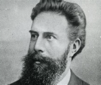
becquerel
1896- first biological effects of x rays/gamma rays
first safety regulation
1921

types of radiation
electromagnetic radiation
particulate radiation
electromagnetic radiation
energy having no mass
ionizing and non-ionizing
made of photons, acts like a wave

particulate radiation
has mass and energy
disintegration of an unstable atom
all types are ionizing

non-ionizing
Atoms or molecules that remain electrically neutral because their electrons stay bound in place.
ionizing
removes electrons from atoms, leads to biological damage

short wavelength
energy is high
frequency is high (short)
long wavelength
energy is low, frequency is low (long)
radiation exposure
amount of ionizing radiation you encounter in the air
Measured as Coulomb (C)/kg air
Traditional unit – Roentgen (R)
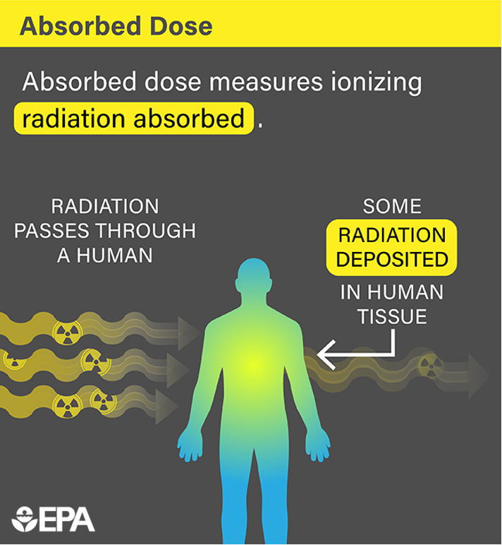
absorbed dose
how much ionizing radiation is absorbed into the object
equivalent dose
measure of how the absorbed dose differs with different types of ionizing radiation on tissue.
regulations of radiation exposure
-Required when radiation workers are likely to receive ≥ 10% of the annual dose limit (5 rem) in one year.
MPD
ALARA
dosimeter
prodromal stage
occurs within hours of 1st dose
nausea, vomiting, diarrhea, fatigue
latent stage
last hours to weeks
no visible symptoms
manifest illness
last from hours to months
acute radiation syndrome
•Large and penetrating dose (≥ 1 Gy)
•External source
•Dose received by entire body
•Dose delivered in a short time (within minutes)
prodromal stage
latent stage
manifest illness
recovery or death
MPD
maximum permisible dose. -Dose equivalent that a person is allowed in a specific period of time resulting in minimal side effects.
ALARA
as low as reasonably achievable
Always minimize radiation dose when possible

safety fundamentals
reduce time
distance
shielding
collimation
minimize restraint and personel
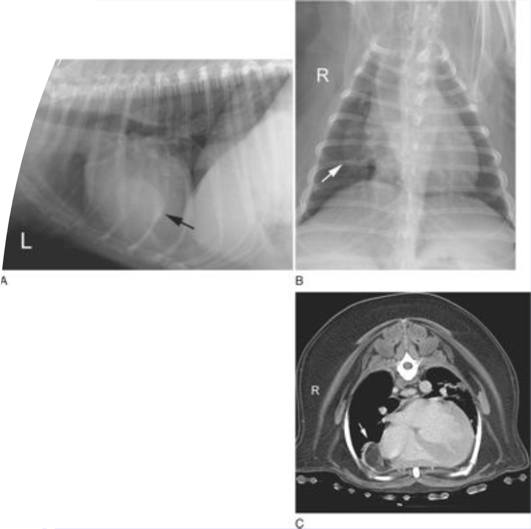
Computed tomography
●Works like traditional x-rays, only 3D
●X-rays strike the patient from multiple angles, creating much larger amounts of radiation
●Each x-ray reaches a detector and gets converted to a Hounsfield Unit (HU), which correlates to different tissue types
MRI
●No radiation exposure
●Combination of magnetic field and application of radiofrequency pulses
●Danger lies in strength of magnetic field