Vision (ch2-2.2 retina)
1/31
There's no tags or description
Looks like no tags are added yet.
Name | Mastery | Learn | Test | Matching | Spaced | Call with Kai |
|---|
No analytics yet
Send a link to your students to track their progress
32 Terms
light
a wave; a stream of photons, tiny particles, that each consist of one quantum of energy
Wavelength
distance between the peaks of an energy wave
wavelengths between 400 nm and 700 nm are visible (nm=10-9 m)
Visible spectrum
wavelength determines colour (hue) perceived (ROYGBIV)
intensity
wave property: height of the peaks of an energy wave (amplitude)
particle property: number of quanta emitted by a light source or reflected off a surface determines brightness perceived → determines brightness perceived
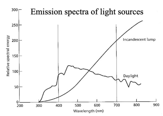
Different light sources emit energy at different wavelengths
Sunlight has more short wavelengths (blue); light bulbs more longer wavelengths (yellow)
once emitted, light can be
absorbed: taken up and not transmitted at all
diffracted (scattered): dispersed in an irregular fashion
by dust or water particles in air
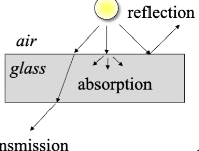
At the interface between 2 media(e.g. Air, water, glass), light may be
transmitted: conveyed from one place to another, usually with refraction (ex. Light entering water)
absorbed: Light energy is taken and converted to heat (ex. Concrete or black shirt absorb light → heated)
reflected: redirected back toward its origin (ex. Mirror reflection)
Color of solid vs translucent object and wavelengths
Solid objects colored cuz of wavelengths reflected off object to eye (light they reflect)
Translucent object colored cuz of wavelengths transmitted through object to eye
The more wavelength reflect, the more you will perceive that color
Blue/violet shortest wavelength
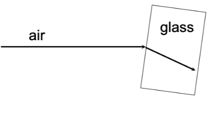
refraction
change in direction (bending) of light ray passing from one transmitting medium into another. Refraction depends on wavelength
shorter WL refract more: Red (long wavelengths) bend (refracted) a little, purple (short wavelengths) bend the most
Anatomy of the human eyeball
cornea - very sensitive to touch, transparent (transmit), tear films, first place light arrives
pupil - hole in iris; light entry
iris - muscular structure, outer colored layer; control size of pupil and amount of light that reaches retina (pupillary light reflex) inner layer of blood vessels
Lens - no blood supply, transparent, light pass after pupil in iris
ciliary muscle - controls shape of lens
aqueous humor - fluid from blood, in anterior chamber; continually replenished, feed and take care of cornea and lens
vitreous humor - fluid in posterior chamber, 80% of internal volume of eye, not replenished; transparent, floaters (small bits of debris drift around)
retina - detects light, initiates neural messages (only half of the light that arrives cornea will reach retina)
sclera - protective white outer surface
choroid - blood vessels; nourishes retina
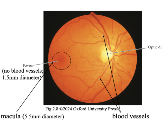
macula - yellow pigmented spot, under fovea, seen thru ophthalmoscope
optic disk - axons leave eye
fovea - where we have best vision
optic nerve (II) – axons of retinal neurons (emerge from the eyeball thru the optic disk and form the optic nerve → signals to brain

Parts of the eye for seeing
Optical apparatus: cornea, aqueous humor, lens, vitreous humor, pupil (iris)
Neural apparatus: retina
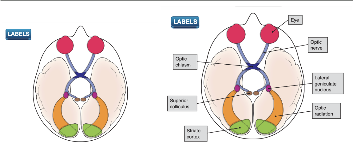
Visual system overview
Optic chiasm: Some of axons of neurons whose cell bodies in the nearest to the nose in optic nerve cross e/o at optic chiasm (ex. Left half of each eye sees the right side of the world)
Superior colliculus: Most axons in optic nerve connect to LGN, some synapse w here.
Eye movements, older than LGN evolutionarily
Lateral geniculate nucleus (LGN)
Primary relay station for info flowing from eye to brain. Axons leaving the retina via optic nerve, then synapse w new neurons
Contains 6 layers of neurons
Optic radiation
Axons from cells that synapse w optic nerve fibers in LGN → optic radiations to primary visual cortex (Contralateral: Radiations on left side of brain are carrying info from right side of the world)
Striate cortex (primary visual cortex): First place in cerebral cortex where visual info is received and processed (40% of cortex is directly involved in processing visual info)
Optical apparatus of the human eye
photoreceptors in retina absorb light
light emitted, transmitted or reflected to eye is transmitted through cornea, aqueous humor, lens and vitreous humor
Image focused on retina
sharpness of image depends on ability of…
cornea (2/3) + lens, aqueous & vitreous humors (1/3) to refract light
accommodation
the process by which the eye changes its focus by changing the shape of the lens
Done through contraction of ciliary muscle (attached to lens)
When focus: Zonules of zinn RELAXED, ciliary muscle contracted, accommodated lens
a fatter lens increases the optical power of the eye to bring images of near objects into focus
in a camera the lens is moved closer or farther from the film/sensors to change the focus

cataract
Opacity of the crystalline lens (harder to move lens, blurry image) of the eye can be congenital (since birth) or develop in later adulthood
Interferes with retinal image quality
Treated by surgical removal and replacement of lens
Pupil and reflexes
Size controlled by iris depending on light level
Direct light reflex: shining a light into one eye causes the pupil of that eye to constrict
Consensual light reflex: shining a light into one eye causes the pupil of the other eye to constrict
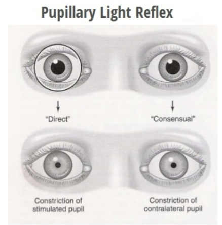
abnormal light reflexes in pupil can indicate..
…brain injury
eg. no direct or consensual reflex in right eye, normal reflexes in left eye
problem with oculomotor nerve (III) in right eye
eg. consensual but no direct reflex in right eye, direct but no consensual reflex in left eye
problem with optic nerve (II): right eye not sensitive to light (cuz II is sensory — no direct reflex on damaged eye, but still consentual, and III is motor — can’t send signals, don’t have reflexes)
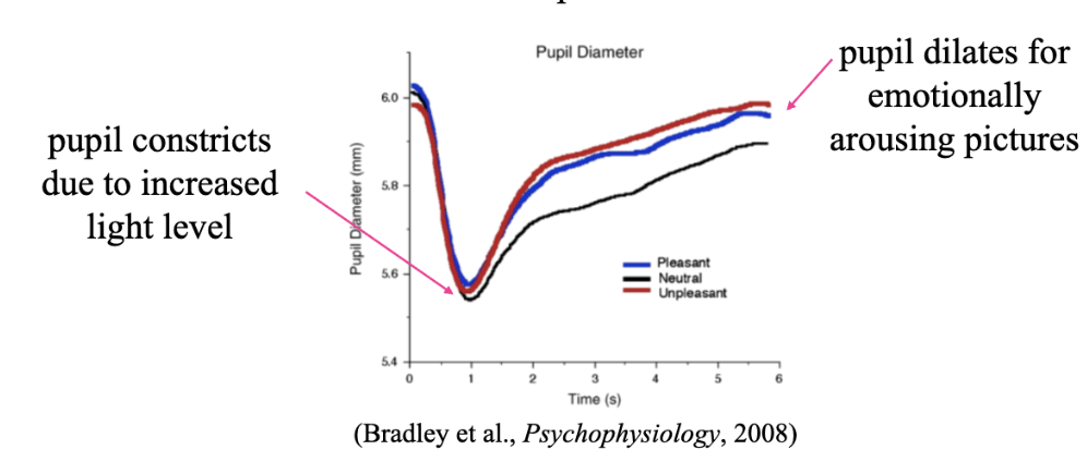
Pupil size
Ranges from 2 to 8 mm; changes with light level & emotional responses
Constricts due to increased light level
Dilates for emotionally arousing pictures
Responses were similar to skin conductance changes, which supports involvement of autonomic nervous system
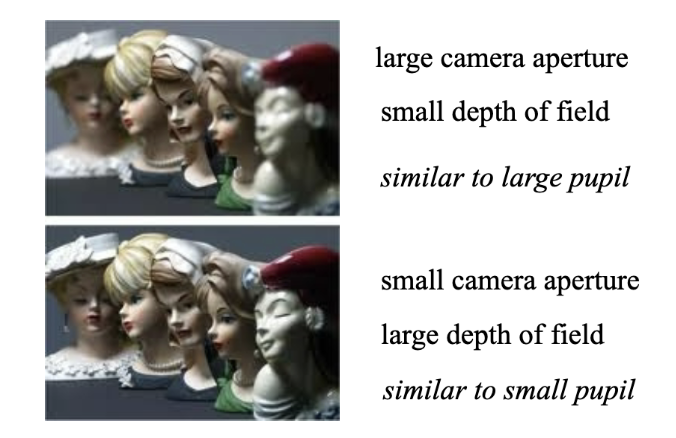
Small pupil provides
Greater depth of field: range of distances over which objects are in focus
Emmetropia
eyeball right length for optical elements; distant objects in focus on retina; focal length 16.8 mm, optical power 59.52 D
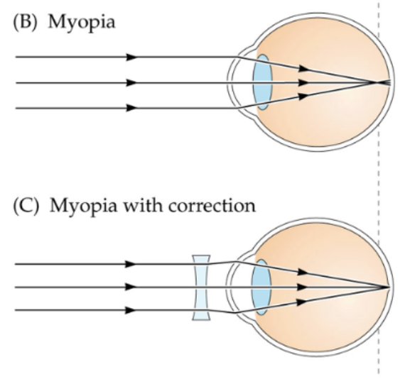
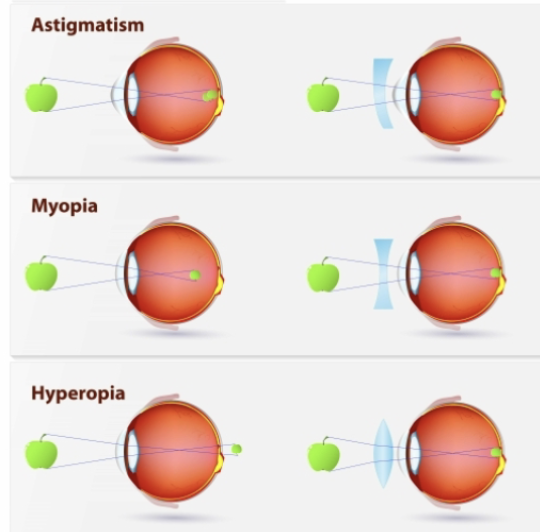
Myopia
Nearsightedness, eyeball too long or optics too strong, distant objects in focus in front of retina
tends to run in families (genetic)
incidence is increasing due to environmental factors
near-work (e.g. reading) especially under low illumination causes eyeball to continue to grow
Need concaved lens
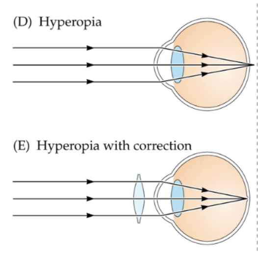
Hyperopia
farsightedness, eyeball too short or optics too weak; distant objects in focus behind retina
easier to see distant objects; accommodation brings them into focus on retina
can’t bring near objects into focus
need positive, convex lens adds optical power to bring both near & distant objects into focus
present in most newborns; many become emmetropic as eyeball grows
What protects against myopia onset in children?
Outdoor activity!
light-induced retinal dopamine may be protective mechanism
2 hrs per day of sunlight is recommended (not clear if this slows myopia progression)
Myopia progression may be slowed by
keeping reading distance of books, phones and tablets >20 cm
low-dose atropine eye drops
overnight orthokeratotic lenses
astigmatism
Unequal curving of eye’s refractive surfaces
visual distortion produced by nonspherical cornea
cause unknown; genetic & environmental factors
astigmatism causes one line to look blacker
Vertical axis stretched in their sight
Correcting vision (lens needed)
Correction with a cylindrical lens with 2 focal points: provide different optical power for 2 axes/orientations because the cornea has a non-spherical shape
needed for distance and near vision
rigid contact lenses for myopia or hyperopia may also correct astigmatism
young people with myopia or hyperopia can also wear their glasses to use accommodation to change the optical power of their eye
Presbyopia
Lose ability to accommodate on near objects
Due to aging
Due to stiffening of lens and loss of mobility in ciliary muscles
near point: closest distance at which objects can be brought into focus (~6.7 cm in young emmetropic eyes [optical power of 15 D])
accommodative amplitude (maximum increase in optical power) decreases with age
dashed line shows amplitude needed to focus at 40 cm (typical reading distance)
corrected with positive, convex lens for near sight (reading glasses)(so same lens as for hyperopia)
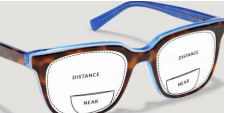
bifocals are glasses with
a near vision correction (for presbyopia) on the bottom to compensate for the loss of accommodation with normal aging
a distance vision correction in the rest of the lens to compensate for refractive errors due to length of eyeball (myopia or hyperopia)
if astigmatism is present, it needs to be corrected in the top and bottom part of the lenses
Neural apparatus of the human eye (retina)
fundus photograph obtained with ophthalmoscope
retinal blood vessels (“vascular tree” in textbook) lie in front of retina
Retinal blood vessels move with the eye and cast shadows on the retina
Usually you don’t see them cuz their images are stabilized
Stabilized image: A retinal image that stimulates the same photoreceptors without moving, and quickly fades from view
A jiggling light makes shadows visible briefly (you can see blood vessels!)
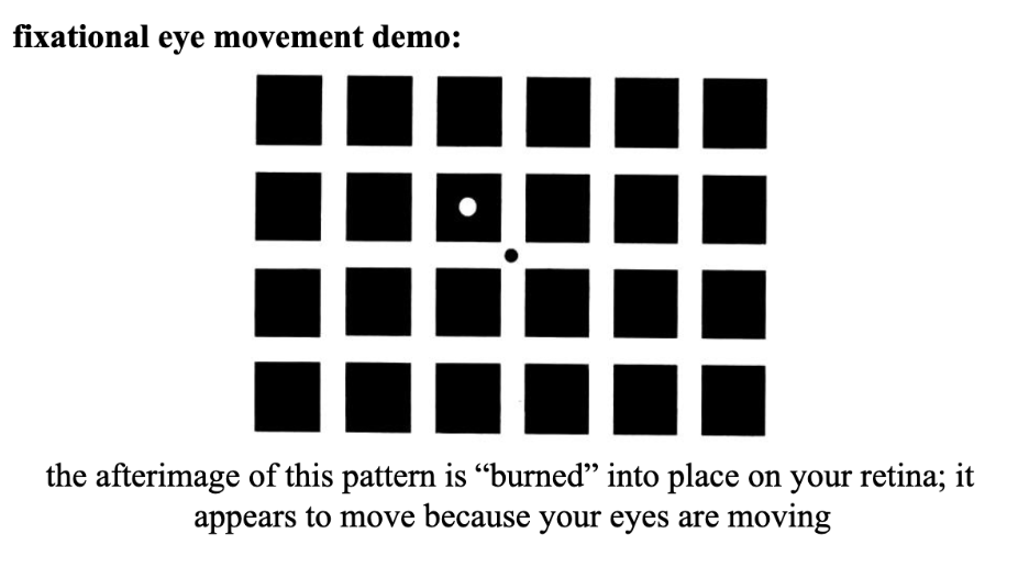
Fixational eye movements
Still see the squares when look away (esp high contrast) cuz your brain still memorize it
Images of objects outside the eye aren’t usually stabilized cuz of involuntary fixational eye movements