Neuroanatomy
 Call Kai
 Learn
Learn Practice Test
 Spaced Repetition
 Match
Match Flashcards
 Knowt Play
1/90

Earn XP
Description and Tags
Name | Mastery | Learn | Test | Matching | Spaced | Call with Kai | Chat |
|---|
No analytics yet
Send a link to your students to track their progress
91 Terms
neuron doctrine
brain composed of separate neurons and other independent structurally, metabolically, and functionally
info transmitted from cell to cell across synapses
synapses
tiny gaps
how many layers of the cerebral cortex
6
average adult brain
3 lbs
15 cm front to back
cortex 3mm thick
86 billion neurons
cortex
outer layer of the brain
number of neurons we are born with
300 billion
soma (cell body)
main body of a neuron
houses nucleus and major organelles
dendrites
composed of a main limb and extensions
input zone of information
axon hillock
generates action potential
“hill” between soma and axon
axon
singular extension
allows action potential/nerve impulse to travel to axon terminal
can split
bifurcating
splitting of an axon
myelin sheath
fatty substance
white
protects axon
insulates the impulse of an axon
not continuously covering an axon
quicker impulse transmission
exist on SOME, not all axons
multiple sclerosis
degenerated myelin
Symptoms
movement issues
pain
migranes
cognitive dysfunction
Source
reactivated Epstein-Barr virus (mono)
action potential
main electrical signal that a neuron makes
accumulation of inputs (excitatory & inhibitory)
axon terminal
houses neurotransmitters
ATP
fuel/energy for all cells
mitochondria
an organelle that is responsible for extracting energy nutrients
presynaptic membrane
releases neurotransmitter
postsynaptic membrane
binds neurotransmitters to transmit nerve signals
synaptic cleft
10-20 nanometers
synaptic vescicles
contain neurotransmitters
move down axon terminal, fuses with presynaptic membrane, and releases neurotransmitters
recycled
Nervous System Cells
sensory neurons
motor neurons
interneurons
sensory neurons
detects changes in external/internal environment
sends motor neuron signal
motor neuron
located within CNS and controls the contraction of a muscle or secretions from glands
interneuron
located within the nervous system
between sensory and motor neurons
Structural Classifications of Neurons
unipolar/monopolar neurons
bipolar neurons
multipolar neurons

unipolar/monopolar
1 axon attached to soma
axon divides (1 branch receives input , 1 branch transmits output)
found in skin / sensory
free nerve ending
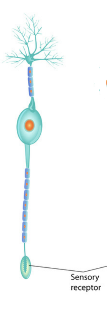
bipolar neuron
1 axon 1 dendrite attached to its soma
segregated sensory neuron (1 axon 1 dendrite)
contain cillia

multipolar neuron
1 axon many dendrites
Supporting Cells
glial cells
astrocytes
oligodendrocytes
glial cells
neuroglia
neural glue
supporting cells
3-4x more abundant than neurons
astrocyte
“star cell”
glial cell
intermediate between blood supply and neurons
cleaning mechanism, constant fluid and chemical (waste and recycling)
modulate neuron function
oligodendrocyte
glial cells
produce myelin
nodes of ranvier
gaps between myelin sheath
allow speed of action potential to speed up
schwann cell
in the PNS
wraps 1 segment of axon with myelin
microtubule
help with structure of an axon
neurofilament
made of actin
anterograde transport
movement of cellular cargo from soma to axon terminal
kinesin
motor protein that “walk” down microtubules and carry cellular cargo (vesicles, organelles, mRNA)
anterograde transport
retrograde transport
movement of cellular cargo from axon terminal to soma
dynein
large cytoskeletal motor proteins that “walk” down microtubules and carry cellular cargo
retrograde transport
neuraxis
imaginary line down through the center of the length of the nervous system
ipsilateral
same side of the body
contralateral
opposite side of the body
transverse plane
frontal/coronal
back from front
sagittal plane
left from right
horizontal plane
top to bottom
transverse plane
slices (like meat)
3 Layers of the Meninges
dura mater
arachnoid mater
pia mater
dura mater
outermost meninx
flexible, unstretchable
tough layer
arachnoid mater
middle meninx
subarachnoid space
between arachnoid mater and pia mater (under arachnoid mater)
space supported by trabeculae
filled with CSF
pia mater
innermost layer
soft layer
cerebrospinal fluid
produced by blood (choroid plexus filters blood)
liquid layer of protection
brain / spinal cord suspended
shock absorber
lasts ~3 hours, replaced every 6 hours
traumatic brain injury (TBI)
damage not always apparent, can accumulate
ventricles
“little bellies”
makes CSF
lateral ventricles, third ventricle, fourth ventricle
lateral ventricles
ventricles that surround the thalamus
third ventricle
ventricle located in the center of the diencephalon
cerebral aqueduct
circulates CSF from third to fourth ventricle
fourth ventricle
located between cerebellum and dorsal pons
rostral frontal lobe
forebrain
areas of executive function (decision making, planning, etc.)
caudal frontal lobe
motor functions
contains precentral gyrus
contains primary motor cortex
temporal lobe
lobe responsible for cognitive function
ex. object recognition, face recognition, language, etc.
primary auditory cortex
located in temporal lobe
central sulcus
transition from frontal to parietal lobe
parietal lobe
lobe responsible for attention and sensory integration
postcentral gyrus
located in the parietal lobe
houses the primary somatosensory cortex
primary somatosensory cortex
recieves information from the body
Diseases Pertaining to the Parietal Lobe
stroke causing visual/hemispatial neglect
Capgras Syndrome (imposter syndrome)
occipital lobe
lobe responsible for vision
occipital pertuberance
bump on skull pertaining to the occipital lobe
calcarine fissure
located in the occipital lobe
houses primary visual cortex
cerebellum
responsible for fine motor movement
creates connection factor when learning motor skills
lateral fissure
fissure under the frontal lobe and above the temporal lobe
grey matter
cell bodies “tucked” in sulci and gyri
white matter
myelin
insular cortex
older part of the brain
pineal gland
in the epithalamus
gland that regulates sleep-wake cycle
midbrain & brainstem
pons & medulla oblongata
pons
attentional awareness
arousal systems (sleep-wake)
neurotransmitter systems - modulate brain activity (dopamine, seratonin, etc.) made here
medulla oblongata
vegetative states (respiratory, cardiac function)
regulates taste information (gag reflex)
motor & sensory (head, neck, shoulders)
cingulate gyrus
limbic cortex
governs limbic system
limbic system
controls physiological states & emotions
hippocampus, amygdala, mamilary body, olfactory bulb, thalamus
hippocampus
spatial recognition; “internal map”
memory (short term —> long term, facts, places, autobiographical)
encodes new information
amygdala
“almond-shaped”
regulates emotions and stress response
sexual arousal
mamilary bodies
pair of nuclei that regulate memory and emotion (ex. help relate memory to smell and reflexes)
olfactory bulb
transfer smell information directly into the cingulate cortex, then to the brain
tightly associated to the amygdala and hippocampus for memory
thalamus
densely packed
relays information (sensory info in —> synapse —> part of the brain, except for smell)
microglia
smallest glial cells
act as phagocytes and protect brain from invading microorganisms
axoplasmic transport
active process of motor proteins carrying cellular cargo down the axon
2 types: anterograde, retrograde
fornix
connects hippocampus with mamilary bodies
above the thalamus