Muscle Physiology
1/78
There's no tags or description
Looks like no tags are added yet.
Name | Mastery | Learn | Test | Matching | Spaced |
|---|
No study sessions yet.
79 Terms
What are the three types of muscle?
skeletal
Cardiac
Smooth
What is skeletal muscle
Used for posture and locomotion. Its the muscle that enables our arms and legs to contract under conscious control
what is the cardiac muscle?
responsible for rhythmic contractions of the heart
what is smooth muscle?
causes involuntary contraction in blood vessels, gut, bronchi and the uterus → controlled by autonomic nervous system
What do muscles do?
they relax and contract
how do muscle contractions allow you to do all these things?
Muscle are attached to connective tissue called tendons → tendons stretched across joints → when muscle contracts, it pulls on the tendon which pulls on the joint causing the joint to change its position
What is skeletal muscle comprised of?
composed of bundles of long thin cells called muscle fibres which are bundled together to make a fascicle surrounded by connective tissue
Then all those bundles make up the muscle
What are muscle fibres made up of?
fibres - myofibrils to be more specific
What is skeletal muscle referred to as?
striated muscle due to the stripes the fibres create
what are the job of muscles
Generate large amounts of force very rapidly → Highly organized structure contributes to this
What are the cells that will eventually turn into muscle fibres called?”
myoblasts → little round cells
What happens over the course of development is that they fuse together to form in mature muscle, a multi nucleated muscle fibre
What are the muscle fibres packed with?
Proteins that allow them to do what they do (contract and relax)
Why is having a lot of nuclei good?
So that you can make more RNA and therefore make more proteins → multiple local spots for protein synthesis all down the length of the muscle fibre
What do skeletal muscle fibres consist of?
filled with long thin fibres called myofibrils
What is the smallest unit that allows muscle fibres to contract?
because the myofibrils contract because the sarcomeres contract
More myofibrils = stronger muscle fibre
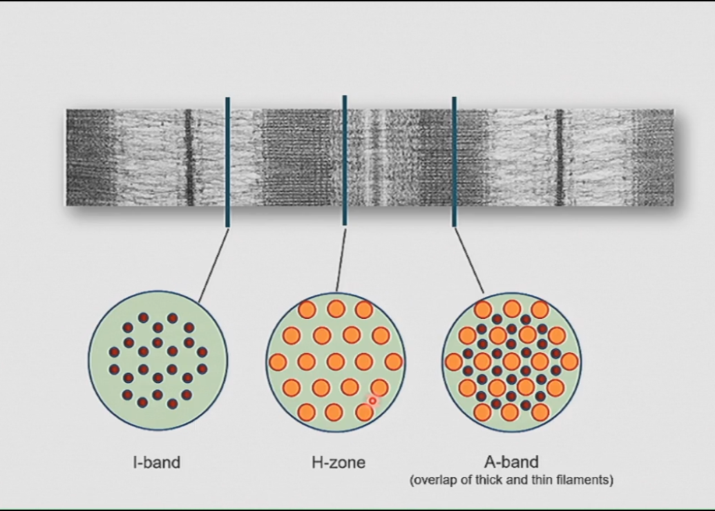
What are the light and dark bands on the myofibrils?
its why the whole muscle fibre has the stripes
Light band = I band
Dark bands = A band
Centre of I band = Z line
Centre of A band = H zone
Centre of H Zone = M line
What do the dark and light bands make up?
Sarcomeres → Z line to Z line
why does a myofibrils contract?
Due to the contraction of sarcomeres
What fibres are each band made of?
I band = thin filaments (actin)
A band = thin and thick filaments
H zone = thick filaments (myosin)
How many thin filaments surround a thick filament? Vise versa?
6 thin surround 1 thick filament
3 thick filaments surround 1 thin
what are the thin filaments made of?
actin - small globular solvable protein that bind to itself forming a helix = actin filament
what are the thick filaments made of?
myosin - a protein that actually is a long filament which has two heads at the end
Thick filaments are bundles of myosin filaments
2 bundles of filaments facing opposite directions
what line are the actin filaments attached to?
the Z line
describe the sliding filament model?
when you want muscle to contract → all down the length of the muscle fibre, all along the myofibrils and the sarcomeres → the head groups on the myosin filaments are going to reach out, grab hold of the thin filaments and pull them overtop of them → they let go → restart this process = contraction (happens in each sarcomere)
Not like a team of rowers → some head groups are pulling, some are grabbing, some are letting go and some are reaching out (they don’t know what any of the other fibres are doing) → this is important because if you were trying to lift something up and they were all doing the same thing at the same time you wouldn’t get anywhere
What is the muscle contraction cycle driven by? (Cross-bridge cycle)
ATP hydrolysis energizes myosin head group → placing head group in active state
Myosin head group binds to actin thin filament → each actin has binding site for myosin
ADP and phosphate are released and the head group pulls on actin thin filament
ATP binds to the myosin head group causing it to let go if the actin thin filament
what are the neurons that control voluntary movements?
there are neurons in the cerebral cortex involved in controlling movement called motor neurons → they have axons that go down to the spinal cord → activate neurons in the spinal cord called motor neurons
Where do motor neurons have their cell bodies, dendrites and axons?
cell bodies and dendrites are in the spinal cord but their axons go out to individual muscle fibres → contraction
What is the role of the motor unit?
the cell bodies of the motor neurons that are going out to the muscle are located in the ventral grey matter → axons leave the spinal cord ventrally (through ventral roots) → each motor neuron can innervate multiple muscle fibres
In small motor units like for the eyes which is small → the motor neurons that innervate the muscles in your eye can innervate up to 5 fibres
Larger motor units like the in the legs have a single motor neuron that can innervate up to 100 fibres
What makes synapses on muscle fibres? Where are the synapses located?
motor neurons
They are located at the centre of the fibre → synapses are pretty large
What is a neuromuscular junction?
where the muscles fibre is innervated by a single motor neuron → the synapse between the motor neuron and the muscle fibre
What are the two parts of the neuromuscular junction? (the two cells)
Presynaptic neuron which is the axon of the motor neuron coming out from the spinal cord and making a synapse in the muscle
Postsynaptic cell (the muscle fibre) → has a specialized region of the muscle fibre that forms the synapse called the end plate
What is the end plate?
the region on the muscle fibres where the synapse takes place
What is the neurotransmitter packaged in the synaptic vesicles of the motor axon?
acetylcholine
What are the receptors for the neurotransmitter on the postsynaptic plate called? Why are they called this?
nicotinic acetylcholine receptors → nicotine can also activate these receptors
How does the synapse between the motor neuron and the end plate work?
An action potential propagates down the motor neuron → think i’m going to move → neurons in the brain send action potentials to the spinal cord → activating the motor neurons → fire action potentials down the motor neuron
When the action potential gets to the presynaptic terminal you get calcium flowing in through channels → synaptic vesicles fuse releasing acetylcholine into the synaptic clef → binds to nicotinic acetylcholine receptors opening up Na+ pores → sodium flows into endplate → depolarization of just end plate = endplate potential (the EPSP of the muscle)
huge EPSP in the muscle fibre → 20-30mv
EPSP is so large that it always triggers and action potential in the muscle fibre
what is the entire muscle fibre covered in? how does it help propagate the action potential?
the whole muscle fibre is covered with voltage-gated sodium channels → endplate region gets depolarized which activates voltage-gated sodium channels on either side of the end plate → action potential in both directions → causes muscle fibre to contract
What does having voltage gated sodium channels all along the fibre allow for?
allows for the message to contract to get all the way to the ends of the fibre as quickly as possible → the whole fibre has to get the message and it has to get it fast → enables entire muscle fibre to contract as a unit
where does the action potential occur?
at the surface of the muscle → at the membrane
what is the difference in membrane potential between the inside of a neuron and the outside?
the difference between the inside of the membrane and the outside of the membrane
what are T-tubules? what do they allow for?
holes in the membrane or tubes in the membrane that start from the outside of the membrane and extend deep down inside the muscle fibre (cell) → inner tube is continuous with the outside of the cell (sarcolema)
enables the outer surface of the membrane to communicate with stuff going on deep down inside the muscle fibre
What is the sarcoplasmic reticulum?
internal compartment inside the cell → storage site for calcium ions
describe excitation-contraction coupling
2 ion channels involved:
in T tubules → voltage gated calcium channel → its a DHP receptor which binds DHP
the role of the channel isn’t actually to let calcium in
in sarcoplasmic reticulum → ryanodine receptors → permeable to calcium ions
What does the action potential do once its reached the T-tubule?
it goes into the T-tubule → wave of depolarization is now in the T tubule and continues this cycle → once in the T tubule though it causes depolarization which activates the voltage gated calcium channels → when DHP receptors are activated they change conformations → transmitted mechanically to the ryanodine receptors which opens them up and a huge amount of calcium in SR flows out into cell → triggers muscle fibre to contract
Where does calcium flow out of?
flows out of the sarcoplasmic reticulum and into the extracellular matrix
what is the signal that causes the muscle fibre to contract?
calcium is the signal that causes the muscle fibre to contract
What are the two important molecules involved in excitation-contraction coupling?
when calcium is released from the SR it binds to troponin on the thin filaments → causes conformational change → moves associated tropomyosin molecule away from the myosin binding site on actin → allows the binding of the heads of the thick filaments
when muscle is relaxed → tropomyosin is blocking the myosin binding sites on the actin filament
when troponin is activated → moves tropomyosin out of the way → allows head groups of myosin to bind
what is a twitch?
fibre doesn’t contract until action potential passes → this is because a bunch of things have to happen → action potential has to trigger all these things like calcium channels to open, ryanidine receptors to open, calcium to flow out then bind to troponin → so there is a lag → and after contracting the the muscle fibre tension returns to the baseline
Why does the twitch relax back to the baseline?
main factor for this is how long it takes to pump the calcium back into the SR
calcium gets released and then immediately starts getting moved back to the SR
*but you need more than just twitching of the muscles!
What is summation and unfused and fused tetanus?
stimulate a motor neuron to fire an action potential → triggers an action potential on the fibre → twitch
if the action potentials are applied more rapidly than individual twitches (once every 100ms), before the twitch falls back to zero, the twitches begin to add together → leads to a sustained contraction of the muscle fibre = unfused tetanus
if you start applying the action potentials even faster (like every 10ms) → the individual twitches are happening so fast that the calcium levels are just go up and not have the chance to come back down → essentially the twitches just fuse together = fused tetanus
how much bigger is a fused tetanus compared to a twitch?
3 times bigger
how do we account for the fact that you muscles can vary the amount of force that they generate over such a broad range?
not through summation because they difference between a twitch and fused tetanus is not that large
what the individual muscle fibres are being used they are creating fused tenants → you can vary the amount of force that you generate by varying how many fibres get activated
the more motor neurons you activate → the more muscle fibres you activate → you can vary the amount of force of contraction
What is recruitment?
used to describe how many fibres get activated
what is summation?
used to describe the much smaller variation from the unfused tetanus to the fused tetanus
what is the energy molecule used by muscle fibres and what is its role?
ATP → binds to the myosin head groups and drives the crossbridge cycling
What happens if a cell runs out of ATP?
all the head groups get locked into a specific position where they are bound to the thin filament and can’t let go because ATP is what allows the head group to let go of the thin filament
how does the concentration of ATP vary in the muscle cell?
it doesn’t → it stays relatively constant
what enables ATP concentrations to be kept high enough so that the muscle continues to contract?
there is another molecule in the muscle fibre called creatine phosphate which transfers a P to ADP to transform it back into ATP while also producing creatine
when the muscle starts to contract → ATP gets converted to ADP → ATP concentration stays stable because of the process explained above (the levels of creatine phosphate decreases)
its a safety mechanism that ensure that at the beginning of contraction there is enough ATP for the muscle to contract
What are the other processes involving glucose that help produce ATP?
Glycolysis and oxidative phosphorylation
how does the glycolysis process work?
does not happen in the mitochondria and does not require oxygen (anaerobic extraction of energy from the glucose molecule)
only generates a few molecules of ATP and used glucose from the blood
faster process but doesn’t generate a lot of ATP
the product of this reaction is pyruvate which is the used in the second phase of this process to get ATP = oxidative phosphorylation
What is oxidative phosphorylation?
happens int he mitochondria and requires oxygen which comes from the blood
generates a lot more ATP but takes longer
in what form do muscles store glucose?
despite the fact that muscles can get glucose from the blood stream → the muscle needs to be able to generate a lot of energy really fast so it would be useful to have its own supply of glucose
stored in the form of glycogen → bunch of glucose molecules chained together
What are the 3 kinds of muscle fibres?
fast glycolytic fibres
slow oxidative fibres
fast oxidative fibres
describe fast glycolytic fibres?
have myosin with high ATPase activity → the muscle can great a large for over a short period of time
use ATP up really fast
get their ATP through glycolysis
no myoglobin (white muscle)
describe slow oxidative fibres
have myosin with low ATPase activity → fir generation of low levels of force over long periods of time
don’t use ATP up as fast
get their ATP through oxidative phosphorylation
use myoglobin (red muscle)
obtain their glucose from the blood and from glycogen
what is fast oxidative fibres
intermediate properties (more oxidative)
fast myosin and oxidative metabolism
what happens when fast glycolytic fibres are used?
since glycolysis is the process being used and lactic acid is a product of glycolysis → if lactic acid builds up inside the muscle fibre → causes muscle fatigue before ATP concentrations go down (another safety mechanism)
What is myoglobin?
it is an oxygen binding protein in red blood cells → facilitates the transfer of oxygen from the blood to key sites for oxidative phosphorylation
*also has a redish colour causing the muscles that have these fibres to be red
what determines the force you generate with your muscle and what is the order of recruitment of the fibres?
the number of fibres that contract is what determines the force
the order of recruitment:
slow oxidative fibres → for endurance, thinnest and generate the least amount of force (not very strong) → when you pick up a feather
fast oxidative fibres → when you pick up a slightly heaver object → get added on top of the slow fibres
fast glycolytic fibres → when you want to generate a large amount of force (have the largest diameter)
Why can the fast glycolytic fibres generate a lot of force?
in part because they are really big fibres
Why might the fibre be bigger?
its because the fibre will have more myofibrils meaning it can generate more force
what are the two types of muscle fatigue?
fatigue in response to hight intensity, short duration activity → takes a shorter time to recover
fatigue that takes a long time to recover → low intensity but long duration
what are the 3 cellular mechanisms responsible for muscle fatigue?
failure of muscle fibre action potential → if you are using a muscle intensely, individual muscle fibres just stop firing action potentials meaning they can no longer contract → a single action potential doesn’t change the concentration gradients very much however when muscle fibres a being used action potentials are continuously being fired so that the muscle stays contracted and overtime the concentration gradients will change causes the pups to be overwhelmed to keep up over a short period of time
the by product of glycolysis which is lactic acid → when muscles are used intensely lactic acid starts to build up (especially with the fast glycolytic fibres) → pH inside muscle fibre goes down causing the mechanisms inside the muscle fibre to stop working because they work at a specific pH range → muscle can’t contract
there’s the fatigue that builds up more slowly and lasts linger → has something to do with internal mechanisms in the muscle fibre that are monitoring energy processes like the storage of glycogen → when levels go down the mechanisms inside the fibre get triggered causing the fibre to fatigue
central command fatigue → when you exercise your muscles a lot, you have to try harder to maintain the same level of activity (CNS)
What is NOT responsible for muscle fatigue?
the concentrations of ATP (they stay stable)→ all the other mechanisms are there to ensure that the muscle gets fatigued before ATP concentrations decrease → don’t want muscles to freeze up and get damaged
What happens to the muscles in response to exercise?
changes in muscle physiology depending on the type of exercise
low intensity, long duration → causes increase in mitochondria and vascularization (fibres become more effective in extracting ATP through oxidative metabolism) → LONG DISTANCE RUNNER
high intensity, short duration → arms get bigger because individual fibres get stronger because more myofibrils are being made = more force can be generated (fast glycolytic fibres) → number of overall fibres stays the same → WEIGHT LIFTER
can there be movement be between fibres? can they change their fibre type?
between fast and slow NO
BUT between fast fibres YES → depending on the type of exercise → if you do more short duration exercise, fast fibres will move more towards the glycolytic phenotype
What is muscle soreness a result of?
a reflection of inflammation
What is smooth muscle and how is it different from the other types of muscle?
involved in involuntary contraction in the digestive tract, blood vessels, uterine contraction, eyes
no striations is smooth → doesn’t have ordered arrangement of thick and thin filaments but does still have actin an myosin and contracts in the same way (doesn’t need to contract as a single unit or have a lot of force)
What is smooth muscle contraction activated by?
signalling molecule is calcium → rise in intercellular calcium that will cause the muscle to contract (sometimes calcium will come from intracellular storage sites or through calcium channels on the external surface of the cell) → regardless calcium flows in triggering contraction
calcium binds to calmodulin rather than troponin → activates the myosin light chain kinase which sticks a phosphate onto the myosin (different kind of myosin) → causes myosin to bind to thin filaments and contract
what can activate the contraction of smooth muscle?
hormones like in uterine contraction you have oxytocin
neurotransmutters/ neuromodulators
mainly activated by metabotropic receptors which when activated activate second messengers on the inside of the cell and initiate biochemical changes on the inside of the cell