ZO 250 Lab - Final Practical
1/47
Earn XP
Description and Tags
Highlighted "terms" are taken from the provided study guide
Name | Mastery | Learn | Test | Matching | Spaced | Call with Kai |
|---|
No analytics yet
Send a link to your students to track their progress
48 Terms
Good science writing
Plain/informal language (little to no jargon)
Narrative building/storytelling
Emotionally intriguing
CSE Citations
In text: Author-Date
Bibliography: Author, date, title, publisher, date accessed, URL
“Adorable ducks tiptoe past dancing unicorns”
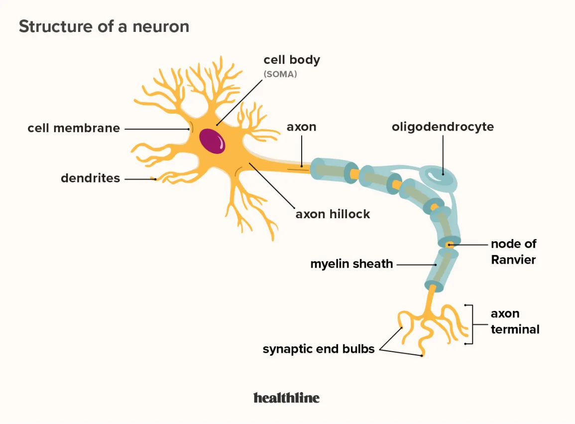
Motor neuron structure
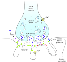
Neuromuscular junction
Space between the synapse and the muscle fiber— ACh is transported by vesicles to the presynaptic membrane, where ACh is released into the neuromuscular junction.
ACh binds to nicotinic receptors on muscle cell, allowing Na+ ions to depolarize the postsynaptic cell. If an AP occurs, Ca+ ions are released and electrical currents are generated → Muscle fiber activation
If ACh does not bind to nicotinic receptors, it is metabolized by AChE into acetate and choline.
EMG indirectly measures the rate of APs/release of acetylcholine into NMJ.
Motor units
A single motor neuron and the muscle fibers it controls
The more units are activated, the greater the force will be.
The number of fibers within a unit (& thus the # of fibers innervated by a single motor axon) varies a ton across muscle types.
Variation results from different degrees of control over contraction force in different structures.
Finger muscles have “many fewer” muscle fibers/motor units than biceps.
Isotonic vs Isometric contractions
Isotonic: Muscle experiences same forces, but shortens and lengthens (“same tension”, pushups)
Isometric: Muscle length remains same throughout (“same measure”, planking)
Electromyography
Directly measures electrical activity of skeletal muscles in mV
Indirectly measures rate of APs/ACh release into NMJ
3 features of skeletal muscle contraction
Muscle Tone: Continuous state of low-level activation (maintains readiness)
Motor Unit Recruitment: Increases in the number of motor units activated within a muscle to generate force
Fatigue: Decreases in contraction strength due to continued activation
Parts of the eye
Cornea: window of the eye, controls/focuses light entry
Anterior chamber/aqueous humor: focuses light on retina/provides nutrition
Iris: restricts light entry (pupil is just a "hole” in iris)
Ciliary body/suspensory ligaments: expand and contract the iris to control light flow into pupil
Vitreous chamber/vitreous humor: provides structure, absorbs shock
Sclera: tough outer coating of eyeball
Retina: photoreceptive layer of eye
Fovea: part of retina containing only cones, allows for sharp central vision
Optic nerve: communicates visual information to the brain
Brain bits - Cerebellum, arbor vitae, gyri/sulci, & olfactory bulb
Cerebellum: Fine motor control, memory of movement patterns
Arbor vitae: transmits info to/from cerebellum
Gyri/sulci: boost surface area— sulci separate regions but gyri do not
Olfactory bulb: smell, limbic system
Brain bits - Pineal gland, corpus callosum, optic chiasm, & ventricles
Pineal gland: Hormone production/regulation
Corpus callosum: communication bw hemispheres
Optic chiasm: optic nerves cross/integration
Lateral ventricle/third ventricles: filled w CSF
Brain bits - Fornix, thalamus, hypothalamus, infundibulum, & pituitary
Fornix: transmits info from hippocampus to mammillary bodies and thalamus
Thalamus: principle sensory info filter, relays to cerebral cortex
Hypothalamus: autonomic nervous system, endocrine system
Infundibulum: pathway for hormones from hypothalamus to posterior pituitary for release into bloodstream
Pituitary: releases/regulates hormones
Brain bits - Midbrain, pons, medulla oblongata, & spinal cord
midbrain: motor movement
Pons: connects midbrain/medulla oblongata
Medulla oblongata: roles vital for life
Spinal cord: pathway for messages bw body/brain
Brain subdivisions
Forebrain: olfaction, sensory integration, behavior coordination
Midbrain: coordinates reflex responses to auditory/visual stimuli
Hindbrain: vital processes
-
Forebrain does sensory integration: “shower beFORE you STINK”
Midbrain coordinates reflex responses: “Many Cats Roar Relentlessly”
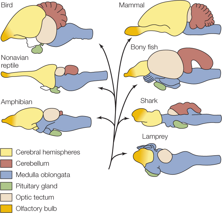
Comparative anatomy of brains
Lamprey: no cerebellum, shortest cerebral hemispheres/olfactory region
Shark: midsized cerebellum that partially covers optic tectum
Amphibian: smallest cerebellum
Bony fish: gap between cerebellum and medulla, largest optic tectum
Nonavian reptile: longest cerebral hemispheres/olfactory region
Bird: Foldy cerebellum, more visible pituitary
Mammal: Even more foldy cerebellum
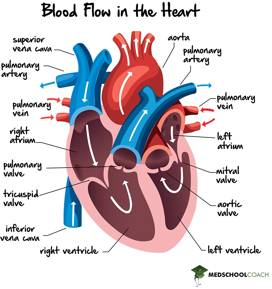
Atria vs Ventricles
Atria accept blood from veins. These are the vena cava in pulmonary circuit on right side of the heart, and pulmonary veins in systemic circuit on left side of heart.
Ventricles pump blood out through the pulmonary artery and aorta. The right ventricle pumps blood to the lungs and is much less forceful than the left ventricle, which pumps blood to the rest of the body.
Atrioventricular vs semilunar valves
Atrioventricular: Between atria & ventricles… tricuspid and bicuspid (mitral)
Semilunar: Between ventricles & artery… aortic and pulmonary
Flow of blood through the heart
Pulmonary: Deoxygenated blood enters right atrium through superior vena cava, passes through AV valve to right ventricle, and out to the lungs.
Systemic: Oxygenated blood enters left atrium through pulmonary vein, passes through AV valve to left ventricle (more muscular), where it is dispersed to body through aorta.
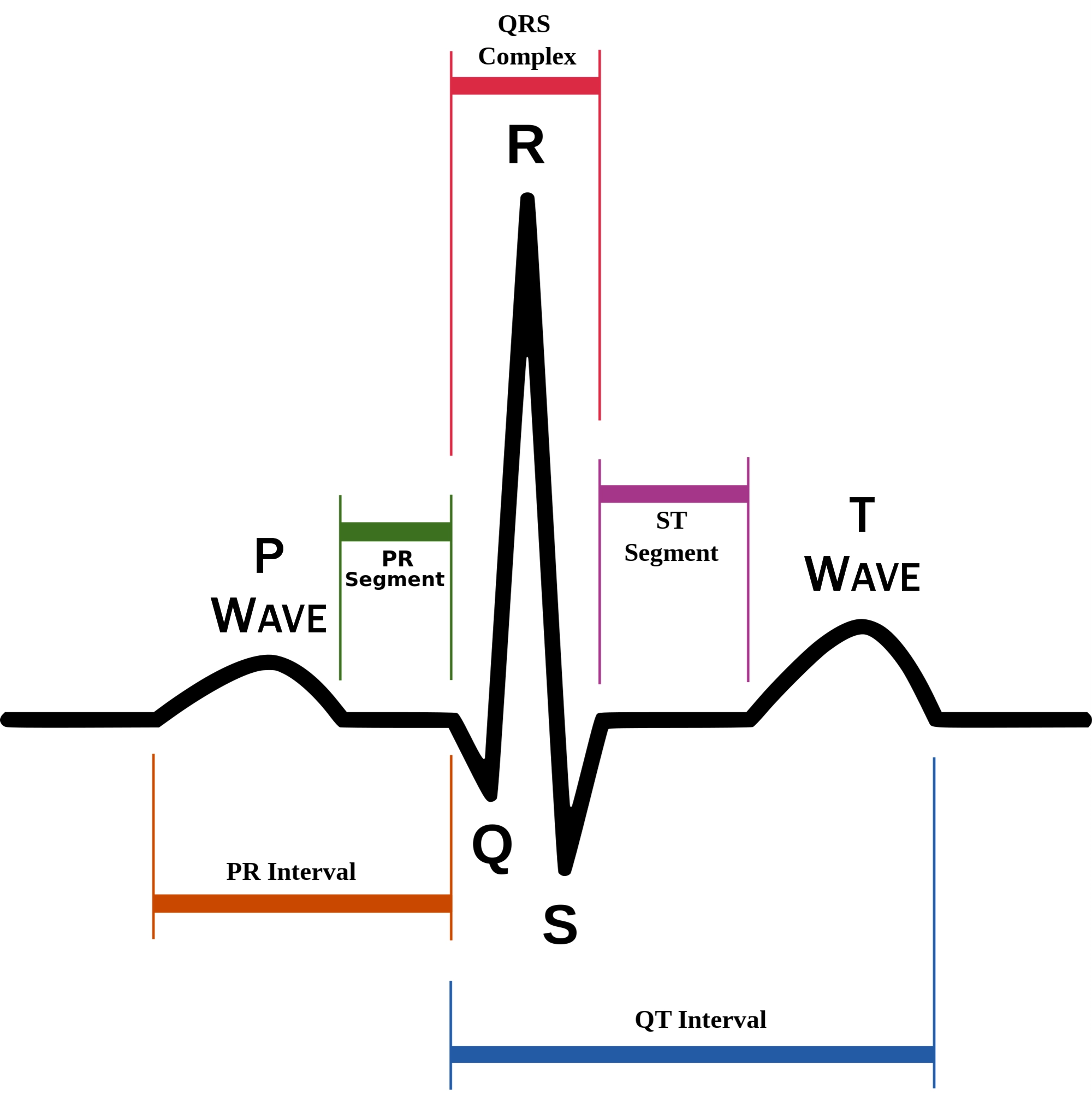
EKG, heart rate, and the cardiac cycle
EKGs directly measure electrical activity in the heart.
P: atrial contraction QRS: ventricular contraction T: ventricular repolarization
Heart rate: 300/(# large squares between R waves)
Lub (systole/ventricular contraction) - PQ segment
Dub (diastole/ventricular relaxation) - ST segment
Sinoatrial node
Initiates an electrical sequence in the heart (natural pacemaker). The signal travels from the SA node to the AV node. Bundle of His carries signal down pathway into the ventricles.
In humans, this occurs ~60-100 times/minute.
Systole & Diastole
Systole (lub): Ventricular contraction (120 in 120/80), AV valves close
Diastole (dub): Ventricular relaxation (80 in 120/80), SL valves close
Spectrophotometry
Dyes absorb specific wavelengths of light, amount of absorption dependent on concentration.
We injected a known amount of dye into known volumes of fluid, then measured absorption of the dye in the roach using the same procedure.
Optical density vs transmittance
Optical density is proportional to concentration, which is proportional to 1/volume.
Density is inversely proportional to transmittance— The less light is transmitted, the greater optical density (light absorbance) will be.
Deriving a standard curve
We created a standard curve by creating a dilution series with vials of known concentrations of the same dye we used in the roach, and we used the curve as a ruler to measure the OD of an unknown sample.
Open vs closed circulatory systems
Open circulatory systems: Fluid freely bathes tissues, is a mix of oxygenated and deoxygenated
Closed circulatory systems: Blood contained in vessels, oxygenated separate from deoxygenated
Waste excretion in open circulatory systems
Malpighian tubules (“simplistic kidney”) — Hemolymph transports waste products here for waste excretion.
Arthropod circulatory system
Heart (long muscular dorsal tube) takes in blood from abdominal hemocoel, empties it into head and thorax.
Hemolymph flows slowly through tissues and back into the abdomen.
Insect circulation lab: Why didn’t we take OD values at zero time?
Malpighian tubules of insect remove dye at the same time it is mixing in the hemolymph, so a “true zero” reading is impossible.
Extrapolating using our standard curve gives us a theoretical value.
3 factors for efficient gas exchange
Large gas exchange surface area
Minimal distance to transport gas
Maintenance of diffusion gradients
3 innovations in mammalian respiration
Diaphragm
Vertically flexed vertebral columns
Loss of abdominal ribs
Active and passive respiration
During active respiration, the diaphragm contracts, ribs expand, and negative pressure forces air IN (inhalation).
During passive respiration, the lungs recoil from stretching and air flows back until rib and lung pressure are equal (exhalation).
Path of air through lungs
Sinuses
Pharynx
Larynx
Trachea
Bronchial tube
Lung > bronchiole > alveoli
Mammalian dive reflex
Slows breathing and heart rate (bradycardia, 10-25%)
Peripheral vasoconstriction (directs blood to vital organs, away from extremities)
Headgut organs
Oral cavity, throat, pharynx
Food procurement and initial processing/transport
Dental Formulae
Incisors (cutting): Canine (gripping): Premolar (grinding): Molar (grinding)
Formulae start from anterior maxilla: “Iguanas can’t play music”
2:1:2:3 in humans
Foregut organs
Esophagus: circular & longitudinal muscle, lined w/ epithelial cells
Stomach: region w/ gastric glandular mucosa which secretes pepsinogen & HCL - sphincters at both ends → one-way flow
Midgut organs
Small intestine, liver, pancreas
Small intestine
Midgut. Lined w/ villi & microvilli.
Major site for digestion of carbs, fat, protein
Divided into duodenum (digestion), jejunum (absorption), and ileum [“donuts don’t just appear”]
Mesentery: thin tissue holding intestines (blood supply & lymph)
Liver
Metabolism (cat/anabolism)
Macromolecules: Stores carbs/fats, processes proteins [“some cool frogs prefer pillbugs”]
Detoxification
Bile production (absorption of fats)
4 functions: “Monkeys March Down Broadway”
Bile
Stored in gallbladder + secreted into dudoenum
Bile interacts w/ fats in duodenum, then fats are absorbed in jejunum and ileum
Pancreas
Exocrine communication: produces bicarbonate to neutralize HCl in small intestine, produces enzymes
Endocrine communication: Glucagon and insulin
Hindgut organs
storage of digesta, retrieval of dietary or endogenous electrolytes & water
microbial fermentation in herbivores
Distinguished from midgut by epithelial morphology, change in diameter, or presence of sphincter
Organ sizes vary by diet type
Large intestine & ceca
Unique characteristics of bird digestive systems
Don’t have true teeth (masticate w/ beak and gizzard)
Crop: An expansion of the esophagus that stores food
Proventriculus: Glandular stomach
Gizzard: Muscular stomach (rocks here sometimes)
Paired ceca to aid in flight
Hindgut vs ruminant fermenters
Hindgut fermenters: fermentation in cecum, proximal colon. Less efficient, so sometimes engage in coprophagy (rabbits)
Ruminant fermenters: ferment in rumen before stomach… More efficient (cows)
Shark digestive tracts
Spiral valve (↑ surface area) and very short tract.
Oily liver (buoyancy — not needed in bony fish bc they have a swim bladder — target for poaching bc it contains squalene).
Rectal gland.
Carnivores, herbivores, and omnivores
Carnivores: Short GI tracts, 10-20x more acidic stomachs than omnivores and herbivores, and stomachs are larger/may have rugae.
Herbivores: Longer GI tracts for fermentation, often greatly expanded ceca, much less acidic stomachs
Omnivores: Intermediate gut length and morphology, much less acidic stomachs than hypercarnivores
Effect of diet on digestive structures
Stomach: Animals prone to consuming large amounts at one time may have gastric folds/rugae to allow for stretching, acidity 10-20x higher in hypercarnivores
Small intestine: Longer in herbivores
Cecum: Long in herbivores, reduced in carnivores. Paired in birds.
Large intestine: Longer in animals that require a lot of fermentation.