MSK TBL 13: Pelvis, Hip, & Gluteal Region Functional Anatomy
 Call Kai
 Learn
Learn Practice Test
 Spaced Repetition
 Match
Match Flashcards
 Knowt Play
1/52

There's no tags or description
Looks like no tags are added yet.
Name | Mastery | Learn | Test | Matching | Spaced | Call with Kai | Chat |
|---|
No analytics yet
Send a link to your students to track their progress
53 Terms
hip bone [os coxae] made up of
ilium, ischium, & pubis
at birth, bones joined by hyaline cartilage
at puberty, separated by Y-shaped triradiate cartilage
fusion begins 15-17 yrs, completes 20-25 yrs
ilium
largest bone of hip
contributes part of upper acetabulum
features
ala [“wing”]
iliac crest
ASIS & AIIS
iliac fossa
*osteoporosis may thin iliac fossa
where iliacus sits
auricular surface
articulates w/ sacrum at sacroiliac joint
gluteal lines
muscle attachment for glute muscles
![<ul><li><p>largest bone of hip</p></li><li><p>contributes part of upper acetabulum</p></li><li><p>features</p><ul><li><p>ala [“wing”]</p></li><li><p>iliac crest</p></li><li><p>ASIS & AIIS</p></li><li><p>iliac fossa</p><ul><li><p>*osteoporosis may thin iliac fossa</p></li><li><p>where iliacus sits</p></li></ul></li><li><p>auricular surface</p><ul><li><p>articulates w/ sacrum at sacroiliac joint</p></li></ul></li><li><p>gluteal lines</p><ul><li><p>muscle attachment for glute muscles</p></li></ul></li></ul></li></ul><p></p>](https://assets.knowt.com/user-attachments/872d00d7-bfed-420f-a087-e737d666fb1f.png)
ischium
forms posteroinferior part of hip bone
main parts
body
ramus
ischial spine
separates greater & lesser sciatic notches
ischial tuberosity
supports body weight when sitting
origin of posterior thigh muscles

iliac tubercle
5-6 cm behind ASIS
marks widest iliac crest point, aligned w/ L5 vertebra
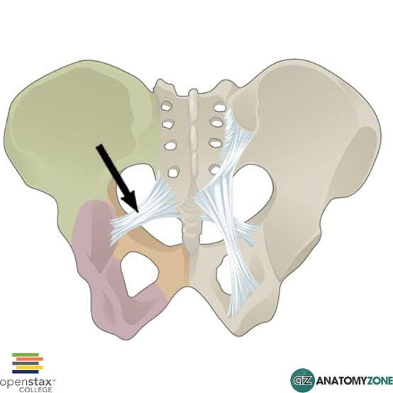
posterior sacro-iliac ligament
continues into the sacrotuberous ligament
sacrotuberous & sacrospinous ligament
convert sciatic notches into greater & lesser sciatic foramen
sacrotuberous ligament
runs from posterior iliac spines, sacrum, & coccyx → medial ischial tuberosity

sacrospinous ligament
lies deep to sacrotuberous ligament
runs from lower sacrum & coccyx → ischial spine
lies edge-to-edge w/ levator ani muscle, forming pelvic floor
key contents of greater sciatic foramen [below piriformis]
POP SI
Pudendal nerve
Obturator internus nerve
Posterior cutaneous nerve of thigh
Sciatic nerve
Inferior gluteal nerve
key contents of greater sciatic foramen [above piriformis]
superior gluteal nerve [& artery & vein]
key contents of lesser sciatic foramen
PIN + Tendon
Pudendal nerve
Internal Pudendal Vessels
Nerve to obturator internus
Obturator internus Tendon
immediately hook forward around ischial spine/ligament to enter perineum via lesser sciatic foramen
*PIN gives spatial relationship from medial → lateral
iliofemoral ligament
reinforces anterior joint capsule
motion restricted
anterior fibers: hyperextension
superior fibers: adduction
inferior fibers: abduction
allows standing w/ minimal muscular effort

pubofemoral ligament
restricts abduction & hyperextension

ischiofemoral ligament
restricts internal/medial rotation & extension

zona orbicularis
restricts femoral head distraction

gluteus maximus
origin
posterior ilium [behind posterior gluteal line], sacrum, coccyx, sacrotuberous ligament
insertion
iliotibial tract [superior fibers]
gluteal tuberosity [deep fibers]
innervation
inferior gluteal nerve [L5, S1, S2]
action
extends hip
lateral rotator
trunk extension when femur fixed
acts w/ TFL via IT tract to stabilize pelvis
trochanteric bursa
btwn superior fibers of gluteus maximus & greater trochanter
most prominent at birth
lateral hip pain w/ bursitis
ischial bursa
btwn inferior glute max & ischial tuberosity
can become inflamed in prolonged sitting [“weaver’s bottom”]
gluteofemoral bursa
btwn iliotibial tract & vastus lateralis
decreased friction during lateral thigh movement
gluteus medius
deep to glute max, superior to minimus
origin
external ilium [btwn anterior & posterior gluteal lines]
insertion
lateral surface of greater trochanter
innervation
superior gluteal nerve [L5, S1]
action
abducts & medially rotates thigh
stabilizes pelvis in gait
clinical
weakness → Trendenlenburg gait
target for deep intramuscular injections [upper outer quadrant]
![<p>deep to glute max, superior to minimus</p><ul><li><p>origin</p><ul><li><p>external ilium [btwn anterior & posterior gluteal lines]</p></li></ul></li><li><p>insertion</p><ul><li><p>lateral surface of greater trochanter</p></li></ul></li><li><p>innervation</p><ul><li><p>superior gluteal nerve [L5, S1]</p></li></ul></li><li><p>action</p><ul><li><p>abducts & medially rotates thigh</p></li><li><p>stabilizes pelvis in gait</p></li></ul></li><li><p>clinical</p><ul><li><p>weakness → Trendenlenburg gait</p></li><li><p>target for deep intramuscular injections [upper outer quadrant]</p></li></ul></li></ul><p></p>](https://assets.knowt.com/user-attachments/4e37e374-1c41-42e3-a0cc-a96ccc729f44.png)
gluteus minimus
origin
lower ilium
margin of greater sciatic notch
insertion
greater trochanter of femur
innervation
superior gluteal nerve [L5, S1]
action
anterior fibers assist w/ hip flexion where primary hip flexors [Iliopsoas, Rectus femoris]
![<ul><li><p>origin</p><ul><li><p>lower ilium</p></li><li><p>margin of greater sciatic notch</p></li></ul></li><li><p>insertion</p><ul><li><p>greater trochanter of femur</p></li></ul></li><li><p>innervation</p><ul><li><p>superior gluteal nerve [L5, S1]</p></li></ul></li><li><p>action</p><ul><li><p>anterior fibers assist w/ hip flexion where primary hip flexors [Iliopsoas, Rectus femoris]</p></li></ul></li></ul><p></p>](https://assets.knowt.com/user-attachments/b3d3a41b-4adc-4653-b3ef-a3fe46dcc815.png)
stance phase [weight bearing limb]
abductors stabilize pelvis & rotate femur medially
aid limb propulsion
swing phase [non-WB limb]
lateral rotators [piriformis, obturator internus]
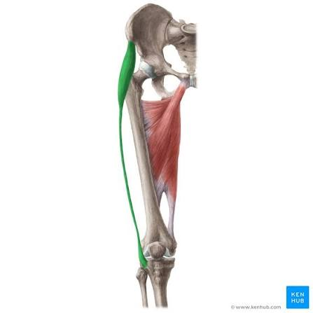
tensor fascia latae [TFL]
anterior part of lateral thigh; superficial layer
origin
ASIS & anterior iliac crest
insertion
iliotibial tract → lateral condyle of tibia
innervation
superior gluteal nerve
action
flexes, abducts, & medially rotates thigh
stabilizes knee via IT tract
clinical
overactive in tight IT band syndrome or runners
Piriformis
deep gluteal muscle; passes thru greater sciatic foramen
origin
middle 3 sacral segments S2-S4
insertion
superior border of greater trochanter
innervation
anterior rami [S1, S2]
action
lateral rotation of extended thigh
abduction when thigh is flexed
clinical
piriformis syndrome
![<p>deep gluteal muscle; passes thru greater sciatic foramen</p><ul><li><p>origin</p><ul><li><p>middle 3 sacral segments S2-S4</p></li></ul></li><li><p>insertion</p><ul><li><p>superior border of greater trochanter</p></li></ul></li><li><p>innervation</p><ul><li><p>anterior rami [S1, S2]</p></li></ul></li><li><p>action</p><ul><li><p>lateral rotation of extended thigh</p></li><li><p>abduction when thigh is flexed</p></li></ul></li><li><p>clinical</p><ul><li><p>piriformis syndrome</p></li></ul></li></ul><p></p>](https://assets.knowt.com/user-attachments/0e793602-e2a2-4114-9f34-1ddb527d75d5.png)
obturator internus [OI]
exits lesser sciatic foramen
origin
pelvis surface of obturator membrane & surrounding bones
insertion
medial surface of greater trochanter [trochanter fossa]
innervation
nerve to obturator internus [L5, S1]
action
laterally rotates extended thigh
abducts flexed thigh
levator ani & OI forms part of lateral wall of both pelvis & perineum
clinical
one of key muscles in triceps coxae
![<p>exits lesser sciatic foramen</p><ul><li><p>origin</p><ul><li><p>pelvis surface of obturator membrane & surrounding bones</p></li></ul></li><li><p>insertion</p><ul><li><p>medial surface of greater trochanter [trochanter fossa]</p></li></ul></li><li><p>innervation</p><ul><li><p>nerve to obturator internus [L5, S1]</p></li></ul></li><li><p>action</p><ul><li><p>laterally rotates extended thigh</p></li><li><p>abducts flexed thigh</p></li></ul></li><li><p>levator ani & OI forms part of lateral wall of both pelvis & perineum</p></li><li><p>clinical</p><ul><li><p>one of key muscles in triceps coxae</p></li></ul></li></ul><p></p>](https://assets.knowt.com/user-attachments/4cebdab9-7837-45a7-91a4-e012bcd9b2da.png)
superior gemellus
origin
ischial spine
insertion
medial surface of greater trochanter [trochanteric fossa]
innervation
nerve to obturator internus
action
work w/ OI to laterally rotate & abduct hip
stabilize joint
clinical
often fused & act in concert
part of triceps coxae
![<ul><li><p>origin</p><ul><li><p>ischial spine</p></li></ul></li><li><p>insertion</p><ul><li><p>medial surface of greater trochanter [trochanteric fossa]</p></li></ul></li><li><p>innervation</p><ul><li><p>nerve to obturator internus</p></li></ul></li><li><p>action</p><ul><li><p>work w/ OI to laterally rotate & abduct hip</p></li><li><p>stabilize joint</p></li></ul></li><li><p>clinical</p><ul><li><p>often fused & act in concert</p></li><li><p>part of triceps coxae</p></li></ul></li></ul><p></p>](https://assets.knowt.com/user-attachments/4ba8a28e-4ab0-4b42-bb12-506426326bc7.png)
inferior gemellus
origin
ischial tuberosity
insertion
medial surface of greater trochanter [trochanteric fossa]
innervation
nerve to quadratus femoris
action
work w/ OI to laterally rotate & abduct hip
stabilize joint
clinical
often fused & act in concert
part of triceps coxae
![<ul><li><p>origin</p><ul><li><p>ischial tuberosity</p></li></ul></li><li><p>insertion</p><ul><li><p>medial surface of greater trochanter [trochanteric fossa]</p></li></ul></li><li><p>innervation</p><ul><li><p>nerve to quadratus femoris</p></li></ul></li><li><p>action</p><ul><li><p>work w/ OI to laterally rotate & abduct hip</p></li><li><p>stabilize joint</p></li></ul></li><li><p>clinical</p><ul><li><p>often fused & act in concert</p></li><li><p>part of triceps coxae</p></li></ul></li></ul><p></p>](https://assets.knowt.com/user-attachments/5cfdfcf2-efca-4e5f-8332-44ee11491842.png)
quadratus femoris
short, flat, rectangular muscle
inferior to obturator internus & gemelli muscles
origin
lateral border of ischial tuberosity
insertion
quadrate tubercle on intertrochanteric crest of femur
innervation
nerve to quadratus femoris [L5, S1]
action
lateral rotation of thigh
stabilize femoral head
clinical
works w/ adj. rotators to maintain joint congruency
![<p>short, flat, rectangular muscle</p><p>inferior to obturator internus & gemelli muscles</p><ul><li><p>origin</p><ul><li><p>lateral border of ischial tuberosity</p></li></ul></li><li><p>insertion</p><ul><li><p>quadrate tubercle on intertrochanteric crest of femur</p></li></ul></li><li><p>innervation</p><ul><li><p>nerve to quadratus femoris [L5, S1]</p></li></ul></li><li><p>action</p><ul><li><p>lateral rotation of thigh</p></li><li><p>stabilize femoral head</p></li></ul></li><li><p>clinical</p><ul><li><p>works w/ adj. rotators to maintain joint congruency</p></li></ul></li></ul><p></p>](https://assets.knowt.com/user-attachments/b5d242e7-8a0d-47aa-988c-57cb65c63c88.png)
obturator externus [OE]
lies deep in proximal thigh, posterior to pectineus & adductors
origin
external surface of obturator membrane & surrounding bone
insertion
trochanteric fossa of femur
innervation
obturator nerve [L3, L4]
unique b/c others mostly innervated by sacral plexus
action
lateral rotation of thigh
most effective when hip is flexed
![<p>lies deep in proximal thigh, posterior to pectineus & adductors</p><ul><li><p>origin</p><ul><li><p>external surface of obturator membrane & surrounding bone</p></li></ul></li><li><p>insertion</p><ul><li><p>trochanteric fossa of femur</p></li></ul></li><li><p>innervation</p><ul><li><p>obturator nerve [L3, L4]</p><ul><li><p>unique b/c others mostly innervated by sacral plexus</p></li></ul></li></ul></li><li><p>action</p><ul><li><p>lateral rotation of thigh </p><ul><li><p>most effective when hip is flexed</p></li></ul></li></ul></li></ul><p></p>](https://assets.knowt.com/user-attachments/898c4469-b0bc-4cea-b3d8-59dfa54b5215.png)
semitendinosus [ST]
long tendinous lower half
lies superficial to semimembranosus
origin
ischial tuberosity
insertion
medial surface of superior tibia [part of pes anserinus]
innervation
tibial division of sciatic nerve [L5, S1, S2]
action
hip extension, knee flexion, medial rotation of leg when flexed
aids in trunk extension when legs are fixed
![<p>long tendinous lower half</p><p>lies superficial to semimembranosus</p><ul><li><p>origin</p><ul><li><p>ischial tuberosity</p></li></ul></li><li><p>insertion</p><ul><li><p>medial surface of superior tibia [part of pes anserinus]</p></li></ul></li><li><p>innervation</p><ul><li><p>tibial division of sciatic nerve [L5, S1, S2]</p></li></ul></li><li><p>action</p><ul><li><p>hip extension, knee flexion, medial rotation of leg when flexed</p></li><li><p>aids in trunk extension when legs are fixed</p></li></ul></li></ul><p></p>](https://assets.knowt.com/user-attachments/0cc35deb-3ed7-453c-8a83-9e1456b579ce.png)
semimembranosus [SM]
flat, broad, membranous muscle
lies deep to ST
origin
ischial tuberosity
insertion
posterior medial condyle of tibia
innervation
tibial division of sciatic nerve
action
extends hip, flexes knee, medially rotates flexed leg
helps stabilize knee joint
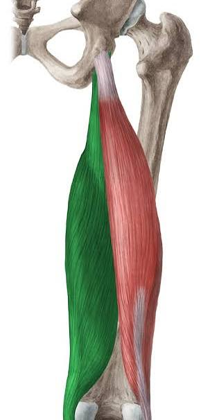
biceps femoris [long head]
lateral muscle of posterior thigh
unites w/ short head lower down
origin
ischial tuberosity
insertion
head of fibula [via common tendon w/ short head]
innervation
tibial division of sciatic nerve [L5-S2]
action
extends hip, flexes knee, laterally rotates flexed leg
![<p>lateral muscle of posterior thigh</p><p>unites w/ short head lower down</p><ul><li><p>origin</p><ul><li><p>ischial tuberosity</p></li></ul></li><li><p>insertion</p><ul><li><p>head of fibula [via common tendon w/ short head]</p></li></ul></li><li><p>innervation</p><ul><li><p>tibial division of sciatic nerve [L5-S2]</p></li></ul></li><li><p>action</p><ul><li><p>extends hip, flexes knee, laterally rotates flexed leg</p></li></ul></li></ul><p></p>](https://assets.knowt.com/user-attachments/c174c772-5e19-4c48-82c5-986fa26b9a08.png)
superior clunial nerves
L1-L3 [posterior rami]
skin over iliac crest & upper gluteal region
middle clunial nerves
S1-S2 [posterior rami]
skin over sacrum & adj. buttocks
inferior clunial nerves
branches of posterior cutaneous nerve of thigh [S2-S3]
skin of lower buttocks toward greater trochanter
Spinal levels of sacral plexus
SIPPS
Sup Glut N to quad fem/inf gem
L4, L5, S1
sometimes only considered L5, S1
Inf Glut N to Obt Int/Sup Gem
L5, S1, S2
Post Cut N of thigh
L1, L2, L3
Pudendal
S2, S3, S4
Sciatic
L4, L5, S1, S2, S3
sciatic nerve
largest nerve in body
formed at inferior border of piriformis
passes midway btwn greater trochanter & ischial tuberosity
splits into
tibial nerve: anterior division
common fibular nerve: posterior division
clinical
piriformis syndrome when common fibular nerve pierces or runs over piriformis [anatomy in 12% of people]
nerve to quadratis femoris exits pelvis…
anterior to sciatic nerve
nerve to obturator internus courses…
posterior to sacrospinous ligament & reenters via lesser sciatic foramen
arterial supply of gluteal region: major branches
superior gluteal artery
inferior gluteal artery
internal pudendal artery
superior gluteal artery
largest branch of interal iliac
superficial branch
glute max & overlying skin
deep branch
glut med, min, & TFL
anastomoses w/ inferior gluteal & medial circumflex femoral arteries
artery to sciatic nerve
gives off slender companion artery of sciatic nerve [arteria comitans nervi ischiadici]
anastomoses w/ superior gluteal, perforating, & circumflex femoral arteries
internal pudendal artery
arises from internal iliac
follows pudendal nerve [greater → lesser sciatic foramen]
supplies perineum
does NOT supply gluteal region or posterior thigh
perforating arteries [from profunda femoris]
pierce adductor magnus → posterior thigh
supply hamstrings & sciatic nerve
fracture of femoral head
medial circumflex femoral & retinacular arteries are torn
femoral head may undergo avascular necrosis [AVN]
Bonnet’s position
flexion, abduction, external rotation
pts w/ intra-articular hip issues [labral tears, femoroacetabular impingement, early osteoarthritis, or capsular tightness] find relied in this position
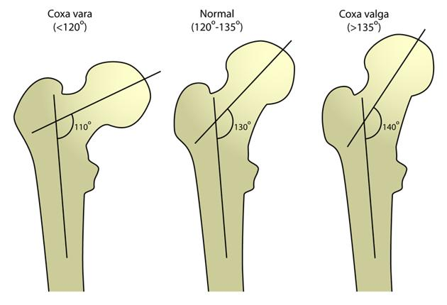
Neck-shaft angle [frontal plane angle]
normal: 120-135 degrees
Coxa valga: >140 degrees
→ genu varum
Coxa vara: <120 degrees
→ genu valgum
alters glute med stability → hip instability
Femoral Version [Transverse plane or Angle of Femoral Torsion]
angle btwn femoral neck & condyles
Normal: 10-20 degrees
Anteversion: >20 degrees
toe-in gait
Retroversion: <10 degrees
toe-out gait
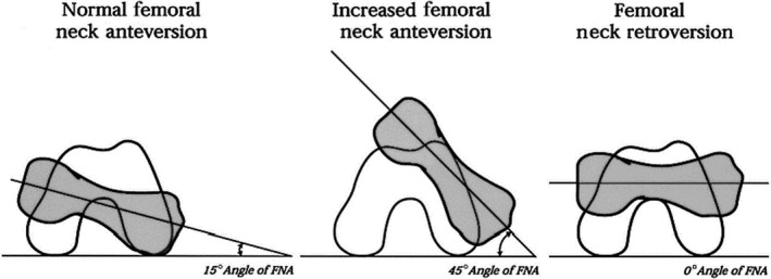
3 main causes of in-toeing [“pigeon toed”] gait
thigh: increased femoral anteversion
in-toe gait & patella turned inward
leg/shin: internal tibial torsion
in-toe gait but patella facing straight
foot: metatarsus adductus
medial deviation of metatarsals
curved, C-shaped lateral foot margin
FAIR test
Flexion, Adduction, Internal Rotation
tests piriformis
FABER test
Flexion, ABduction, External Rotation
tests hip flexors [iliopsoas], adductors, sartorius
Ober’s test
tests TFL