Y1-055 Anatomy of the Thoracic Cavity: Heart, Lungs, Ribs, Sternum & Musculature
 Call Kai
 Learn
Learn Practice Test
 Spaced Repetition
 Match
Match Flashcards
 Knowt Play
1/194

There's no tags or description
Looks like no tags are added yet.
Name | Mastery | Learn | Test | Matching | Spaced | Call with Kai | Chat |
|---|
No analytics yet
Send a link to your students to track their progress
195 Terms
18th day
What day are the cardiogenic cords in the embryo formed?
21st or 22nd
What day do cells canalize to form endocardia heart tubes and fuse together to form the pericardial cavity in an embryo?
23rd day
What day does the heart form the ventricular and atrial regions in an embryo?
cardiovascular system
What is the first functioning organ system in an embryo?
4-8th weeks
What weeks of embryonic development are associated with formation of the interatrial and interventricular septa and division of the truncus arteriosus into the aorta and pulmonary trunk?
Left-to-right blood flow and increased blood flow in the right atrium can cause pulmonary hypertension, congestive heart failure, atrial arrhythmias, and increased risk of stroke.
What are the abnormal blood flow patterns and potential complications associated with an atrial septal defect (ASD)?
Left-to-right blood flow and increased blood flow in the right ventricle can cause heart failure, pulmonary hypertension, and arrhythmias
What are the abnormal blood flow patterns and potential complications associated with a ventricular septal defect (VSD)?
Ventricular septal defect (VSD), overriding aorta, pulmonary stenosis, and right ventricular hypertrophy
What are the four cardiac defects associated with Tetralogy of Fallot?
Oxygenated and deoxygenated blood mix, causing both oxygenated and deoxygenated blood to flow to the body
What is the resulting effect on blood flow in Tetralogy of Fallot?
Superior- above the heart, Anterior- in front of the heart, Middle- contains the heart and pericardial sac, posterior- behind the heart.
What are the four divisions of the mediastinum, and where is each located relative to the heart?
anterior mediastinum
B

posterior mediastinum
E
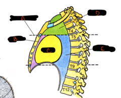
superior mediastinum

middle mediastinum
C

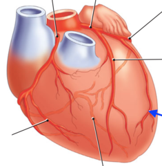
atrioventricular groove

anterior interventricular groove

posterior interventricular groove
2

right (acute) marginal artery, supplies oxygenated blood directly to the muscle tissue across the front and outer surface of the right ventricle
label and identify importance

left (obtuse) marginal artery, supplies oxygen-rich blood to the lateral wall of the left ventricle
label and identify importance
R atria

L atria

R auricle, increases the holding capacity of the right atrium to handle larger volumes of deoxygenated blood
label and identify importance
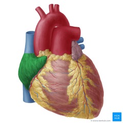
L auricle, increases the holding capacity of the left atrium to handle incoming oxygenated blood from the lungs
label and identify importance

ventricles
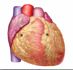
epicardium

epicardial fat- provides energy, regulates inflammation, and protects coronary arteries
label and identify importance
pericardium- fibrous outer sac, parietal layer- lines the fibrous sac, visceral layer- covers the heart and great vessels.
What are the layers of the pericardium, and where is each located?
pericardium
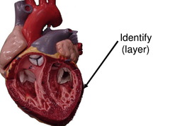
parietal layer
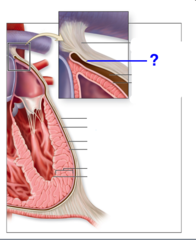
visceral layer

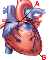
apex of the heart, marks the location of the left ventricle
B, label and identify importance

base of the heart, marks the location of the left atrium and connects the great vessels that pump blood through the body
A, label and identify importance
right margin, marks the boundary formed by the right atrium and ventricle
label and identify importance

left margin, marks the location of the left ventricle
label and identify importance

inferior margin (formed by the right ventricle)
2
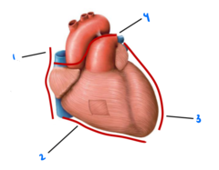
superior margin, connects the atria to the great vessels
label and identify importance
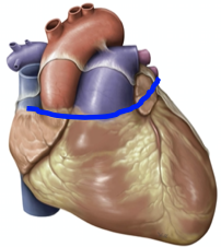
left coronary artery- left atrium, the left ventricle, and the interventricular septum
label and identify which areas of the heart it supplies

right coronary artery- right atrium, right ventricle, inferior and posterior left ventricle, and the SA and AV nodes
label and identify which areas of the heart it supplies

small cardiac vein, posterior surfaces of the right atrium and the right ventricle
label and identify which areas of the heart it drains
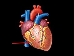
middle cardiac vein- posterior walls of the left and right ventricles, the interventricular septum, and the apex of the heart
label and identify which areas of the heart it drains

great cardiac vein, anterior surface of both ventricles and the anterior part of the interventricular septum
label and identify which areas of the heart it drains

superior vena cava, returns deoxygenated blood from the upper body to the right atrium
label and identify its importance in circulation

inferior vena cava, returns deoxygenated blood from the lower body to the right atrium
label and identify its importance in circulation

pulmonary artery, carries deoxygenated blood from the right ventricle to the lungs
label and identify its importance in circulation

pulmonary veins, carries oxygenated blood from the lungs to the left atrium
label and identify its importance in circulation

aorta, carries oxygenated blood from the left ventricle to the body
label and identify its importance in circulation

right atrium, receives deoxygenated blood from the body and pumps it into the right ventricle
label and describe function

right ventricle, pumps blood into the lungs
label and describe function

left atrium, receives oxygen-rich blood from the lungs and pumps it into the left ventricle
label and describe function

left ventricle, pumps oxygenated blood to the body
label and describe function

fossa ovalis and limbus, marks the closed fetal shunt
label and describe function

sinus venarum

crista terminalis, separates the sinus venarum from the pectinate muscles
label and describe function
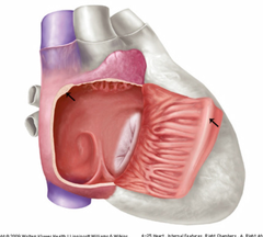
pectinate muscles, increase the power of atrial contraction
label and describe function

conus arteriosus, directs blood from the ventricle into the aorta while preventing backflow
label and describe function
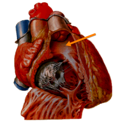
pulmonary trunk, transports deoxygenated blood from the right ventricle of the heart to the lungs for oxygenation
label and describe function

coronary sinus and valve, collects deoxygenated blood from the heart muscle and drains it into the right atrium
label and describe function
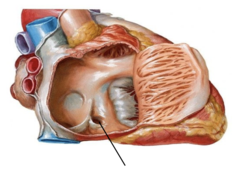
SA node, the heart's natural pacemaker, initiating electrical impulses to regulate the heartbeat at an intrinsic pace of 60-100 bpm
label and describe function

AV node, back-up pacemaker with an intrinsic rate of 40-60 bpm, delaying the electrical signal so the ventricles can fill with blood before pumping
label and describe function

tricuspid valve (AV valve), allows blood to flow from the right atrium into the right ventricle and closes tightly to prevent backward flow of blood
label and describe function

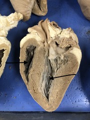
trabeculae carneae, prevents the walls of the heart's ventricles from sticking together and suctioning during contractions
label and describe function

posterior papillary muscles, anchors the chordae tendineae to the mitral and tricuspid valve leaflets
E, label and describe function

moderator band (septomarginal trabecula), carries electrical signals to help the right ventricle contract evenly
label and describe function

chordae tendineae, anchors the AV leaflets to prevent backflow of blood
label and describe function
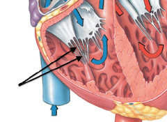
anterior cusp of (R) tricuspid valve

septal cusp of (R) tricuspid valve

posterior cusp of (R) tricuspid valve
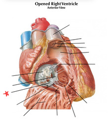
Septal papillary muscle (medial papillary muscle)

Tricuspid, pulmonary, mitral (bicuspid), and aortic valves
What are the four valves of the heart?
Open- tricuspid and mitral valves, Closed- pulmonary and aortic valves
Which heart valves are open and closed during diastole?
Open- pulmonary and aortic valves, Closed- tricuspid and mitral valves.
Which heart valves are open and closed during systole?
The coronary arteries branch from the aorta and supply blood to the heart muscle
What is the relationship between the aorta and the coronary arteries?
foramen ovale, shunts blood from the right atrium to the left atrium
label #5 and describe its role in prenatal circulation

ductus venosus, shunts some umbilical vein blood directly to the inferior vena cava
label #4 and describe its role in prenatal circulation

ductus arteriosus, shunts blood from the pulmonary artery to the aorta, bypassing the lungs
label #8 and describe its role in prenatal circulation

it closes and becomes the fossa ovalis
What happens to the foramen ovale after birth?
fossa ovalis
3

Ligamentum teres
What does the umbilical vein (#2) become after birth?

ligamentum arteriosum
2
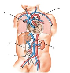
ligamentum venosum
What does the ductus venosus become after birth?
ligamentum venosum
5

Ligamentum arteriosum
What does the ductus arteriosus become after birth?
ligamentum arteriosum
4

It decreases
What happens to right atrial pressure when placental circulation is occluded at birth?
It increases above right atrial pressure
What happens to left atrial pressure at birth?
Higher left atrial pressure closes the foramen ovale
How does the pressure change between the atria contribute to closure of the foramen ovale?
It decreases
What happens to pulmonary vascular resistance when the lungs aerate at birth?
T1-T4
What is the spinal nerve origin of sympathetic control of the heart?
Cervical ganglia
Where does sympathetic innervation of the heart synapse before reaching the cardiac plexus?
Increases heart rate and force of contraction
What is the effect of sympathetic stimulation on the heart?
The vagus nerve (CN X), it decreases heart rate
What cranial nerve provides parasympathetic control of the heart, and what is its effect?
nasal cavity

nasal pharynx

oral pharynx

laryngeal pharynx
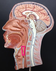
larynx
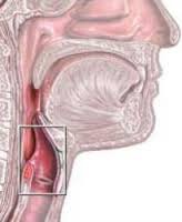
primary bronchi

right primary bronchi

left primary bronchi

intermediate bronchi
