Assorted Skull Facts
 Call Kai
 Learn
Learn Practice Test
 Spaced Repetition
 Match
Match Flashcards
 Knowt Play
1/11

There's no tags or description
Looks like no tags are added yet.
Name | Mastery | Learn | Test | Matching | Spaced | Call with Kai | Chat |
|---|
No analytics yet
Send a link to your students to track their progress
12 Terms
How were the bones of the skull formed?
Intramembranous ossification - frontal, parietal, occipital
Endochondral ossification - sphenoid, temporal
Sutures? Where do they meet?
Coronal suture - frontal/parietal
Sagittal suture - two parietal
Lambdoid suture - parietal/occipital
Coronal + sagittal = bregma
Sagittal + lambdoid = lambda
Name the air sinuses of the skull
Frontal
Maxillary
Sphenoid
Ethmoid
Mastoid (temporal)
What is the pterion?
Point where frontal, parietal, temporal and sphenoid bones meet. Under it runs the middle meningeal artery. Damage -> extradural haemorrhage.
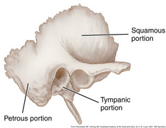
Parts of the temporal bone?
Squamous, petrous.
Tympanic. Zygomatic process, styloid process.
Signs of petrous temporal fracture?
> CSF otorrhea
> Bruising over mastoid process (Battle sign)
Signs of anterior cranial fossa fractures?
> CSF rhinorrhea
> Bruising around eyes ("raccoon eyes")
What is in the cavernous sinus? How are they arranged?
> Internal carotid
> Carotid plexus (sympathetic)
> Abducens VI
In lateral wall:
> Oculomotor III
> Trochlear IV
> Ophthalmic trigeminal V1
> Maxillary trigeminal V2
Lateral wall, superior to inferior - O TOM
Sinus contents, medial to lateral - CAT
Name 3 types of haemorrhage.
Extradural, subdural, subarachnoid
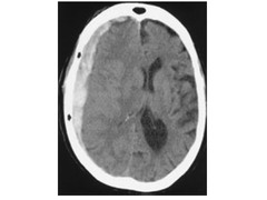
Extradural haemorrhage
Between inner surface of skull and dura mater.
Biconvex in shape, limited by cranial sutures.
Frequently arterial & associated with head trauma (eg. pterion trauma -> middle meningeal artery)
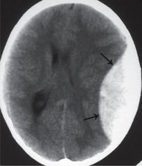
Subdural haemorrhage
Between dura mater and arachnoid mater.
Typically crescent-shaped and not limited by cranial sutures.
Often venous - hard trauma to head can tear superior cerebral veins as they enter superior sagittal sinus. Can actually develop very slowly.
Risk factors: alcohol misuse and old age, as this causes cerebral atrophy & consequential stretching of veins.
Subarachnoid haemorrhage
In the subarachnoid space.
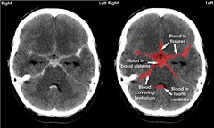
Usually as a rupture of a 'berry' aneurysm at the base of the brain. ~85% are anterior circle of Willis, ~15% vertebrobasilar.
Sudden, very severe "thunderclap" headache. Nausea, vomiting, stiffness.
Can either surgically clip, or endovasular embolisation.