physics 1-2 exam
 Call Kai
 Learn
Learn Practice Test
 Spaced Repetition
 Match
Match Flashcards
 Knowt Play
1/152

There's no tags or description
Looks like no tags are added yet.
Name | Mastery | Learn | Test | Matching | Spaced | Call with Kai | Chat |
|---|
No analytics yet
Send a link to your students to track their progress
153 Terms
ionizing radiation
radiation w/enough energy to free electrons from atoms forming ions, may cause cancer (ex. gamma, X-rays, UV).
energy capable of penetrating matter, AND eject an orbital electron from an atom's shell
natural (background)
Radiation that is contained in the environment:
-cosmic
-gases
-internal
-terrestrial
Man-made (artificial) radiation
ionizing radiation created by humans for various uses
-medical procedures
-nuclear industry
-consumer products
natural radiation and sources
-background radiation
-occur spontaneously in nature
-NOT affected by human nature
sources:
cosmic- sun/stars
gases-radon/thoron (37%)
internal - potassium 40 (5%)
terrestrial- uranium/thorium (3%)
Man-Made Radiation Sources
- computed tomography (24%)
-nuclear medicine (12%)
-interventional fluroscopy (7%)
- conventional radiography/ fluroscopy (5%)
- consumer prodcuts (2%)
-industrial (
Consumer products
-smoke detectors
-clocks/watches
-camera lens
-ceramics
-glass
-fertillzer
-exit signs
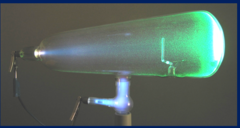
Crookes tube
by William Crookes.
glass tube filled with a small amount of gas. Electrodes (cathode/anode) in the tube allow for the passage of electricity through the gas.
-fluorsced when energized
Dr. Wilhelm Roentgen
Discovered X-rays on November 8, 1895.
"x-light"
x represents the unknown
director of physics institute
Rontgen Experiment
studying the flurescent properties of cathode rays
1.covered cathode ray tube with black cardboard
2. energized cathode ray tube
witnessed nearby piece of cardboard fluoresce (covered with barium platinocyanide)
7 weeks investigation
12 properties of x-rays
1) highly penetrating, invisible
2) electrically neutral, not affected by electric or magnetic fields
3) can be produced over a wide variety of energies and wavelengths (polyenergic/heterogenous)
4) produce secondary/scatter radiation
5) travel at the speed of light, 3x10^8 miles per second
6) travel in straight lines
7) canNOT be focused by a lens
8) cause flourosence (emission of light) of certain crystals
9) can affect photographic film
10) can ionize matter
11) release small amounts of heat when passing through matter
12) produce chemical/biological changes in matter through
Rontgens first radiograph
-first radiograph of body part
bones of his wife's hand (bertha rontgen)
-used barium platinocyanide screen
-recorded on photographic paper
early medical use
inefficient and varied x-ray output
- low kVp
-long exposure times (20-30min) could take 2 hours
indications for early medical x-ray
- Gallstones
- Bullets & Broken Bones
- Kidney Stones
Dangers of Early X-ray
-thought to be harmless
equipment was not enclosed, shielded or grounded
-skin damage, hair loss, anemia
-thought burns were caused by the heat/glow of the cathode ray tube
Thomas Edison
Invented the light bulb/ fluoroscope
-1st to document serious dangers with x-rays
-suffered radiation burns to his face and injury to his left eye during his experiments
Clarence Dally
-Thomas Edison's Assistant.
-1st documented fatailty from x-ray exposure
-death within 8 years
Dr. William Coolidge
-Developed "hot cathode" tube (coolidge tube)
-x-ray intensity/energy set separately
-intensity controlled by milliamperage (mA)
-energy controlled by kilovoltage peak (kVp)
Kilovoltage peak (kVp)
Highest voltage of x-ray tube used during an exposure
QUALITY
Milliamperage (mA)
QUANTITY of exposure
Snook apparatus
portable machine consisting of
-rotary converter
-transformer
-high-tension rectifier
connects with coolidge tube or any ordinary tube
ALARA Principle
Keep radiation exposure as low as reasonably achievable.
the 3 cardinal rules
Time, distance, shielding
time
MINImize the length of time exposed to ionizing radiation for patient, technologist
-length of exposure
-number of times the patient is exposed for a radiologic exam
-time a radiographer spends in a procedure involving fluroscopy
distance
MAXImize the distance from the source of ionzing radiation during an exposure
-inverse sqaure law
Inverse Square Law
the intensity of radiation is inversely proportional to the square of the distance from the source of radiation
-as one increases the distance from an ionzing radiation source, the intensity (quanitity) decreases

Shielding
use of radiopague materials to reduce radiation exposure to
-patient anatmoy that is not important to exam being done
-radiographers during exam
-personnel who are not able to leave exam area
most common material - lead
Protective Apparel
must be worn
must be at least 0.25 Pb
0.5 mm Pb is commonly used
reduction of occupational radiation exposure
will not exceed 1 mSv/yr
-useful beam never diercted toward operating console
-exposure cords on mobile units must be at least 2 meters long
-protective aprons
-structural lead shielding
primary barriers
Any barrier that has the potential of being struck by the primary beam
- lead 1/16 in. of lead (Pb) 7 ft. high
-perpendicular to the line of travel of the primary x-ray beam
-floor beneath the x-ray table
-wall behind upright bucky

Secondary Barriers
only be struck by scatter radiation
parallel to the line of travel or the primary x-ray beam
covers areas only exposed to scattered/leakage radiation
-wall separating the control/exam rooms
-ceiling
1/32 in Pb
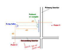
factors that determine protective barrier thickness
-distance
-time of occupancy
-workload
-use (u)- % of time that x-ray beam is energized and aimed
Time Occupancy
amount of time an area is occupied by people
controlled time
-occupied by radiation
-designed to reduce exposure rate to less than 100 mrem/wk
Uncontrolled time
occupied by non-radiation personnel (general public)
-deigned as either full, partial, occasional
reduce exposure less than 10 mrem/wk
workload
radiation level activity in that area
-the more exams performed, the thicker the barrier needed
-accounts for weekly average tube current/tube
measured mA-min/wk
Collimator
Restricts the size and shape of the x-ray beam
Low kVp
high contrast
patient's tissue abosrbs ALL photons and don't reach receptor = WHITE image
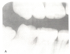
High kVp
low contrast
photons pass through patient's tissue and reach receptor= DARK image
beam energy/ penterating abilty increases

Low mAs
not enough photons to record the body part on the receptor= White image
high mAs
too many photons reach receptor= Dark image
increases x-ray quantity
15% rule
A 15% increase in kVp causes a halving mAs

output intensity intensity in air
-British: Roentgen(R)- Quantify radiation intensity
-SI Metric System: coulomb/kilogram(C/kg)- Measure of the # of electrons liberated by ionization per kilogram of air
SI unit of radiation exposure
radiation absorbed dose (rad)
gray (Gy)
dose equivalent/ effective dose
radiation equivalent man (rem)
sivert (Sv)
acivity
curie (ci)
becquerel (Bq)
Coulomb per kilogram (C/kg)
measure of the # of electrons liberated by ionization per kilogram of air
Roentgen (R)
The amount of radiation that ionizes one cubic centimeter of air
Gray (Gy)
quanitity of radiation energy absorbed by tissues being irradiated
radiaton abosrbed dose (rad)
quantify the biologic effects of radiation on humans and animals
-gives measure to the amount of energy depostied by ionzing radiation
1 Gy = ___ rad
100 rad
1 rad = ___ Gy
0.01 Gy
Sievert (Sv)
addresses the different biologic effects of different types of ionizing radiation to which a radiation worker may be exposed
Radiation Equivalent Man (rem)
standard unit for occupational exposure
1 Sv = ___ rem
100 rem
1 rem= _____ Sv
0.01 Sv
Becquerel (Bq)
quantity of radioactive material
-not the effect of the radiation emitted from it
-is quantifying the # of indivdual atoms decaying per second
Curie (Ci)
traditional unit of radioactivity
kerma
kinetic energy released in matter
describes the quantity of radiation energy delivered to a given point
unit: joules/kg or Gy
air kerma
kinetic energy released per unit mass of air
1 R = air kerma
1 sv = ___ mSv
1000 mSv
1 rem = ___ mSv
10 mSv
1 rem = ___ rad
1 rad
annual dose limits
5 rem (50 mSv)
cumulative dose limit
10 mSv/ 1 rem x age (ex: 25 yo -> 250 mSv)
lens of eye dose limit
150 mSv/yr (15 rem)
skin, hands, feet dose limit
500 mSv/year (50 rem)
continous dose limit
1 mSv (0.1 rem)
2 types of personnel monitors
thermoluminescent (TLD) & optically stimulated luminscence (OSL)
OSL dosimeter
most common
consists aluminum oxide, unaffected by heat, moisture, and pressure
can measure as low as 10 uGya
can be worn for intervals up to 1 year
worn on torso

TLD dosimeter
contains lithium fluoride , has crystals, the more light= the more exposure
can measure exposure as low as 50uGya
can be worn up to 1 year
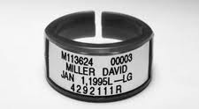
extremity TLDs
worn on fingers of dominate hand
when wearing gloves should be worn under gloves
label should be palm-side of hand
Dosimetry report
Provides radiation readings to occupational radiation workers

SL
minimal dose or dose under 20 mrem
electromagnetic theory
- has no mass
-carries energy in waves
- travels speed of light (186,000 mps or 3x10^8)
Transverse Waves
displacement is PERPENDICUALR to the direction the wave is traveling
MAGENTICE FIELD

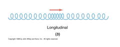
Longitudinal Waves
displacement is PARALLEL to the direction the wave is traveling
ELECTRIC FIELD
origin of EMR
EM waves are emitted when changes in atoms occur
-electrically charged particles which are moving, generate electrical and magnetic fields that are perpendicular to each other
velocity
how FAST the wave is moving; constant
ALL EM ENERGY TRAVEL AT THE SPEED OF LIGHT IN A VACCUM

Amplitude
Height of a wave
TALLER waves carry more magnitude or more mass
intensity of the wave- controlled by mA and quantity

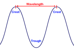
wavelength
length of a wave from one peak to the next peak
INVERSELY PROPORTIONAL TO ENERGY/ FREQUENCY
Frequency
# of cycles per second
DIERCTLY PROPORTIONAL TO ENERGY
INVERSELY PROPORTIONAL TO WAVELENGTH
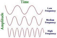
wavelength and frequency relationship
inversely related
wavelength formula
velocity= freq x wavelength
Photon Energy
Directly proportional to frequency
controlled by Kvp
Attenuation
total # of photons
photon energy and frequency will
DECREASE
photon wavelength will
INCREASE
increase Kvp
increase energy, frequency,
decrease wavelength
decrease Kvp
increase wavelength
decrease photon energy, frequency
increase thickness in matter
decrease energy/frequency
increase wavelength
decrease thickness in matter
increase energy/frequency
decrease wavelength
quantum theory
x-ray photon is a discrete bundle of energy
quantum of EM energy
photon energy is direclty proportional to its frequency
E= hf
low to high frequency radiation
radio waves
microwaves
infrared radiation
visible light
ultraviolent
x-rays
gamma rays
radio waves
low energy
long wavelength
MRI
cannot ionize atoms
idenfited by frequncy
Microwaves
shortest wavelengths
highest frequencies
commonly used to transmit cell phone signals and heat food
do not ionize atoms

infrared light
-Sometimes used to "beam" information between electronic devices
-Does not ionize atoms
heat

visible light
represents the colors we see in the world around us
it is a tiny portion of the electromagnetic spectrum
does not ionize atoms
identifed by wavelength

Ultraviolet Light
- Has energies approaching those of x-rays and gamma rays
- Commonly used in tanning beds
- Can be harmful
- Stimulates melanin production in skin cells
- Does not ionize atoms

x rays and gamma rays are similar bc
high frequency
short wavelength
ionizing elec, radiation
Wave-Particle Duality
electrons and light can behave as both a wave and a particle