UNIT 2: Cells
1/211
There's no tags or description
Looks like no tags are added yet.
Name | Mastery | Learn | Test | Matching | Spaced | Call with Kai |
|---|
No analytics yet
Send a link to your students to track their progress
212 Terms
What is a eukaryotic cell?
A eukaryotic cell is a cell with a nucleus and membrane bound organelles.
What is the function of the cell-surface membrane?
The cell surface membrane regulates the movement of substances in and out of the cell.
What is the structure of the cell-surface membrane?
The cell-surface membrane has a phospholipid bilayer with embedded proteins.
It has receptor molecules to respond to chemicals like hormones.
It is selectively permeable.
What is the function of the nucleus?
The nucleus controls the cells activity though transcription on mRNA.
What is the structure of the nucleus?
The nucleus is surrounded by a nuclear envelope (double membrane) with many pores.
The nucleus contains chromosomes, made from protein-bound linear DNA, and one or more nucleoli.
What is the function of the nuclear pores?
The nuclear pores allow substances (e.g. RNA) to move between the nucleus and cytoplasm.
What is the function of DNA?
DNA contains instructions to make proteins.
What is the function of the nucleolus?
The nucleolus makes ribosomes.
What is the function of the mitochondria?
The mitochondria are the sites of aerobic respiration, where ATP is produced.
What is the structure of the mitochondria?
The mitochondria have a double membrane.
The inner membrane is folded to form cristae, and inside is the matrix.
What is the function of cristae?
Cristae provide a large surface area for the attachment of enzymes and other proteins involved in respiration.
What is the function of the matrix in the mitochondria?
The matrix contains enzymes involved in respiration.
What is the function of the chloroplasts?
The chloroplast is the site of photosynthesis in plant and algal cells.
What is the structure of the chlroroplasts?
The chloroplast is flattened and surrounded by a double membrane.
It has membranes inside called thylakoid membranes, which are stacked up to form grana.
The grana are linked together by lamellae.
Explain why the grana is where light absorption takes place.
The grana is where light absorption takes place because the thylakoid membranes contain chlorophyll.
Explain why the stroma is where sugar synthesis takes place.
The stroma is where sugar synthesis takes place because it is a fluid filled matrix with many enzymes and starch grains.
What is the function of the Golgi apparatus?
The Golgi apparatus processes and packages new lipids and proteins.
It also makes lysosomes.
What is the structure of the Golgi apparatus?
The Golgi apparatus has fluid-filled membrane-bound flattened sacks.
There are vesicles on the edges of the sacs.
What is the function of the Golgi vesicle?
The Golgi vesicle stores lipids and proteins made by the Golgi apparatus.
It transports them out of the cell via the cell-surface membrane.
What is the structure of the Golgi vesicle?
The Golgi vesicle is a small fluid-filled sac in the cytoplasm surrounded by a membrane.
What is the function of the lysosomes?
The lysosomes releases hydrolytic enzymes to digest pathogens or break down worn out cell components.
What is the structure of the lysosomes?
The lysosomes is a type of Golgi vesicle and is surrounded by a membrane, to isolate the hydrolytic enzymes.
What is the function of the ribosome?
The ribosome is the site of protein synthesis.
What is the structure of the ribosome?
The ribosome is made of proteins and RNA, and is not membrane-bound.
It has a small subunit and large subunit.
What are the two types of ribosomes and where are they found?
The two types of ribosomes are 80S and 70S.
80S - eukaryotic cells
70S - prokaryotic cells, chloroplasts, mitochondria
What is the function of the rough endoplasmic reticulum?
The rough endoplasmic reticulum folds and processes proteins that have been made at the ribosomes.
What is the structure of the rough endoplasmic reticulum?
The rough endoplasmic reticulum is a system of membranes enclosing a fluid-filled space.
It is covered with ribosomes.
What is the function of the smooth endoplasmic reticulum?
The smooth endoplasmic reticulum synthesises and processes lipids.
What is the structure of the smooth endoplasmic reticulum?
The smooth endoplasmic reticulum is a system of membranes enclosing a fluid-filled space.
It has no ribosomes.
What is the function of the eukaryotic cell wall?
The cell wall is rigid and surrounds cells, supporting them and preventing them from changing shape.
What is the structure of the eukaryotic cell wall?
The celll wall is made of cellulose in plants and algae.
The cell wall is made of chitin in fungi.
What is the function of the cell vacuole?
The cell vacuole helps maintain pressure in the cell and keeps it rigid, preventing wilting.
It isolates unwanted chemicals in the cell.
What is the structure of the cell vacuole?
The cell vacuole contains cell sap, a weak solution of sugar and salts.
It is surrounded by a membrane called the tonoplast.
What happens to eukaryotic cells in complex multicellular organisms?
The eukaryotic cells become specialised for specific functions.
Order the four keywords from smallest to largest:
Tissue, Organ system, Specialised cell, Organ
Specialised cell
Tissue
Organ
Organ system
What is a prokaryotic cell?
A prokaryotic cell is a cell without a nucleus and membrane bound organelles.
True or False: Prokaryotic cells have bigger ribosomes.
False. Prokaryotic cells have smaller ribosomes.
What does a prokaryotic cell have instead of a nucleus?
A prokaryotic cell has a single circular DNA molecule instead.
It is free in the cytoplasm and is not associated with any proteins.
What is the cell wall in a prokaryotic cell made of?
The cell wall in a prokaryotic cell is made of murein, a glycoprotein.
What is a plasmid?
A plasmid is a small loop of DNA, which contains genes that allow the prokaryotic cell to survive in adverse conditions.
It is not part of the main circular DNA molecule.
What is the capsule?
The capsule is a slime layer that surrounds the cell, protecting the bacteria.
It also allows groups to join together, allowing further protection.
What is the flagellum?
The flagellum is a long, hair-like structure that rotates to make the prokaryotic cell move.
What is a virus?
A virus is an acellular and non-living particle.
What is the genetic material of a virus? What is it surrounded by?
The genetic material is nucleic acids, such as DNA and RNA, which are surrounded by protein.
This protein coat is called the capsid.
What are attachment proteins on a virus?
The attachment proteins are on the edge of the capsid, and allow the virus to identify and bind to receptors on a host cell.
How do optical microscopes form an image?
Optical microscopes use light to form an image.
Which organelles can be seen by with an optical microscope? Which ones cannot?
The mitochondria and the nucleus can be seen, but not in perfect detail.
Ribosomes, the endoplasmic reticulum, and lysosomes cannot be seen.
What are limitations of an optical microscope?
Limitations of an optical microscope include a low magnification and low resolution.
What are advantages of an electron microscope?
Advantages of an electron microscope include a higher magnification and a higher resolution.
How do transmission electron microscopes (TEMs) form an image?
TEMs use electromagnets to focus a beam of electrons, which is transmitted through the specimen.
The denser parts of the specimen absorb more electrons, making them look darker.
Evaluate the use of a transmission electron microscope.
TEMs give high resolution images.
However, they can only be used on thin specimens.
How do scanning electron microscopes (SEMs) form an image?
SEMs scan a beam of electrons across the specimen.
This knocks off electrons from the specimen.
The electrons are gathered in a cathode ray tube to form an image of the surface.
Evaluate the use of a scanning electron microscope.
SEMs give a 3D image and can be used on thick specimens.
However, the resolution of the images is lower than those of a TEM.
Describe how to prepare a temporary mount of a specimen on a slide.
Pipette a small drop of water onto the slide.
Use tweezers to place a thin section of the specimen on top of the water drop.
Add a drop of stain to highlight objects in the cell.
Add the cover slip.
Describe the technique to add the coverslip.
When adding the coverslip, stand it upright on the slide, next to the water droplet.
Carefully tilt and lower it onto the specimen, ensuring no air bubbles are trapped.
What is magnification?
Magnification is how much bigger the image is when compared to the object.
What is resolution?
Resolution is the ability to distinguish between two points that are close together.
What is the formula for magnification?
magnification = size of image / size of real object
What is cell fractionation?
Cell fractionation is the process of breaking cells up and separating their organelles.
What are the three steps for cell fractionation?
The three steps for cell fractionation are:
1. Homogenisation
2. Filtration
3. Ultracentrifugation
Describe two ways homogenisation can be done.
Homogenisation can be done by vibrating or blending the cells.
What does homogenisation do?
Homogenisation breaks up the plasma membrane, opening the cell and releasing the organelles into solution.
This produces a homogenate.
What conditions must the solution be under during cell fractionation?
The solution must be ice-cold to slow enzyme activity, preventing the digestion of organelles.
The solution must be buffered to maintain pH so that the enzymes and proteins are not denatured.
The solution must have the same water potential as the tissue to prevent osmosis so there is no lysis or shrinkage of the organelles.
Why is the homogenate filtered during cell fractionation?
The homogenate is filtered to remove debris and complete cells.
What is needed to separate the organelles from the homogenate?
A centrifuge is needed to separate the organelles.
Describe the process of ultracentrifugation to isolate a nucleus.
Pour the cell fragments into a tube.
Place the tube in a centrifuge and spin at a low speed.
Heaviest organelles get flung to the bottom of the tube.
The pellet (thick sediment) forms at the bottom of the tube.
The supernatant stays suspended in fluid above the pellet.
What is done to the supernatant?
The supernatant is drained off and poured into another tube.
This is spun at a higher speed, so the second heaviest organelles form a pellet at the bottom of the tube.
The new supernatant is drained and the process repeats.
True or False: The ribosomes need a low speed during ultracentrifugation.
False. Separating ribosomes requires a high speed.
True or False:
In multicellular organisms, all cells retain the ability to divide.
False.
In multicellular organisms, not all cells retain the ability to divide.
What do eukaryotic cells that retain the ability to divide show?
Eukaryotic cells that retain the ability to divide show a cell cycle.
What is mitosis?
Mitosis is the part of the cell cycle where a eukaryotic cell divides to produce two daughter cells.
What does each daughter cell produced by mitosis have?
Each daughter cell has identical copies of DNA produced by the parent cell during DNA replication.
What is mitosis needed for?
Mitosis is needed for growth of multicellular organisms and for repairing damaged tissues.
Which phase occurs before mitosis?
The interphase occurs before mitosis.
Describe what happens during each phase of the interphase.
Gap Phase 1 - the cell grows, ATP content increases and new organelles and proteins are made
Synthesis Phase - the DNA is unravelled and replicated
Growth Phase 2 - the cell keeps growing and proteins needed for cell division are made
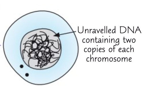
Describe how chromosomes are after the interphase.
After the interphase, the chromosomes are made of two strands joined in the middle by a centromere.
The separate strands are called chromatids.
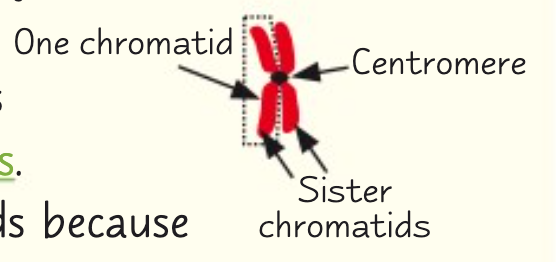
Why are there two strands in each chromosome after the interphase?
There two strands in each chromosome after the interphase because an identical copy has been made.
What are the four phases of mitosis?
The four phases of mitosis are Prophase, Metaphase, Anaphase, Telophase (PMAT)
Penguins Meet After Tea
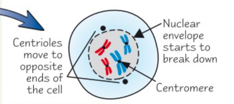
Describe what happens to the cell during the prophase.
During the prophase, the chromosomes condense, getting shorter and fatter.
Centrioles move to opposite ends of the cell, forming the spindle protein fibres.
The nuclear envelope breaks down and chromosomes lie free in the cytoplasm.
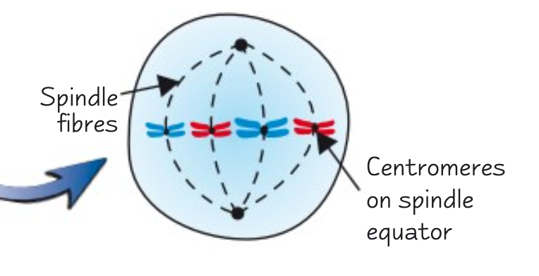
Describe what happens to the cell during the metaphase.
During the metaphase, the chromosomes, with two chromatids, line up along the equator of the cell.
They become attached to the spindle by their centromere.
Describe what happens to the cell during the anaphase.
During the anaphase, the centromeres divide, separating each pair of sister chromatids.
The spindles contract, pulling chromatids to opposite poles of the spindle, from the centromere.
This makes the chromatids appear v-shaped.

Describe what happens to the cell during the telophase.
During the telophase, the chromatids reach the opposite poles on the spindle. They uncoil, becoming long and thin again.
They are now called chromosomes again.
The nuclear envelope forms around each group of chromosomes, forming two nuclei.
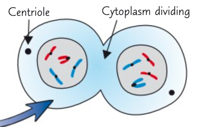
Describe what cytokinesis is.
Cytokinesis is the division of the cytoplasm.
This starts in the anaphase and ends in the telophase.
What do the sister chromatids end up as when mitosis is over?
When mitosis is over, the sister chromatids end up as one-strand chromosomes in the daughter cells.
What is mitosis controlled by?
Mitosis is controlled by genes; the cells stop dividing after they have made enough new cells.
What results from uncontrolled cell division?
Unconttolled cell division can lead to the formation of tumours and cancers.
What do many cancer treatments do?
Many cancer treatments are directed at controlling the rate of cell division.
Describe the process of binary fission in prokaryotic cells.
During binary fission, the circular DNA and plasmids replicate.
The cytoplasm divides to produce two daughter cells, each with a single copy of the circular DNA, and a variable number of copies of plasmids.
Why don't viruses undergo cell division?
Viruses do not undergo cell division because they are non-living and acellular.
How do viruses replicate themselves?
Viruses replicate themselves by injecting their nucleic acid into the infected host cells.
This host cell replicates the virus particles.
True or false:
The basic structure of all cell membranes of eukaryotes is the same.
True.
The basic structure of all cell membranes, including cell-surface membranes and the membranes around the cell organelles of eukaryotes is the same.
Which model is used to describe the arrangement of molecules in the cell-surface membrane?
The fluid mosaic model is used to describe the arrangement of molecules in the cell-surface membrane.
Describe the arrangement of phospholipids in the cell-surface membrane.
In the cell-surface membrane, phospholipids are arranged in a bilayer.
The hydrophilic heads point to the outside of the membrane.
The hydrophobic tails point into the centre of the membrane.
Describe three functions of the phospholipid bilayer in the cell-surface membrane.
In the cell-surface membrane, the phospholipid bilayer allows lipid-soluble substances to enter/leave the cell.
It prevents water-soluble substances from entering/leaving the cell.
It makes the membrane flexible and self-sealing.
Describe what a glycoprotein is and how it is arranged in the cell-surface membrane.
A glycoprotein consists of a carbohydrate chain attatched to the extrinsic proteins.
In the cell-surface membrane, it occurs on the surface of the bilayer, and doesn't extend across it.
Describe three functions of glycoproteins in the cell-surface membrane.
In the cell-surface membrane, the glycoproteins act as receptors for hormones and neurotransmitters.
They help cells to attach to one another and form tissues.
They enable cell recognition.
Describe what a glycolipid is and how it is arranged in the cell-surface membrane.
A glycolipid consists of a carbohydrate bonded with a lipid.
In the cell-surface membrane, the carbohydrate portion extends from the bilayer into the watery environment outside the cell.
Describe three functions of glycolipids in the cell-surface membrane.
In the cell-surface membrane, glycolipids act as receptors for chemicals.
They help cells to attach to one another and form tissues.
They help maintain stability of membrane.
Describe what cholesterol is and how it is arranged in the cell-surface membrane.
Cholesterol is a very hydrophobic lipid which may occur within the bilayer.
Describe three functions of cholesterol in the cell-surface membrane.
In the cell-surface membrane, cholesterol restricts lateral movement of other molecules, including phospholipids.
It makes the membrane less fluid at high temperatures.
It prevents leakage of water and dissolved ions from cell.