Case 9: Terrance K. - Aortic Stenosis + Regurgitation
 Call Kai
 Learn
Learn Practice Test
 Spaced Repetition
 Match
Match Flashcards
 Knowt Play
1/33

There's no tags or description
Looks like no tags are added yet.
Name | Mastery | Learn | Test | Matching | Spaced | Call with Kai | Chat |
|---|
No analytics yet
Send a link to your students to track their progress
34 Terms
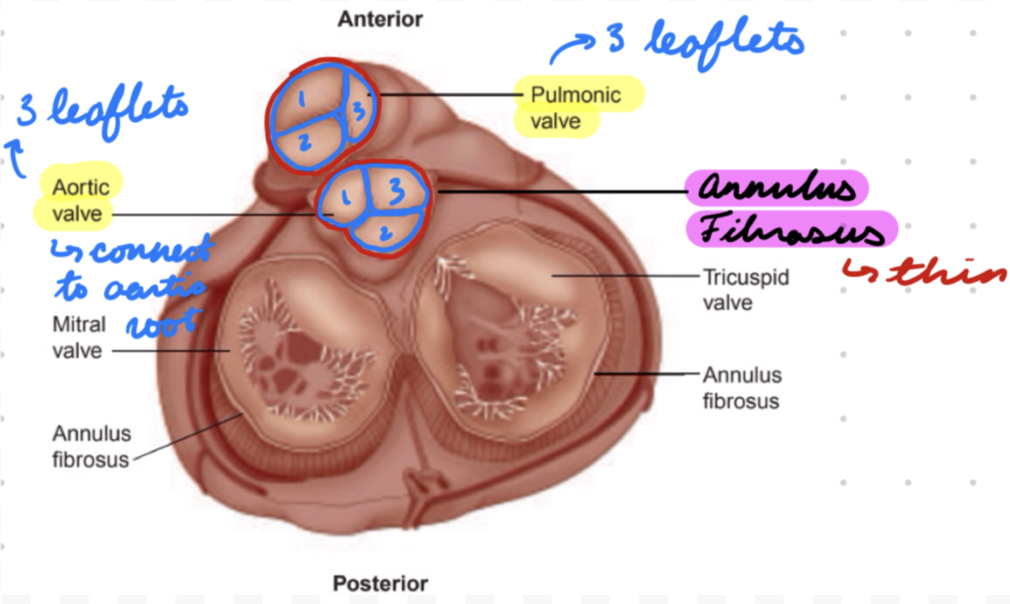
SL Valves Anatomy: Leaflets
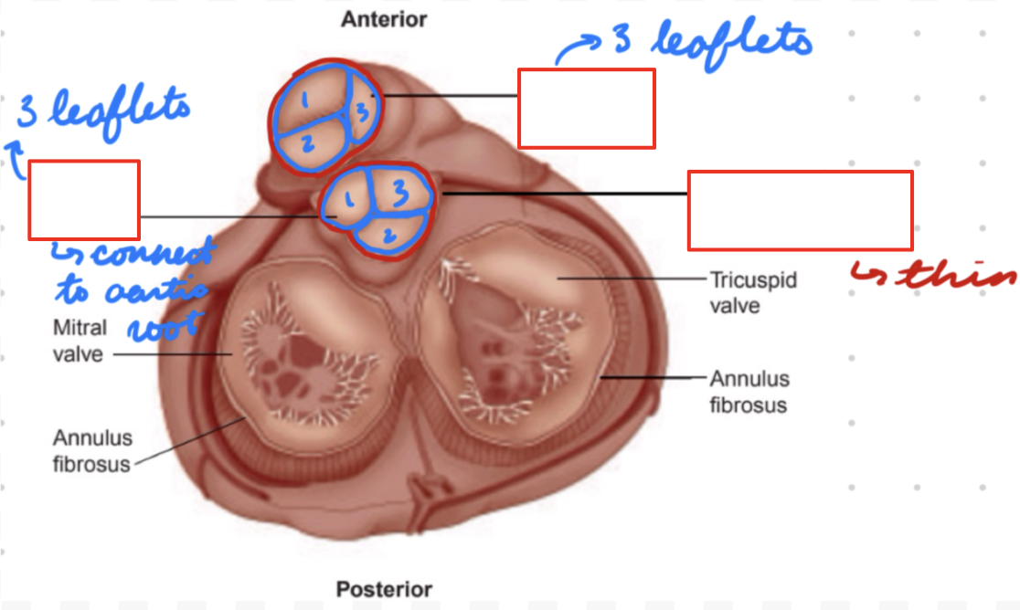
Attach to annulus fibrosus (fibrous ring) connecting to cardiac skeleton (connective tissue)
3 in aortic (L) and pulmonary (R)
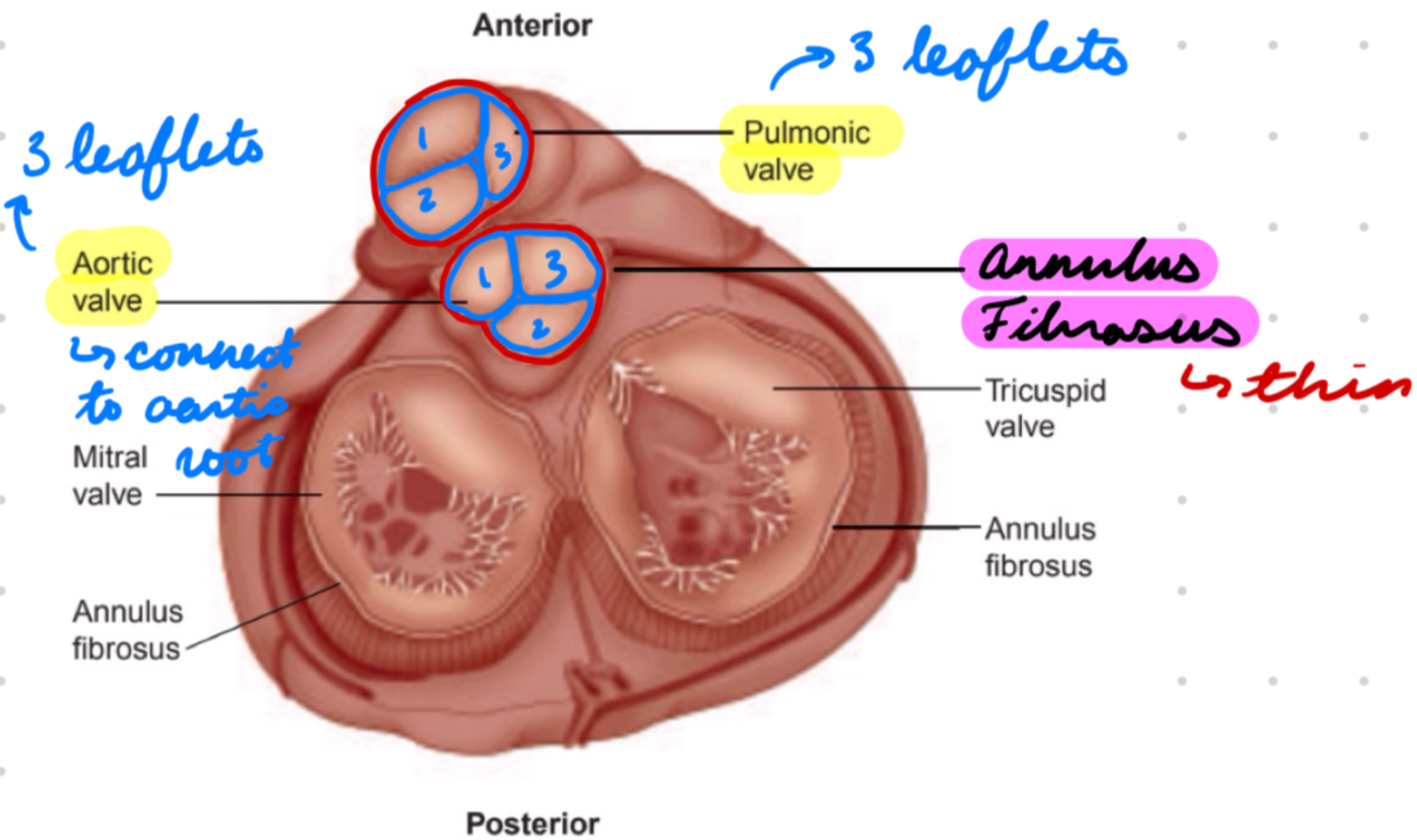
SL Valves Anatomy: Annulus
Thinner than AV
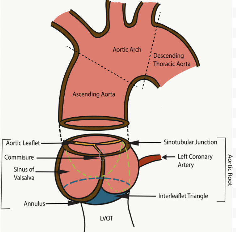
SL Valves Anatomy: Aortic Root
Proximal segment of ascending aorta
Include annulus, sinuses of Valsalva, sinotubular junction
SL Valve Histology: Leaflets
Covered by single endothelial cell layer
Contain valve interstitial cells
3 layers
Fibrosa: Great artery side
Contain: Collagen
Provide tensile strength
Spongiosa: Middle layer
Contain: Proteoglycans
Increase compressibility
Absorb shock
Ventricularis: Ventricle side
Contain: Elastin fibres
For elasticity → Rapid recoil + coaptation
SL Valves Physiology: Leaflets
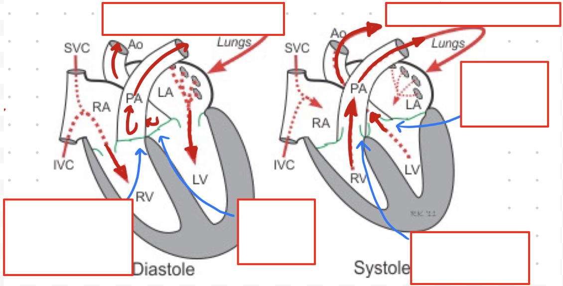
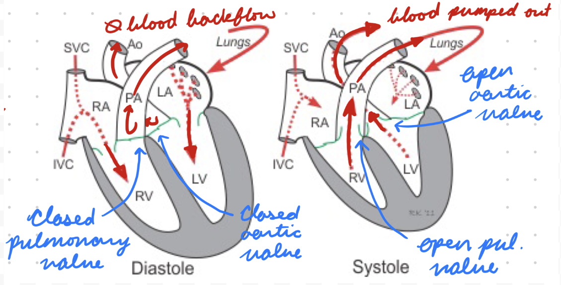
Separate ventricles from great arteries (pulmonary artery and aorta)
Diastole: Closed = Prevent regurg
Great artery elastic recoil = Blood back to heart = Distend leaflets together
Systole: Open = Blood flow through
SL Valves Physiology: Aortic Root
Connect LV and systemic circulation (ascending aorta)
Maintain valve structure for coaptation
Accommodate pressure and volume changes during systole and diastole
For coronary artery perfusion
Aortic Stenosis (AS): Description
Aortic valve narrowing with leaflet thickening and fibro-calcific remodelling
Evolve to obstruction = Restrict CO
AS: Epidemiology
Risk factors:
Older age
Metabolic syndrome
High von Willebrand factor in valve tissues (increase calcification)
Athersclerotic risk factors
Male
Smoking
Hypertension
Hypercholesterolemia
AS: Etiology
Aortic Valve Sclerosis: Leaflet calcification and fibrosis (most common)
Bicuspid Aortic Valve: Leaflet fusion in utero (congenital)
Rheumatic Fever: Common in low-income
AS: Types
Depend on echo findings
Normal:
Aortic valve area (AVA) = 3-4 cm²
Mean transvalvular pressure (MTP) < 5 mmHg
High-Gradient:
AVA ≤ 1 cm²
MTP ≥ 40 mmHg
Low-Flow Low-Gradient with Reduced LVEF:
AVA ≤ 1 cm²
MTP < 40 mmHg
LVEF < 50%
Stroke Volume Index (SVI) ≤ 35 mL/m²
Low-Flow Low-Gradient with Preserved LVEF:
AVA ≤ 1 cm²
MTP < 40 mmHg
LVEF ≥ 50%
SVI < 35 mL/m²
Normal-Flow Low-Gradient with Preserved LVEF:
AVA ≤ 1 cm²
MTP < 40 mmHg
LVEF ≥ 50%
SVI > 35 mL/m²
AS: Pathogenesis
Lipid accumulation, inflammation, calcification (interstitial cells → Osteoblasts) of aortic valve = Stiffening
Fibro-calcific leaflet remodelling over time
Response to injury
Narrow aortic valve orifice = Increase LV load
Increased systolic pressure = LV concentric hypertrophy
Decrease coronary flow = Unable to match O2 demand
AS: Investigations
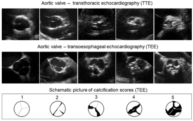
Echo (TTE and transesophageal/TEE)
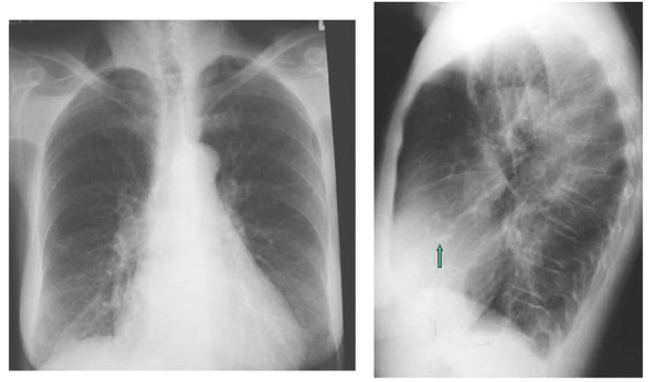
CXR
Cardiac catheterization
AS: Echo
Assess aortic valve structure, function, and stenosis severity
Assess LV
AS: CXR
Determine presence of pulmonary edema
Calcifications in aortic valve = More severe
LV hypertrophy
AS: Cardiac Catheterization
Evaluate aortic valve area, CO, and MAP gradient
AS: Clinical Presentation
Mostly asymptomatic
Dyspnea
Angina
Dizziness and syncope
Physical:
Low BP
Weak and delayed distal pulse
Auscultation:
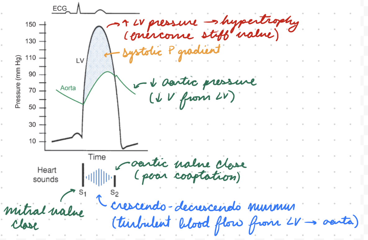
Crescendo-decrescendo late systolic ejection murmur → Bilateral radiation to carotids
Soft S2
S4
AS: Treatment/Mangement
Surgery: Surgical aortic valve replacement (AVR)
Transcatheter AVR
Wedge replacement valve into old valve
Percutaneous balloon valvuloplasty
Inflate balloon in valve = Stretch open stenotic valve
Aortic Regurg (AR): Description
Blood regurg through aortic valve into LV during diastole
AR: Epidemiology
Risk factors:
Older age
Appetite suppressants
AR: Etiology
Acute:
Malcoaptation from leaflet abnormality (infective endocarditis, LV dilation)
Aortic annulus or root abnormality (aortic dissection)
Chronic:
Malcoaptation from leaflet abnormality (calcific aortic valve, bicuspid aortic valve, rheumatic disease)
Aortic annulus or aortic root abnormality (dilation)
AR: Pathogenesis
Aortic valve dysfunction = Regurg from aorta → LV
Chronic AR = LV dilation + eccentric hypertrophy = Compensatory SV increase
AR: Investigations
Echo (TTE)
CXR
AR: Echo
Assess aortic valve structure, function, regurg severity
Abnormal leaflets
Anterior mitral valve leaflets fluttering
Aorta dilation
Visualize LV
Dilation
Hypertrophy
AR: CXR
Assess pulmonary edema
Large aortic root/arch
Chronic: Large cardiac silhouette
AR: Clinical Presentation
Acute:
Sudden severe dyspnea
Pulmonary edema
Chronic:
Palpitations
High pulse pressure (diff between systolic and diastolic)
Water Hammer Pulse: Rapid upstroke then vessel collapse (bounding)
LHF symptoms
Exertional dyspnea
Angina
Orthopnea
Fatigue
Syncope
Auscultation:
S3
High-pitched decrescendo early diastolic murmur
Austin Flint murmur (low-pitched mid-diastolic murmur at apex)
AR: Treatment/Management
Surgery: Aortic valve replacement
Pharmacological: Treat HF symptoms
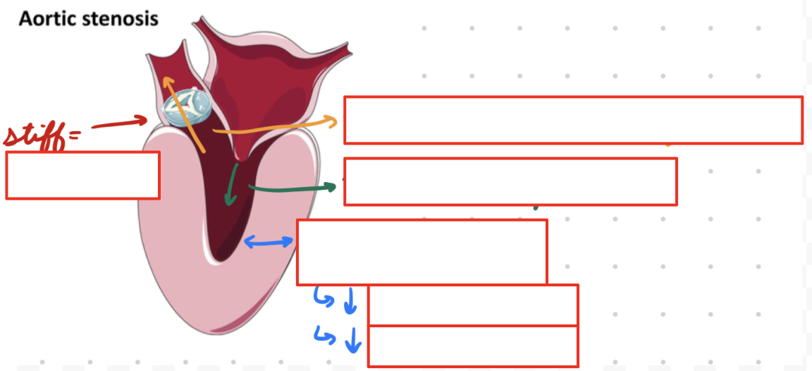
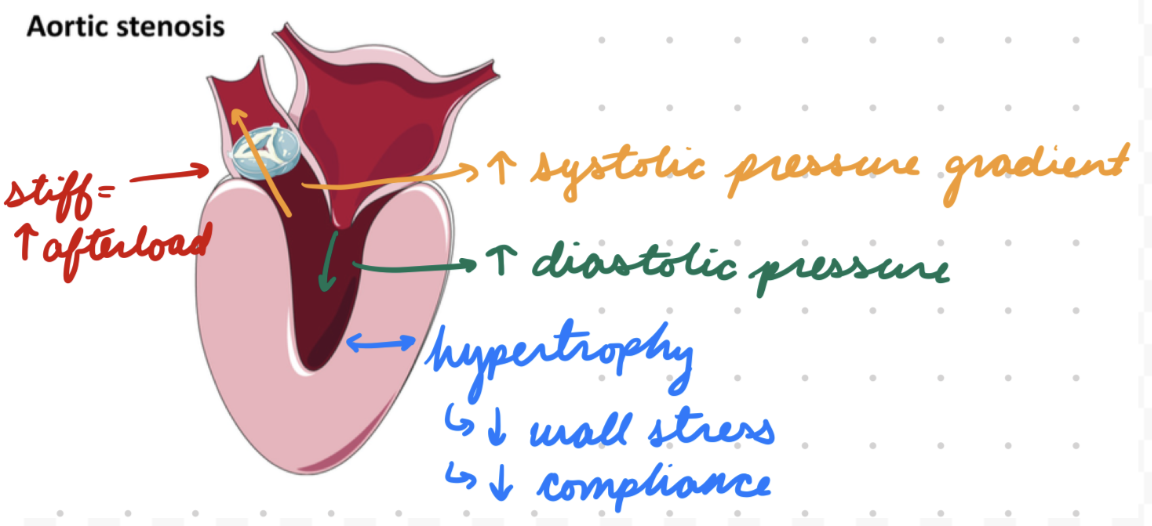
Pressure Changes in AS
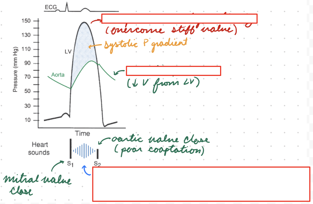
High afterload = Decrease blood flow across aortic valve in systole = Increase LV systolic pressure
Systolic pressure gradient between LV and aorta
LV concentric hypertrophy over time = Decrease wall stress + compliance
Increase LV diastolic pressure = LA hypertrophy (overcome high LV pressure)
Volume Changes in AS
High afterload = Decease blood flow across aortic valve in systole = Increase LV ESV
LV dilation over time = Increase compliance
Accommodate increased blood volume
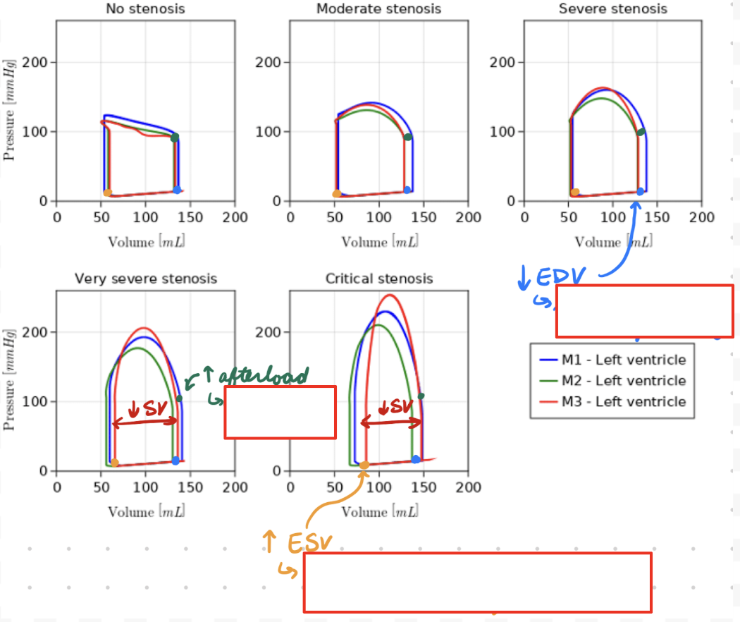
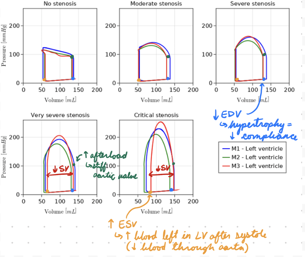
AS: P-V Loop Changes
Increased ESV
Low blood emptying from LV → Aorta
Decreased EDV
Hypertrophy = Decrease compliance (blood entering)
Decreased SV
LV cannot compensate for increased afterload (stiff aortic valve) = Decrease blood outflow
Increase systolic ejection pressure